
 is a fungal infection that affects the nails. OM can involve any component of the nail...)
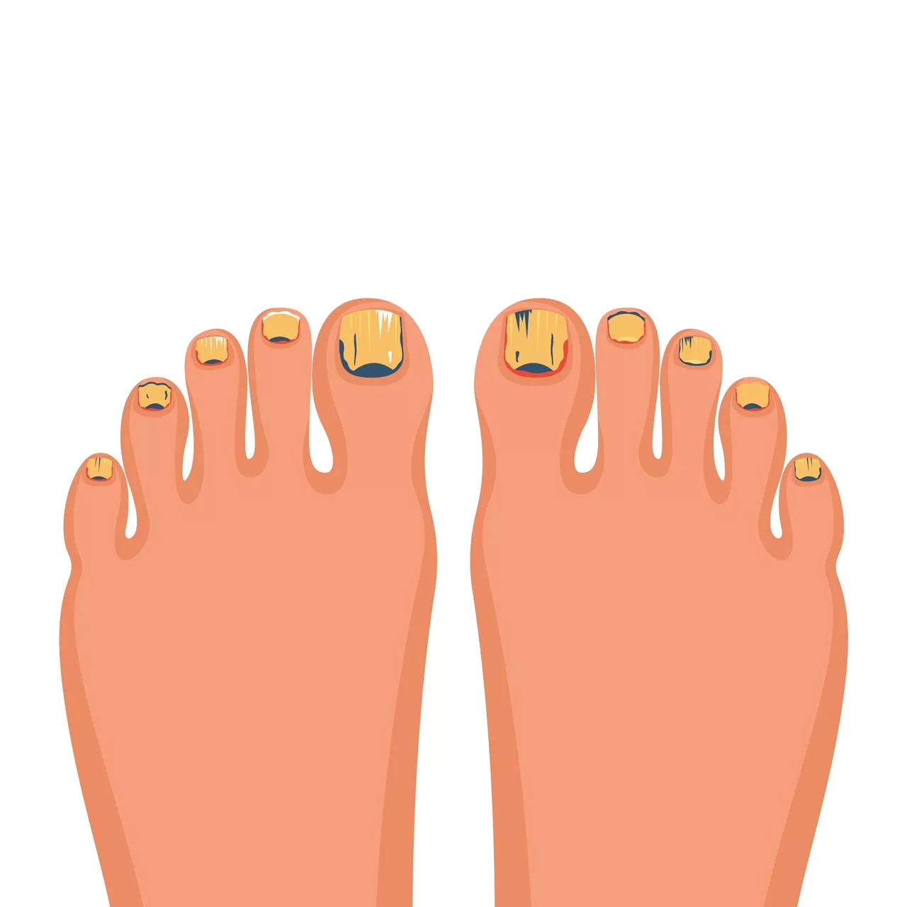
Onychomycosis Symptoms Treatment Onychomycosis (OM) is a fungal infection that affects the nails. OM can involve any component of the nail unit, including the nail matrix, bed, or nail bed of the toe or fingernails. However, OM of the toenail is much more prevalent. OM includes infections caused by dermatophytes (the most frequent group), non-dermatophyte molds, and yeasts; when dermatophytes are the culprit, it is also termed tinea unguium. OM causes nail discoloration, thickening, and dystrophy, and can produce pain. Although the condition is not life-threatening, it may lead to severe physical and occupational limitations.
This common infection is increasing day by day. Prevalence varies worldwide, with estimates ranging from about 1% to 13% in adults, and commonly 1% to 8% in Europe and North America.[1] [2] Notably, around half of abnormal-looking nails are not fungal, making proper diagnosis essential.
Causes of Onychomycosis: Onychomycosis Symptoms Treatment
The most common cause of OM is dermatophytes. Researchers have identified them in 50% of the fingernail OM and 90% of the toenail as Trichophyton rubrum.[3]
- Epiderophyton floccosum
- Trichophyton mentagrophytes
The yeast Candida albicans accounts for 2% of the OM (mainly in fingernails).[4]. Non-dermatophytic mold that is responsible for 8% of the toenail OM includes:
- Aspergillus
- Scytalidium
- Fusarium
- Acremonium
- Scopilariopsis brevicaulis
Pathophysiology of Onychomycosis
The pathophysiology of onychomycosis involves fungal infection of the nail unit, which usually begins after the hyponychial seal of the nail breaks due to microtrauma or a moist environment. This breakage allows fungi to invade the nail bed. Dermatophytes produce keratinases that degrade keratin. Degradation facilitates fungal penetration into the nail bed and nail plate. The infection often starts in the distal or lateral edge of the nail and spreads proximally through several stages. At first, there is a low-grade inflammatory response in the nail bed with nail dystrophy. There can also be onycholysis (separation of the nail plate) and subungual hyperkeratosis. Later, the chronic infection damages the nail matrix. The damage then leads to thickened, distorted, and discolored nails, typical of total dystrophic onychomycosis.[5]
Signs & Symptoms of Onychomycosis
Many patients first notice changes in nail appearance rather than symptoms. Patients with OM can present:
- Cosmetic concerns due to nail discoloration and deformity
- After disease progression, there can be effects on standing, walking, and exercising.
- Pain
- Discomfort
- Loss of dexterity in fingernail disease
- Paresthesia
- Loss of self-esteem
- Reduced self-esteem and social withdrawal
Patients may have a combination of these subtypes. Total dystrophic OM, which is the most advanced form of OM, presents as yellow-brown, thickened, and opaque nails. The symptoms can vary according to the five subtypes of the OM.
The Clinical Subtypes of OM
The subtypes of OM are:
Distal Lateral Subungual Onychomycosis (DLSO):
DLSO spreads from the plantar skin via the hyponychium (the thickened layer of skin located under the free edge of the nail). Features of DLSO are:
- Subungual hyperkeratosis (the buildup of skin cells and a chalky white substance under the nail bed) and onycholysis
- Yellow streaks on nails
- Thickened, brittle nail plate

Distal and lateral subungual onychomycosis (DLSO): Whitish discoloration, onycholysis, and subungual hyperkeratosis. Image Courtesy: Onychomycosis: A Review by Pirracini et al. 2015,doi.org/10.3390/jof1010030, available via https://www.mdpi.com/2309-608X/1/1/30, CC BY 4.0.
White Superficial Onychomycosis (WSO):
WSO is confined to the toenails. The features of WSO are:
- There can be small, speckled, white, or powdery patches on the surface of the nail plate of the patients.
- The nails of the patient become crumbly or rough.
- Deeper invasion in cases caused by molds.[6]

White superficial onychomycosis (WSO): white opaque friable patches of the nail plate. Image Courtesy: Onychomycosis: A Review by Pirracini et al. 2015,doi.org/10.3390/jof1010030, available via https://www.mdpi.com/2309-608X/1/1/30, CC BY 4.0.
Proximal Subungual Onychomycosis (PSO):
As the name indicates, PSO invades the proximal nail fold. The features include:
- Area of leukonychia (white discoloration of the nails, appearing as spots, lines on some part or entire nails).
- Marked periungual inflammation (especially in OM caused by molds).[7]
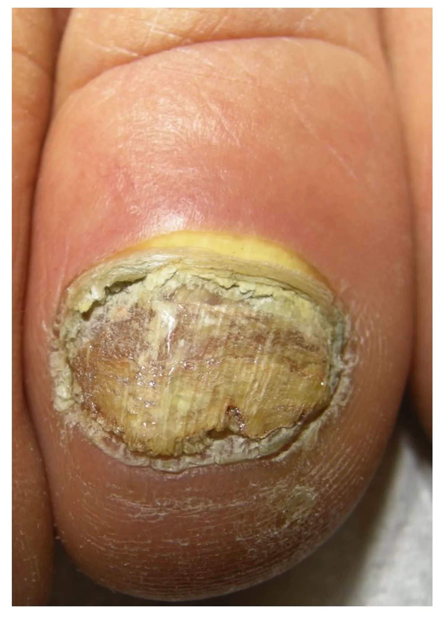
Proximal subungual onychomycosis (PSO): white discoloration of the proximal nail plate. Image Courtesy: Onychomycosis: A Review by Pirracini et al. 2015,doi.org/10.3390/jof1010030, available via https://www.mdpi.com/2309-608X/1/1/30, CC BY 4.0.
Endonyx Onychomycosis (EO):
EO is an unusual variant of OM that involves the inner surface of the nail plate. Features include:
- White or milky discoloration of the nail plate.
- No subungual hyperkeratosis or onycholysis.

Endonyx onychomycosis: white discoloration of the nail plate that is firmly attached to the nail bed. Image Courtesy: Onychomycosis: A Review by Pirracini et al. 2015,doi.org/10.3390/jof1010030, available via https://www.mdpi.com/2309-608X/1/1/30, CC BY 4.0.
Candidal Onychomycosis:
Candida infections are less common. They can usually be secondary to the altered immunity as developed in patients with chronic mucocutaneous candidiasis or immunodepression. Features of Candida OM are as follows:
- Total nail involvement with periungual inflammation
- Digits often take on a drumstick or bulbous appearance
- Affects several or all the digits
Diagnosis of Onychomycosis
Doctors can establish the diagnosis by following this procedure:
History & Physical Examination:
Onychomycosis (OM) is often asymptomatic, and many patients first present for cosmetic concerns rather than physical discomfort. On examination, clinicians look for nail discoloration, thickening, onycholysis, and brittleness. However, clinical appearance alone is not always reliable, since nail disorders such as psoriasis, lichen planus, or traumatic nail dystrophy can mimic fungal infection. A thorough medical and occupational history helps identify predisposing factors such as repeated moisture exposure, occlusive footwear, or immunosuppression. While visual inspection can strongly suggest OM, confirmatory testing is usually required before treatment..[8]
Laboratory Tests:
The clinical features of OM can mimic various other nail disorders, so laboratory diagnosis is a must before initiating treatment.
Culture
Fungal culture remains the gold standard for species identification, although its sensitivity is limited and results can take weeks. A negative culture does not fully exclude OM, but a positive result provides definitive evidence.
Direct Microscopy
The nails are cleaned and clipped with an alcohol swab to remove debris and bacteria. A 20% potassium hydroxide (KOH) preparation in dimethyl sulfoxide (DMSO) is useful for ruling out the presence of fungi. The preparation does not require heating or prolonged incubation.
- In DLSO, the healthcare provider obtains a specimen from the nail bed by curettage. They remove the onycholytic nail plate and obtain the sample at the site most proximal to the cuticle.
- In the case of PSO, they take a sample from the ventral nail plate.
- In WSO, they use a No. 15 blade to remove the specimen from the nail surface.
- In suspected candida OM, the examiner takes the sample from the affected nail bed closest to the lateral and proximal edges.
The examiner can crush the larger pieces of nail plates using a nail micronizer, as the nail fragments must be small enough for examination under the low power.
Other Tests:
- Polymerase chain reaction (PCR) detects fungal DNA from infected nails. There is a highly nested PCR assay with species-specific primer pairs based on the 28S ribosomal RNA gene that permits detection of both dermatophytes and non-dermatophytes.
- Dermoscopy can be used as a complement to culture and PCR assays. It can be a means of screening for laboratory sampling.[9]
- Histopathology (PAS staining): Nail biopsy with PAS staining highlights fungal elements within keratin. While it confirms infection, it does not determine the fungal species.[10]
Management & Treatment of Onychomycosis
Management and treatment of OM depend on the clinical subtype, severity of the condition, and number of affected nails. Distal or superficial infections may respond to topical therapy, while more extensive involvement (including the lunula or nail matrix) usually requires systemic treatment. Combination therapy often improves cure rates, though recurrence remains common.
Oral Therapy:
You need to take prescribed oral antifungal medications for the treatment. The oral antifungal options include:
- Terbinafine (first-line, fungicidal against dermatophytes)
- Itraconazole (broad-spectrum, including non-dermatophytes and yeasts)
- Fluconazole (used off-label, often in cases resistant to first-line drugs)[11]
You need to take these medicines for several months on a daily basis. However, oral antifungals are not for everyone as they can affect your liver and interact with other medications. Your healthcare provider can do blood tests to check for the potential side effects of the medicines.
Topical Medication for OM:
You can regularly apply topical agents to your nail, and it can treat the fungus over time. Topical treatment alone generally can not cure OM because of its insufficient nail penetration. Hence, topical medications are the most effective when paired with oral therapy.
- The topical options include Amorolfin and cicloprox. They can penetrate deep through the nail layers, but may have low efficacy when used alone. Daily application for a longer duration can give better results.
- Tavaborole is an effective topical antifungal for treating OM.
- Other topical options include Efinaconazole, which is very effective in treating yellow streaks and dermatophytoma, which are usually resistant to oral antifungals.
- The healthcare providers can also combine laser treatment with topical agents.
Laser Treatment:
Laser and photodynamic therapies use heat or light to temporarily improve nail clarity. However, evidence for long-term cure is limited, and they are not considered first-line treatments. They may be used as adjuncts with topical medications.
Surgical Care:
Surgical approaches to OM treatment include mechanical, chemical, or surgical nail avulsion. Removal of the nail plate is an adjunctive treatment in patients with oral therapy. Chemical removal using the urea compound is a painless method for thick nails. A combination of oral, topical, and surgical therapy can increase the efficacy of the treatment.
Prognosis and Recurrence
Onychomycosis is difficult to cure completely, and recurrence is common even after successful treatment. Reported cure rates vary depending on the drug, treatment duration, and patient compliance, but long-term relapse can occur in up to 20–50% of cases. Reinfection is especially likely in individuals with diabetes, peripheral vascular disease, immunosuppression, or those frequently exposed to moist environments. The prognosis of OM varies depending on several factors. These factors include:
- Type and severity of the infection
- Status of your immune system
- Underlying health conditions
- Response to the treatment
- Compliance with the provided treatment[12]
Differential Diagnosis
Some similar nail pathologies can mimic the OM. The differential diagnosis of OM includes:
- Drug-induced nail dystrophy
- Lichen planus
- Chronic paronychia
- Yellow nail syndrome
- Nail malignancy
- Hypothyroidism-related nail changes
- Contact dermatitis
- Psoriatic nails
Complications
OM is not a severe condition and is not fatal. However, some of the complications observed in the patients include:
- Recurrent cellulitis (especially in diabetics)
- Secondary bacterial infections
- Pain and tissue damage
- Rarely, osteomyelitis
- Permanent nail loss or dystrophy
Onychomycosis versus Onychodystrophy
OM and onychodystrophy are related conditions, but are distinct nail conditions, and their key differences are given in Table 1.
Table 1: Key differences between Onychomycosis versus Onychodystrophy
| Featues | Onychomyosis | Onychodystrophy |
|---|---|---|
| Definition | Nail dystrophy caused by fungal infection. | Nail dystrophy from various nonfungal causes |
| Causes | Dermatophytes Yeasts Molds | Brittle, thickened, discolored, and deformed nails. |
| Symptoms | Clinical exam, Biopsy Lab tests | Thickened, discolored, onycholysis, and subungual debris. |
| Diagnosis | KOH, culture, PAS, PCR | Clinical exam, Biopsy, Lab tests |
| Treatment | Depends on the cause. | Antifungal medications |
A Quick Review
OM is a common infection that can be difficult to treat. It is not usually painful. However, it can make you feel self-conscious about your feet. Talk to your healthcare provider if it bothers you. A trained specialist can provide your guidance while protecting your overall health.
References
[1] Bodman, M. A., & Krishnamurthy, K. (2017). Onychomycosis.
[2] Jaworek, A. K., Hałubiec, P., Wojas-Pelc, A., & Szepietowski, J. C. (2025). Analysis of Causative Factors and Potential Predictors of Onychomycosis: A Retrospective Single-Center Study in Poland.Journal of Fungi,11(2), 131. https://doi.org/10.3390/jof11020131
[3] Appelt, L., Nenoff, P., Uhrlaß, S., Krüger, C., Kühn, P., Eichhorn, K., … & Bauer, A. (2021). Terbinafin-resistente Dermatophytosen und Onychomykose durch Trichophyton rubrum.Der Hautarzt,72(10), 868-877. Other dermatophytes include:
[4] Bodman, M. A., & Krishnamurthy, K. (2017). Onychomycosis
[5] Zaikovska, O., Pilmane, M., & Kisis, J. (2014). Morphopathological aspects of healthy nails and nails affected by onychomycosis.Mycoses,57(9), 531-536.
[6] Piraccini, B. M., & Tosti, A. (2004). White superficial onychomycosis: epidemiological, clinical, and pathological study of 79 patients.Archives of dermatology,140(6), 696-701.
[7] Mehta, M., Sharma, J., & Bhardwaj, S. B. (2020). Proximal subungual onychomycosis of digitus minimus due to Aspergillus brasiliensis.Pan African Medical Journal,35(1).
[8] Gupta, A. K., & Studholme, C. (2016). Novel investigational therapies for onychomycosis: an update.Expert opinion on investigational drugs,25(3), 297-305.
[9] Litaiem, N., Mnif, E., & Zeglaoui, F. (2023). Dermoscopy of onychomycosis: a systematic review.Dermatology Practical & Conceptual,13(1), e2023072.
[10] Nikitha, S., Kondraganti, N., Kandi, V., & Kondraganti IV, N. (2022). Total dystrophic onychomycosis of all the nails caused by non-dermatophyte fungal species: a case report.Cureus,14(9).
[11] Aggarwal, R., Targhotra, M., Kumar, B., Sahoo, P. K., & Chauhan, M. K. (2020). Treatment and management strategies of onychomycosis.Journal de mycologie medicale,30(2), 100949.
[12] Lipner, S. R., & Scher, R. K. (2019). Onychomycosis: treatment and prevention of recurrence.Journal of the American Academy of Dermatology,80(4), 853-867.