


Spina bifida Prevention Spina bifida is a congenital defect of the spine and spinal cord that occurs when the neural tube does not close properly during fetal development. The name literally means “split spine,” referring to the incomplete closure of the backbone and membranes around the spinal cord. Depending on the location and severity of the spinal opening, this condition may lead to physical disability to a certain degree.[1]
What is Spina Bifida?: Spina bifida Prevention
The human spinal cord is a complicated structure, and it develops at an early stage of pregnancy. In the first month of pregnancy, a part of the embryo known as the neural tube slowly folds to create the brain and the spinal cord. The neural tube fails to close completely in the case of patients with spina bifida, which causes an opening in the spinal column and tissues surrounding it. This incomplete sealing may occur in any section of the spine, but most of the time it occurs in the lower back.Neural tube defects. Annual Review of Neuroscience, 37, 221–242.” style=”position:relative;color:#309b65;cursor:help;border-bottom:1px dotted #309b65;font-weight:bold”>[2]
The nerves and the spinal cord that go through it control the movements and sensations in the body. When a spine may not close properly, both the spinal cord and nerves may be damaged or fail to develop properly, and a range of physical and neurological complications may occur. The condition is usually permanent, but with different treatments and interventions, spina bifida and its symptoms can be managed, which improves the quality of life.[3]
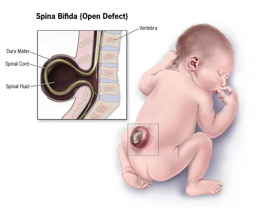
Illustration of open spina bifida (myelomeningocele) in an infant. Credit / License: Public domain (U.S. federal government, CDC / HHS)
Global Incidence of Spina Bifida
The incidence of folic acid deficiency in the global population varies depending on the region, history, and the status of folic acid fortification. The neural tube defects, such as spina bifida, are usually high, at 1–3 cases per 1,000 live births. This rate is lowered in those countries where the folic acid fortification programs have been put in place.[4]
Folic acid fortification reduced the rate by 5.04 to 0.77 per 10,000 live births in the United States. Conversely, there are still higher rates in parts of Africa and South Asia where the supplementation programs are still not stable.[5]
What Causes Spina Bifida?
The precise cause of spina bifida is not clearly known, but studies have shown that it is due to a combination of both genetic and environmental factors. Contrary to genetic disorders, which are inherited, spina bifida is not normally inherited through conventional patterns. Rather, it is a complicated interplay between several genes and environmental factors.
Folic Acid Deficiency:
Poor intake of folic acid during pregnancy, especially in the first trimester, is one of the greatest identified risk factors. Folic acid is a B vitamin, which is necessary in DNA synthesis and cell division, and is important in adequate neural tube development.[6] Various studies have shown that women who intake inadequate levels of folic acid before and during the initial pregnancy period are at a considerably elevated risk of giving birth to a child with spina bifida.[7]
Environmental Factors:
- Maternal diabetes, obesity, and the use of some drugs like anticonvulsants or retinoids are other environmental risk factors.
- Some studies indicate that maternal fever in early pregnancy could also be a risk factor.
- Moreover, some populations have a greater prevalence than others, like Hispanic and Native American populations.[8]
Genetic Factors:
There are also genetic influences affecting the condition. In case of spina bifida in a parent or a sibling, there is a high risk of occurrence in subsequent pregnancies. Nevertheless, spina bifida is not seen in all pregnancies in families with a positive history.[9]
Types of Spina Bifida
Spina bifida has a number of various manifestations, each having different clinical images and the degree of the disease. It is important to understand the various types to understand the variety of consequences that come as a result of this condition.
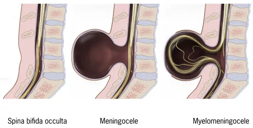
Types of spina bifida: occulta, meningocele, myelomeningocele.” Credit / License: Public domain (U.S. federal government, CDC / HHS)
Myelomeningocele:
Myelomeningocele is the most severe form of spina bifida, affecting approximately 80 percent of cases. In this type, both the meninges (membranes that cover the spine) as well as the spine itself come out of the hole in the spine, and a sac is formed on the surface of the skin. Almost all cases of myelomeningocele cause some form of paralysis and neurological impairment below the lesion, and loss of bladder and bowel control. This condition is life-threatening and needs early intervention.[10]
Meningocele:
Meningocele is a less severe form that takes place in about 10 to 15 percent of cases. In the meningocele, the meninges are only thrust out of the opening of the spine, whilst the spinal cord is in its normal position. Due to the lack of involvement of the spinal cord, patients with meningocele do not have serious neurological problems as compared to myelomeningocele, yet the patient may suffer some nerve damage.
Spina Bifida Occulta:
The least serious condition is Spina Bifida Occulta, where one or more of the vertebrae fail to be fully closed without any protrusion of the spinal cord or the meninges. Interestingly, the word “occulta” is derived from hidden because more often there is no outward manifestation of the condition. Most cases of spina bifida occulta can be asymptomatic. They may only be detected by imaging tests when investigating for some other condition. However, other people can have some mild neurological symptoms, including lower back pain, foot weakness, or problems with the bladder.[11]
Recognizing Spina Bifida: Signs & Symptoms
Spina bifida has a wide range of symptoms based on the nature and severity of the disorder. In spina bifida occulta, many cases do not manifest symptoms in life. But in more serious forms, the symptoms may be very strong and have a great effect on normal functioning.
Spina Bifida Symptoms:
The signs and symptoms of spina bifida include the following:[12] [13]
Physical & Neurological Symptoms of Spina Bifida
- Visible opening or dimple on the spine or lower back region. In myelomeningocele, you can see a fluid-filled sac on the back, which shows the herniated spinal cord and meninges.
- Weakness or paralysis of the legs and feet, the severity depends on the site and area of the spinal defect. Those patients who have lesions of higher levels usually have more serious paralysis.
- Bladder and bowel control are affected, leading to the inability to remove urine and feces. This happens in myelomeningocele.
- Paresthesia or numbness of the lower extremities. Individuals may not feel pain, temperature, or other sensations in affected areas.
Secondary Effects & Associated Conditions
- Accumulation of cerebrospinal fluid around the brain, a condition called hydrocephalus. This occurs in approximately 80 percent of individuals with myelomeningocele and requires surgical intervention to prevent brain damage.
- Clubfoot, or orthopedic deformity, e.g., dislocation of the hip or stooped spine. These abnormalities usually happen secondary to muscle weakness and imbalance.
- A tethered spinal cord is a condition in which the spinal cord is attached to other surrounding tissue, leading to the progressive deterioration of the nervous system.
- Intellectual disability is avoidable, though it harms learning and cognitive development. Some spina bifida patients have normal or near-normal intelligence.
- Chiari malformation is a condition in which the brain tissue extends into the spinal canal, and this could cause neurological complications.
How is Spina Bifida Diagnosed?
Diagnosis of Spina bifida in the prenatal period, before birth or after birth, depends on the severity and manifestation of the condition.
Prenatal Diagnosis:
Prenatal screening usually starts with the measurement of the maternal serum alpha-fetoprotein (AFP) levels in the second trimester. The high levels of AFP may reflect neural tube defects and should be investigated. The imaging technique used in prenatal diagnosis is mostly ultrasound, which shows the abnormalities of the spine, fluid buildup, or any other typical appearances, like the head shape changes, a characteristic of spina bifida.[14]
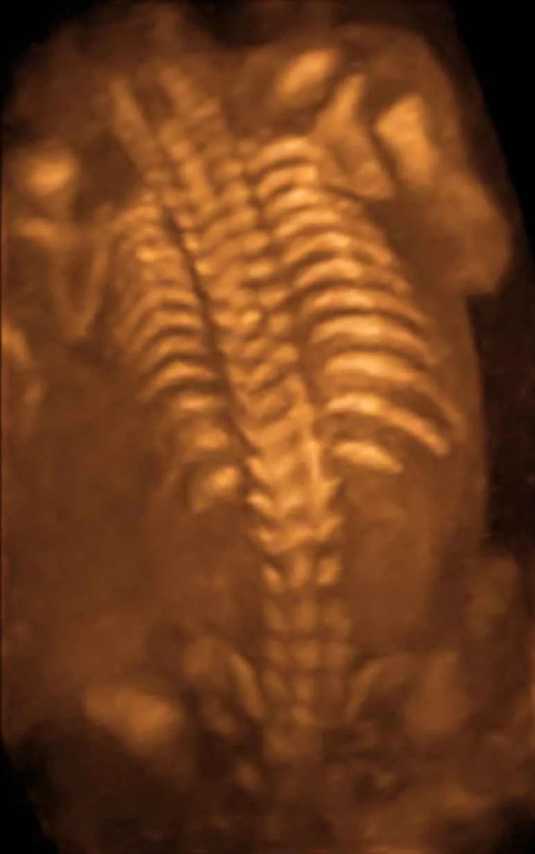
3D sonogram of fetal spine (approximately 20 weeks of gestation)”. Credit / License: Wolfgang Moroder, CC BY-SA 3.0 (via Wikimedia Commons)
In case of uncertainty in the ultrasound results, use advanced types of imaging, including magnetic resonance imaging (MRI), to provide an in-depth evaluation of the nature and position of the defect.
Postnatal Diagnosis:
Following birth, diagnosis can be evident in case the infant has an apparent hole or bulge on the back of the body. Physical examination enables the medical personnel to determine the neurological functioning, such as the reflexes, muscle strength, and sensation. The imaging studies (ultrasound, CT scan, or MRI) identify the extent of spinal cord involvement.[15]
In a few instances, laboratory tests (such as genetic testing) determine related genetic abnormalities or to give prognostic data.
Understanding Spina Bifida Treatment Options
The treatment of spina bifida involves a multidisciplinary team of experts, such as pediatricians, neurosurgeons, orthopedic surgeons, urologists, and rehabilitation experts. Therapeutic interventions are dependent on the form and the extent of spina bifida.
Surgical Management:
In myelomeningocele, surgeons usually close the spinal defect surgically within 24–72 hours of birth to prevent further destruction of exposed nerve tissue and reduce the risk of infection. The surgeon seals the opening in the spine with care, covering the spinal cord and the meninges with the layers of skin and tissue.
Hydrocephalus is treated with a ventriculoperitoneal (VP) shunt. They insert a tube into the brain to drain the excess cerebrospinal fluid into the abdomen, where the body absorbs it. Placing the shunts helps avoid the elevation of intracranial pressure and brain injuries.
The orthopedic surgery can treat structural abnormalities, like clubfoot, hip dislocation, or spine curvature that is secondary to muscle weakness and imbalance.[16]
Medical Management:
Bowel management and bladder management are essential for spina bifida. Clean intermittent catheterization enables normal bladder emptying and prevents urinary tract infections and renal damage. Medication, diet, and bowel training programs control bowel functioning.[17]
Depending on the degree of lower extremity dysfunction, doctors prescribe assistive devices, such as braces, crutches, wheelchairs, or walkers, to improve mobility and autonomy.
Occupational therapy and physical therapy are critical in maximizing independence and mobility. Therapy exercises enhance the muscle groups and teach coping skills in performing daily tasks.
Ongoing Management:
Regular medical follow-ups check complications such as a tethered spinal cord, formation of a syrinx, or progressive neurological loss. To monitor these complications, imaging studies can be carried out on a regular basis.
Complications Associated with Spina Bifida
Patients with spina bifida are prone to a lot of complications that need regular medical care and treatment.[18]
- Hydrocephalus
- A tethered spinal cord
- Syrinx formation (fluid-filled cavity within the spinal cord)
- Thelatex allergy
- Urinary tract infections and damage to the kidneys
- Skin breakdown and pressure ulcers
- Orthopedic complications such as scoliosis, contractures, and joint deformities develop progressively because of muscle weakness and imbalance.[19]
Prevention of Spina Bifida:
Prevention of spina bifida is mostly approached by the consumption of folic acid during early and prenatal pregnancy. Folic acid aids in the synthesis of DNA and the formation of the neural tube within the first month of the gestation period. Women who intend to conceive are supposed to take folic acid at least a month before conception and continue through the first trimester.[20]
The World Health Organization (WHO) and Centers for Disease Control and Prevention (CDC) suggest 400 micrograms (4 mg) of folic acid daily as a dose to women of reproductive age. The higher-risk group (e.g., with a history of pregnancy but neural tube defects or under anticonvulsant therapy) is recommended a higher dose, 4 mg/day, with medical supervision.[21]
Compulsory fortification of grains and flour with folic acid has shown a remarkable decline in the incidence of spina bifida in several countries, proving that this is a cheap and safe preventive measure.
Prognosis & Long-Term Outcomes
The outlook of patients with spina bifida has significantly improved in the last few decades. The reason for this success is the improvement in surgical procedures, medical care, and other rehabilitation services. Nonetheless, the results may be different according to the nature and degree of the condition.[22]
The prognosis of persons with spina bifida occulta is often good with little or no functional impairments. A significant number of individuals with this variant do not have any symptoms or know about the disease by chance.
Meningocele patients usually have a good outcome when managed properly, although some may still experience neurological involvement.
The prognosis for myelomeningocele is variable. Some people become severely disabled, but a good number of them manage great independence through proper interventions. The survival rates have drastically increased, and life expectancy has come close to the general population for those with proper medical treatment.[23]
Factors affecting prognosis are:
- the location and extent of the spinal defect,
- the existence and the extent of related abnormalities like hydrocephalus
- availability of full medical and rehabilitative services.
Early intervention and continued multidisciplinary care have a great impact on quality of life and outcomes.
Conclusion
Spina bifida is a complicated birth defect that has different manifestations and consequences based on the severity and type. Doctors cannot permanently cure the condition, but they can use modern medical care, surgery, and rehabilitation to manage it effectively. These treatments help most people with spina bifida achieve a good level of independence and quality of life. Early diagnosis and, when necessary, proper surgical intervention, full medical care, and follow-up rehabilitative care are crucial to the maximization of outcomes. In case of a suspected case of spina bifida or a problem with the spine that requires a proper diagnosis and management plan, then it would be important to seek the services of qualified healthcare professionals.[24]
References
[1] Mitchell, L. E., & Adzick, N. S. (2021).Epidemiology and pathogenesis of spina bifida. The New England Journal of Medicine, 384(24), 2280–2292.
[2] Greene, N. D. E., & Copp, A. J. (2014).Neural tube defects. Annual Review of Neuroscience, 37, 221–242.
[3] Sadler, T. W. (2019).Langman’s Medical Embryology(14th ed.). Wolters Kluwer.
[4] Kancherla, V., Wapner, R., & Oakley, G. P. (2023). Global burden of neural tube defects and the impact of folic acid fortification: A systematic review.Birth Defects Research, 115(7), 543–558.
[5] Zaganjor, I., Sekkarie, A., Tsang, B. L., Williams, J., Razzaghi, H., Mulinare, J., & Rosenthal, J. (2016). Describing the prevalence of neural tube defects worldwide: A systematic literature review.MMWR Morbidity and Mortality Weekly Report, 65(1), 1–6.
[6] Czeizel, A. E., & Dudas, I. (1992).Prevention of the first occurrence of neural-tube defects by periconceptional vitamin supplementation. The New England Journal of Medicine, 327(26), 1832–1835.
[7] Blom, H. J., Shaw, G. M., den Heijer, M., & Finnell, R. H. (2006).Neural tube defects and folate: Case far from closed. Nature Reviews Neuroscience, 7(9), 724–731.
[8] Detrait, E. R., George, T. M., Etchevers, H. C., Gilbert, J. R., Vekemans, M., & Speer, M. C. (2005).Human neural tube defects: Developmental biology, epidemiology, and genetics. Neurotoxicology and Teratology, 27(3), 515–524.
[9] Greene, N. D. E., & Copp, A. J. (2009).Development of the vertebrate central nervous system: Lessons from genetic and developmental studies of neural tube defects. Human Molecular Genetics, 18(R2), R113–R120.
[10] Oakeshott, P., & Hunt, G. M. (2003).Long-term outcome in open spina bifida. British Journal of General Practice, 53(493), 632–636.
[11] Bowman, R. M., McLone, D. G., Grant, J. A., Tomita, T., & Ito, J. A. (2001).Spina bifida outcome: A 25-year prospective. Pediatric Neurosurgery, 34(3), 114–120.
[12] Copp, A. J., Stanier, P., & Greene, N. D. E. (2013).Neural tube defects: Recent advances, unsolved questions, and controversies. The Lancet Neurology, 12(8), 799–810.
[13] Northrup, H., & Volcik, K. A. (2000).Spina bifida and other neural tube defects. Current Problems in Pediatrics, 30(10), 313–332.
[14] Wald, N. J., Cuckle, H. S., Densem, J. W., et al. (1992).Maternal serum alpha-fetoprotein measurement in antenatal screening for anencephaly and spina bifida in early pregnancy. Journal of Medical Screening, 1(1), 22–30.
[15] Stevenson, R. E., & Hall, J. G. (2015).Human Malformations and Related Anomalies(3rd ed.). Oxford University Press.
[16] Bowman, R. M., McLone, D. G., Grant, J. A., Tomita, T., & Ito, J. A. (2001).Spina bifida outcome: A 25-year prospective. Pediatric Neurosurgery, 34(3), 114–120.
[17] Adzick, N. S., Thom, E. A., Spong, C. Y., et al. (2011).A randomized trial of prenatal versus postnatal repair of myelomeningocele. The New England Journal of Medicine, 364(11), 993–1004.
[18] Kaufman, B. A., & Terzis, J. K. (2002).Neurosurgical management of spina bifida: The evolving role of the pediatric neurosurgeon. Neurosurgical Focus, 16(1), 1–8.
[19] Sawin, K. J., Bellin, M. H., Roux, G., et al. (2015).The experience of self-management in adolescent women with spina bifida. Journal of Pediatric Nursing, 30(5), 692–702.
[20] De-Regil, L. M., Peña-Rosas, J. P., Fernández-Gaxiola, A. C., & Rayco-Solon, P. (2015). Effects and safety of periconceptional folate supplementation for preventing birth defects.Cochrane Database of Systematic Reviews, (12), CD007950.
[21] Czeizel et al., 1992; De-Regil et al., 2015, Cochrane Database of Systematic Reviews.
[22] Bowman, R. M., McLone, D. G., Grant, J. A., Tomita, T., & Ito, J. A. (2001).Spina bifida outcome: A 25-year prospective. Pediatric Neurosurgery, 34(3), 114–120.
[23] Hopson, B., & Kancherla, V. (2017).Long-term outcomes in spina bifida: A review of functional and psychosocial aspects. Developmental Medicine & Child Neurology, 59(11), 1117–1123.
[24] Copp, A. J., & Greene, N. D. E. (2022).Neural tube defects and spina bifida: Advances in research and care. Nature Reviews Neurology, 18(5), 285–298.