
, historically called Reiter syndrome, is characterized by joint pain and inflammation...)
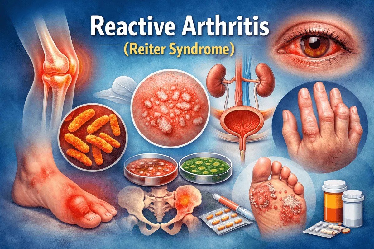
Reactive Arthritis Reiter Reactive arthritis (ReA), historically called Reiter syndrome, is characterized by joint pain and inflammation following an infection in another part of the body. The infection typically starts in the gut, genitals, and urinary tract. The condition typically affects the ankles, knees, and feet. It can also cause inflammation of the eyes, skin, and urethra. It is often described as a classic triad of arthritis, urethritis, and conjunctivitis, although most patients do not present with all three features simultaneously. The name Reiter syndrome was given by Hans Reiter, who first described it. However, this term is no longer widely used because of Reiter’s association with unethical human experimentation during the Nazi era, and the condition is now more appropriately referred to as reactive arthritis.
The disorder was believed to occur due to an abnormal immune response following certain bacterial infections. ReA is relatively rare. The incidence in population-based studies is approximately 0.6 to 27 cases per 100,000 people annually. It is more common in adult males and typically occurs in the second and third decades of life.
Causes of Reiter Syndrome: Reactive Arthritis Reiter
ReA itself is not contagious. However, the bacteria that cause it can spread through sexual contact or through food. Several types of bacteria can trigger this disease. ReA is known to be triggered by a bacterial infection, particularly:
The genitourinary bacteria:
- Chlamydia trachomatis
- Occasionally Ureaplasma urealyticum
- Occasionally, Mycoplasma hominis.
It spread through sexual contact. The infection typically starts in the bladder, urethra, or vagina.
The Gastrointestinal (GI) bacteria:
These bacteria infect the gastrointestinal tract and cause symptoms such as diarrhea, abdominal pain, and fever.
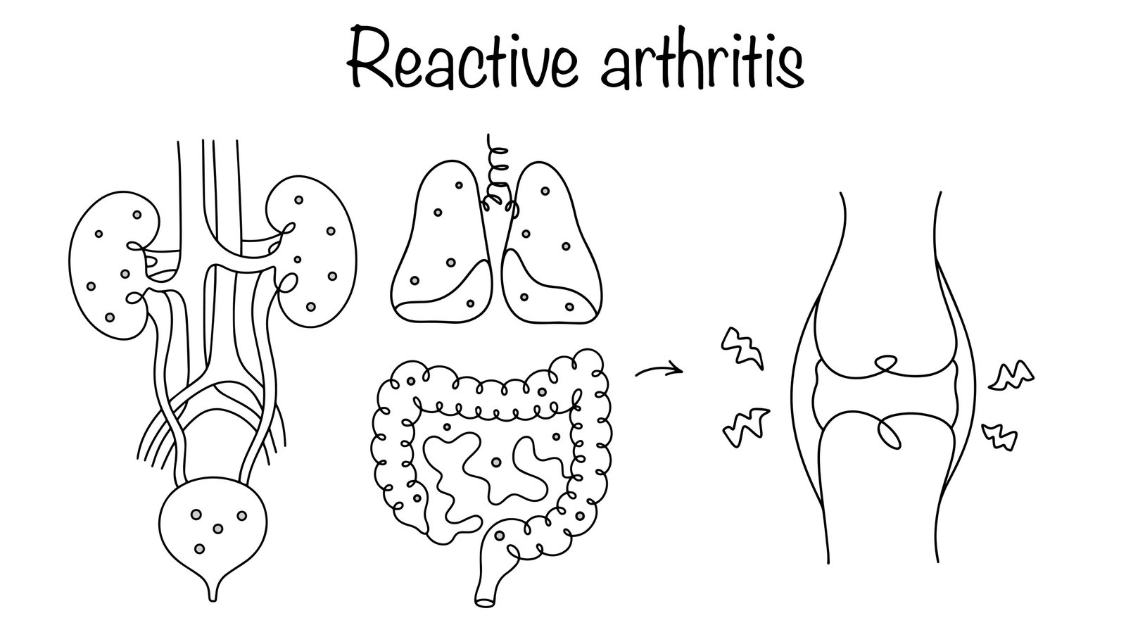
Reactive arthritis: inflammatory joint disease triggered by infections in the genitourinary, intestinal, or respiratory tract.
Risk Factors for Reactive Arthritis
The factors that increase the risk of ReA are:
- Adults between the ages of 20 and 40 years.
- Recent foodborne or gastrointestinal infections
- Males are more likely to develop it after a sexually transmitted infection.
- Genetic factors, such as a specific genetic marker (HLA-B27), increase the risk. However, the people with this marker never get ReA.
- Having HIV can also increase the risk of developing ReA.
Pathophysiology: How does it occur?
ReA is an immune-mediated syndrome triggered by a recent infection. According to current theories, bacterial components such as nucleic acids and lipopolysaccharides can persist in the body after the initial infection and trigger an immune response.
These bacterial antigens may activate T lymphocytes, which then target the synovial tissue and other structures. This process may occur through molecular mimicry, in which immune cells mistakenly attack host tissues that resemble bacterial antigens. It is believed that anti-bacterial cytokine response is also impaired in reactive arthritis (resulting in decreased bacterial elimination).
The presence of the HLA-B27 genetic marker potentiates ReA by presenting bacterial antigens to T cells, thereby altering the host immune system’s self-tolerance. This may contribute to persistent inflammation in joints and surrounding tissues.
However, the exact mechanisms underlying reactive arthritis are still not fully understood.
Signs and Symptoms of Reiter Syndrome
The symptoms of ReA typically start within four weeks after you have a triggering infection. The common symptoms of this condition are:
- Pain and stiffness are often in the knees, ankles, and feet. You may also experience pain in your lower back, buttocks, and heels.
- ReA can cause eye inflammation that can lead to red, irritated, or painful eyes, as well as blurred vision.
- Increased urinary output. Burning and discomfort while urinating. Inflammation of the cervix or prostate glands.
- Inflammation of tendons and ligaments (enthesitis), especially at the heel
- Inflamed toes or fingers (dactylitis, sometimes called “sausage digits”)
- Pain in the lower back that may worsen at night or in the morning.
- Skin issues like mouth sores, rashes on the palms or soles (keratoderma blennorrhagica), or painless ulcers on the penis (circinate balanitis)
- Nail dystrophy.
Diagnosis of Reiter Syndrome
There is no single test that doctors can use to diagnose ReA. Other types of spondyloarthritis can exhibit similar symptoms and may need to be ruled out. The process doctors follow to diagnose ReA includes the following:
Medical History and Physical Examination
The healthcare provider performs a detailed history and physical examination to investigate the recent illness, such as diarrhea or urethritis. For sexually acquired ReA, there is a history of sexual intercourse, usually with a new partner, within 3 months of arthritis symptoms. Individuals with HIV who develop ReA often develop severe psoriasiform dermatitis on the palms, flexures, and scalp.
Two more of the following symptoms and the involvement of the skeletal system establish the diagnosis:
- Sausage-shaped finger, toe, or heel pain
- Conjuntivitis or iritis
- Asymmetric oligoarthritis is usually of the lower extremities
- Urethritis or genital ulcers
- Acute diarrhea
Laboratory Tests
The physicians may order the following laboratory tests to diagnose this condition:
Bacterial Cultures
Culturing stool and urine specimens can reveal bacteria that frequently trigger ReA. However, a negative result is not conclusive because in most cases, patients no longer have detectable bacteria by the time joint symptoms appear.
HLA-B27
This blood test looks for the presence of HLA-B27. It is a genetic risk factor for ReA. Having this marker is consistent with having arthritis, but it is not definitive, as people who test negative can still have ReA. Additionally, not everyone who tests positive has the condition.
Erythrocyte Sedimentation Rate and C – reactive protein
These blood tests measure inflammation, but they are not specific for ReA. These tests measure inflammation. A positive test result can indicate any inflammatory disorder. A negative test does not rule out ReA because these markers are usually not elevated in the chronic form of the condition.
Joint Fluid Test (Synovial fluid analysis)
This test assesses the level of inflammation in the joint and rules out other causes for the pain (such as joint infection or other conditions, such as gout. The doctor will draw fluid from a joint using a needle and syringe (arthrocentesis)
Imaging
The doctor may order these scans:
X-Rays
X-rays reveal the status of the joints, including signs of ReA, such as inflammation of the sacroiliac joints (the joints located in the lower back). They can also help rule out other causes of joint pain. X-rays often do not pick up abnormalities until later in the course of ReA.
Ultrasounds, MRI & CT
These imaging techniques are useful for detecting early joint inflammation, tendon involvement, and enthesitis.
Some Other Tests
In patients suspected of having reactive arthritis, physicians may order certain autoimmune markers to rule out other rheumatologic conditions.
- Antinuclear antibody (ANA) test, which is associated with systemic lupus erythematosus.
- Antibody Tests for Rheumatoid Factor (RF)
- Another commonly used test is anti-cyclic citrullinated peptide antibodies (anti-CCP). It is highly specific for rheumatoid arthritis.
Management and Treatment of Reactive Arthritis
There is no cure for ReA, so treatment focuses on relieving the symptoms. Treatment is individualized based on the severity of symptoms and the joints involved. Medications include:
Antibiotics
If an active bacterial infection such as Chlamydia trachomatis is still present, antibiotics such as doxycycline or azithromycin may be prescribed to treat the underlying infection. However, antibiotics generally do not treat the arthritis once the immune response has already developed.
Non-Steroidal Anti-Inflammatory Drugs (NSAIDs)
NSAIDs are usually the first-line treatment. Medications such as Indomethacin or naproxen help reduce pain and inflammation in affected joints.
Disease Modifying Anti-Rheumatic Drugs (DMARDs)
These medicines (sulfasalazine and indomethacin) suppress the immune system broadly, helping to block inflammation in joints and other tissues. They usually help when NSAIDs do not work.
Corticosteroids
Corticosteroids may be used to reduce severe inflammation.
They may be administered as:
- Topical creams for skin symptoms
- Injections into inflamed joints
- Short courses of oral corticosteroids in severe cases
Physical Therapy
Physical therapy can help ease the pain and improve joint function. The therapists teach techniques to strengthen the muscles surrounding a joint, providing support and improving joint flexibility.
Differential Diagnosis
The most common differential diagnoses of ReA include:
- Rheumatoid arthritis
- Gonococcal arthritis
- Psoriatic arthritis
- Gouty arthritis
- Septic arthritis
- Still disease
- Ankylosing spondylitis
- Rheumatic fever
- Immunization-related arthropathy
- Tubercular arthritis
- Secondary syphilis
Reactive Arthritis vs Psoriatic Arthritis
Reactive arthritis and psoriatic arthritis are both types of spondyloarthritis that can cause joint inflammation, skin changes, and tendon involvement, but they differ in their triggers, patterns, and associated features. The table below summarizes the key differences:
| Feature | Reactive Arthritis | Psoriatic Arthritis |
|---|---|---|
| Cause | Immune response triggered by infection | Autoimmune disease associated with psoriasis |
| Trigger | Gastrointestinal or genitourinary infection | Chronic inflammation in people with psoriasis |
| Skin Findings | Keratoderma blennorrhagica, circinate balanitis | Psoriatic plaques,nail pitting,onycholysis |
| Joint Pattern | Asymmetric oligoarthritis, usually in the lower limbs | Variable; may involve distal interphalangeal joints, can be symmetric or asymmetric |
| Timing | Appears 1–4 weeks after infection | Chronic and progressive |
| HLA Association | Often HLA-B27 positive | HLA-B27 may be positive in some cases, more variable |
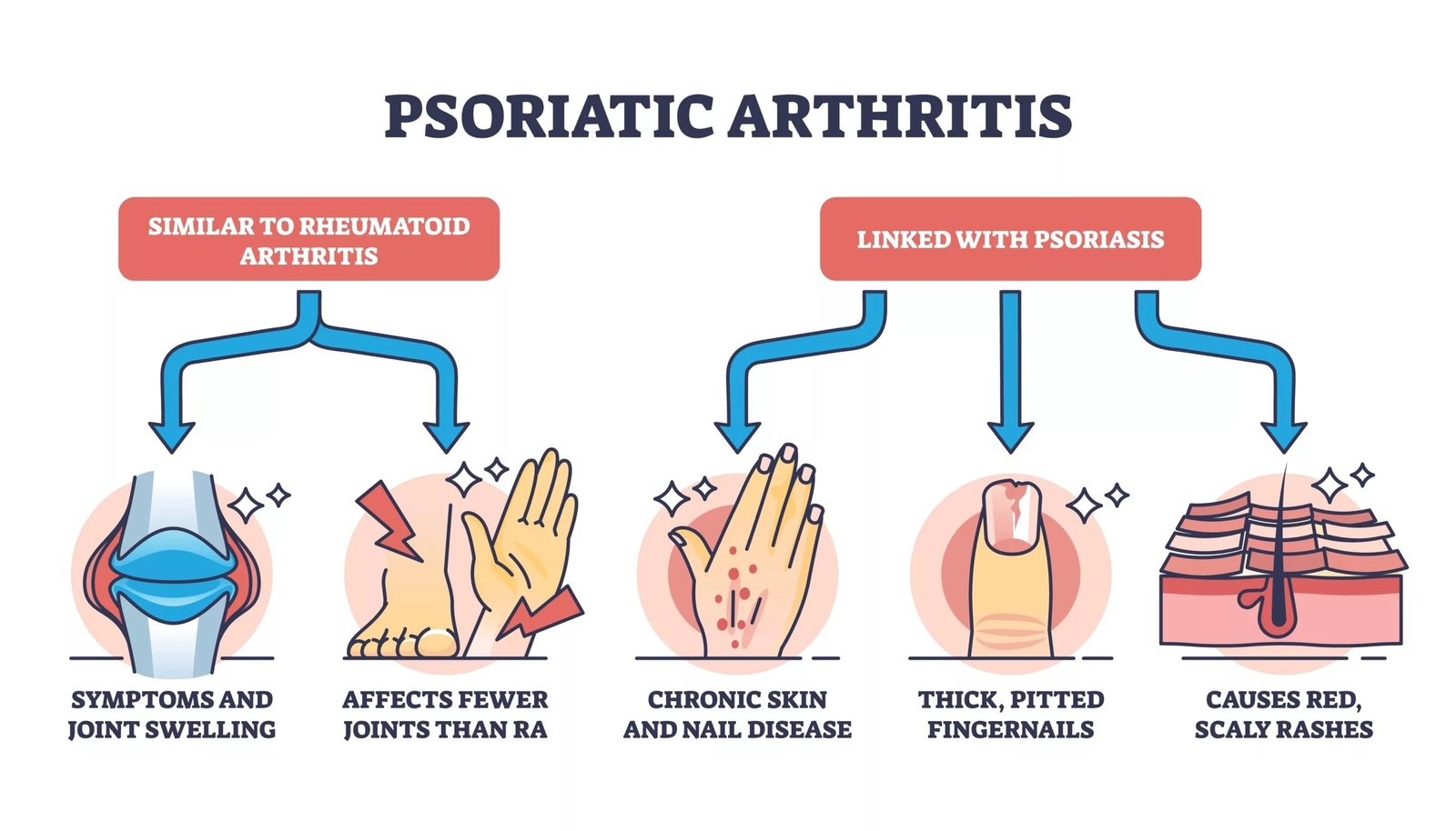
Educational diagram of psoriatic arthritis showing chronic inflammatory joint swelling, skin plaques, and nail abnormalities typical of the disease.
Prognosis
ReA usually has a self-limited disease course. The symptoms resolve within three to six months. Symptoms that persist beyond six months indicate a chronic component of the disease. Patients who are HLA-B27-positive have a higher risk of ReA recurrence. Approximately 15 to 30% of the patients with ReA can develop long-term arthritis or joint abnormalities.
Complications of Reactive Arthritis
The complications of ReA are:
- Chronic arthritis or sacroiliitis
- Recurrent arthritis
- Progression to ankylosing spondylitis or other forms of spondyloarthritis
- Chronic eye inflammation, such as uveitis
- Cardiovascular complications, such as aortic root inflammation leading to aortic regurgitation
- Urethral stricture
Final Remarks
When you thought your recent infection was over, ReA appears as an unexpected and unwelcome condition. It can cause the symptoms of the original infection to return, along with other symptoms (particularly joint inflammation). Whether it affects you a little or a lot, it’s hard to live like this, not knowing when it will end. In most cases, the treatment accomplishes the desired effect, and the condition resolves.
References
[1] Cheeti, A., Chakraborty, R. K., & Ramphul, K. (2023). Reactive arthritis. InStatPearls [Internet]. StatPearls Publishing.
[2] Muilu, P., Rantalaiho, V., Kautiainen, H., Virta, L. J., Eriksson, J. G., & Puolakka, K. (2019). Increasing incidence and shifting profile of idiopathic inflammatory rheumatic diseases in adults during this millennium.Clinical rheumatology,38(2), 555-562.
[3] Cheeti, A., Chakraborty, R. K., & Ramphul, K. (2023). Reactive arthritis. InStatPearls [Internet]. StatPearls Publishing.
[4] Hamdulay, S. S., Glynne, S. J., & Keat, A. (2006). When is arthritis reactive?.Postgraduate medical journal,82(969), 446-453.
[5] Ding, Y., & Xue, L. (2022). HLA-B27 negative reactive arthritis versus HLA-B27 positive reactive arthritis: A retrospective study.Medicine,101(35), e30383.
[6] Colmegna, I., Cuchacovich, R., & Espinoza, L. R. (2004). HLA-B27-associated reactive arthritis: pathogenetic and clinical considerations.Clinical microbiology reviews,17(2), 348-369.
[7] Cheeti, A., Chakraborty, R. K., & Ramphul, K. (2023). Reactive arthritis. InStatPearls [Internet]. StatPearls Publishing.
[8] Wu, I. B., & Schwartz, R. A. (2008). Reiter’s syndrome: the classic triad and more.Journal of the American Academy of Dermatology,59(1), 113-121.
[9] Selmi, C., & Gershwin, M. E. (2014). Diagnosis and classification of reactive arthritis.Autoimmunity reviews,13(4-5), 546-549.
[10] Bojović, J., Strelić, N., & Pavlica, L. (2014). Reiter’s syndrome-disease of young men: Analysis of 312 patients.Medicinski pregled,67(7-8), 222-230.