
 is a benign vascular birthmark that presents itself...)
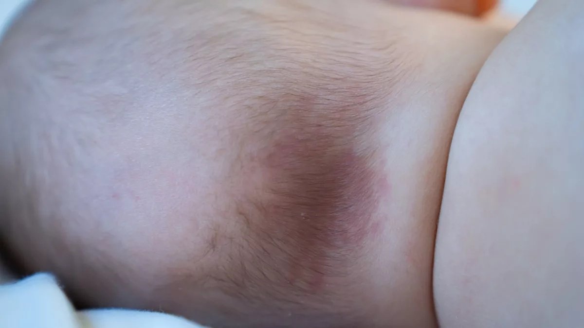
Nevus Simplex Salmon Nevus simplex (also called a salmon patch, stork bite, or angel kiss) is a benign vascular birthmark that presents itself during the infancy of a baby. It is the most common vascular birthmark, seen in about 20–44% of newborns (estimates vary by population). These marks are flat, pink to red patches on the skin caused by clusters of dilated blood vessels close to the surface. In contrast to other birthmarks, nevus simplex usually vanishes with time, but some nape lesions may persist into adulthood. The name “nevus simplex” stands for simple birthmark and shows that it is simple and harmless in nature. Although such marks are of parental concern, they are not medically significant, so they don’t need any treatment.
Understanding Vascular Birthmarks
The skin has a vast system of capillaries (little blood vessels) which supply the skin with oxygen and nutrients. When they fail to form properly in the process of developing the fetus, they may leave behind noticeable prints on the skin. Nevus simplex represents a mild developmental variation where these superficial capillaries remain more visible due to dilation.
What Happens in Nevus Simplex?: Nevus Simplex Salmon
There is a complicated process of skin cell development that occurs during embryonic development. Nevus simplex is a consequence of failure of this maturation after the migration and merging of cells in the skin nervous system (the neuroectoderm). The result of this developmental delay is the dilation of dermal capillaries, which is characteristic of the condition. Due to being stretched (ectatic) and superficial, the enhanced blood extraction to the region is noticeable in the form of pink or red marks through the thin, fragile skin of newborns.
Common Locations and Names
Most commonly occurring locations of the nevus simplex have a nickname associated with them:
Stork Bite (Nevus Flammeus Nuchae)
These marks are found at the back of the neck (nape of the neck) and were named such by the folklore that storks gave birth and transported the babies around by the neck. More than 81 percent of cases of nevus simplex contain stork bites.
Angel’s Kiss
These marks exist on the forehead, between the eyebrows (glabella), or upper eyelids (45 percent prevalence). The romantic name shows their position on the face as kissed by an angel.
Other Locations
The N. simplex may also occur on:
- Upper lip
- Nose bridge
- Posterior scalp
- Lower back (less common)
- Eyelids
Symmetrical patterns of the marks are normally seen on both sides of the face, and they stand out as different compared to other vascular birthmarks, which are normally unilateral.
Appearance and Characteristics
The typical features that aid in the identification of nevus simplex are the following:
- Color: Pink, red, or light purplish patches
- Shape: Flatness of skin surface (no elevation)
- Borders: Borders are poorly defined, feathery, and with a fusion into other skin.
- Texture: Smooth, same texture as normal skin
- Size: Variable, from small spots to larger patches
Dynamic Color Changes
It is one of the characteristics of nevus simplex that it can temporarily alter its appearance:
- Brightened with the crying of the baby, straining with bowel movements, warmed up.
- Temporarily fades when pressure is applied (blanching)
- More prominent in an increase or decrease in temperature or increased blood circulation.
The basis of these color variations is that the enlarged blood vessels react to the increased blood flow and thus the mark is made more visible. This is quite normal and does not signify exacerbation of the condition.
Causes of Nevus Simplex
Knowledge of the etiology of nevus simplex can reduce the anxiety of parents:
Primary Cause: Developmental Delay
This is mainly because the maturation of the skin cells is retarded during embryonic development. This is due to the migration and fusion of cells within the neuroectoderm, which causes dermal capillaries to be dilated.
Blood Vessel Formation
Nevus simplex occurs when blood vessels enlarge (dilate) in the course of fetal development. These capillaries form incorrectly and remain enlarged after birth. The condition is the residual of the fetal circulatory system that failed to develop sufficiently.
What Doesn’t Cause Nevus Simplex
It’s important to understand that nevus simplex is NOT caused by:
- Everything that the mother did or did not do during pregnancy.
- Food consumed when pregnant.
- Stress during pregnancy
- Medications (in most cases)
- Extrapartum trauma or pressure at birth.
- Genetic mutations
The condition is a spontaneous occurrence during the normal growth of the fetus and cannot be avoided.
Who Gets Nevus Simplex?
Prevalence
- Incidence: 30-60% of all newborns
- This is one of the most widespread birthmarks.
- More common in Caucasian infants
- Affects both male and female infants.
Risk Factors
Nevus simplex does not have certain risk factors, unlike many conditions. It occurs in fetal development spontaneously, and affects infants of all genders, including:
- Ethnic backgrounds
- Socioeconomic groups
- Geographic locations
- Family histories
Nevus Simplex Symptoms and Features
Primary Symptom: Visible Mark
The pink or red spot on the skin is the primary sign. Importantly, nevus simplex:
- Causes no pain or discomfort
- Does not itch
- No related swelling (as is the case with other birthmarks)
- Causes no change in texture to the skin
- No systemic symptoms
Blanching Response
One of the unique characteristics is that, when pressure is exerted, the mark becomes temporarily light and, on releasing the pressure, it goes back to its original colour. This blanching reaction proves that the mark is of vascular nature.
Behavior Over Time
- Present at birth (congenital) or in the first few weeks
- It may be more salient within the initial months.
- The onset of the fading is normally at 1-2 years of age.
- The majority of the facial marks disappear by age 18 months.
- There are higher chances that neck marks (stork bites) would remain.
How is Nevus Simplex Diagnosed?
Clinical Examination
It is easily diagnosed, and often only a physical examination is necessary. Your doctor will do:
- Visual analysis of the color, size, and position of the mark.
- Palpation: to assess blanching.
- Medical History: Find out the character of marks and change.
- Family History of family members, whether they have similar marks.
When Additional Tests Are Required
No testing is required in most cases. Imaging may, however, be prescribed when:
- The mark is either unusually large or it does not exist where it should
- It is situated above the spine (to eliminate the possibility of spinal abnormalities)
- The vascular marks are multiple
- Symptoms related to them exist
- The diagnosis is uncertain
Imaging Options:
- Doppler ultrasound is used to demonstrate blood flow.
- MRI (Magnetic Resonance Imaging): gives detailed views of blood vessels
- CT scan: This is done when bone involvement is suspected
When to See a Doctor?
Consult your child’s healthcare provider if the mark:
- Grows rapidly in size
- Raised or develops bumps
- Bleeds easily or frequently
- Changes texture to a greater extent
- Causes functional problems (affects vision, breathing, etc.)
Nevus Simplex vs. Nevus Flammeus (Port-Wine Stain)
These two states are frequently confused with each other; however, they possess significant distinctions:
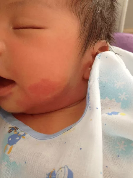
Port wine stainor naevus flammeus red birthmark on the face, skin, and neck of a baby boy
| Feature | Nevus Simplex | Nevus Flammeus (Port-Wine Stain) |
|---|---|---|
| Prevalence | 30-40% of newborns | 0.3% of newborns |
| Location | Midline (neck, forehead, eyelids) | Anywhere, often unilateral |
| Appearance | Pink to light red, flat | Dark red to purple, flat |
| Border | Poorly defined, feathery | Well-defined, sharp borders |
| Symmetry | Often bilateral/symmetric | Usually unilateral |
| Natural Course | Fades over time (1-2 years) | Continues and grows dark with age. |
| Texture Changes | Remains flat and smooth | May thicken, develop bumps |
| Treatment Need | Rarely needed | Often treated for cosmetic/medical reasons |
| Associated Syndromes | None | May suggest Sturge-Weber syndrome |
| Genetic Cause | None identified | GNAQ gene mutation |
Nevus Simplex vs. Strawberry Hemangioma (Infantile Hemangioma)
One more significant difference is between nevus simplex and the strawberry hemangiomas:
| Feature | Nevus Simplex | Strawberry Hemangioma |
|---|---|---|
| Classification | Vascular malformation | Vascular tumor |
| Timing | Present at birth | Appears weeks after birth |
| Growth Pattern | There is no growth, merely a change in visibility. | Rapid growth phase (0-9 months) |
| Texture | Flat, smooth | (strawberry-like) raised, bumpy. |
| Color | Pink to light red | Bright red to deep red |
| Natural History | Fades gradually | Grows then shrinks (involution) |
| Complete Resolution | Half before the neonatal period, the majority before 18 months. | 90% by age 10 |
| Residual Changes | None | May leave loose skin or discoloration |
| Risk of Complications | None | Ulceration, bleeding, functional impairment |
| Treatment Frequency | Rarely needed | May require intervention |
| Medication | Not applicable | Beta-blockers (propranolol) can be taken. |
Nevus Simplex Treatment Options
Conservative Approach: Observation
In the vast majority of cases of nevus simplex, no treatment is the most effective approach. This is an active non-intervention approach and involves:
- Realizing that the condition is benign.
- Most marks fade away naturally.
- Keeping track of the changes.
- Good skin care: Use cleansers and moisturizers.
It is more likely since 95 percent of the marks fade in the first 2 years. Natural resolution gives the most aesthetic cosmetic result.
Laser Therapy for Persistent Marks
Laser therapy is most effective and widely used in the treatment of marks that do not fade and remain a cosmetic concern.
Pulsed Dye Laser (PDL):
The Pulsed Dye Laser is the best method of treating vascular birthmarks such as nevus simplex. It functions by releasing a certain color of light, which is taken up by the red blood cells within the small vessels and makes them warm up and seal off without hurting the skin around the area.
It works by emitting a beam of light, which is absorbed by the red blood cells in the small vessels and warms them up, and closes them off without injuring the skin surrounding the area.
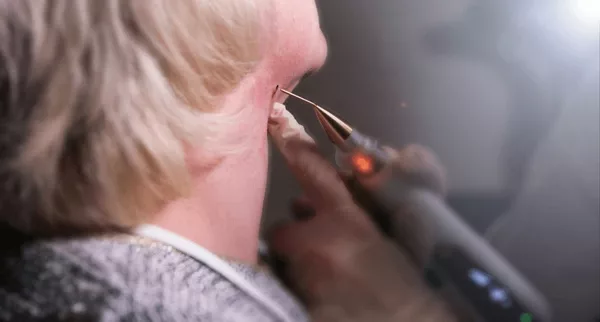
Cosmetic procedure, removal of a birthmark with a laser.
Expectations of the Treatment:
Doctors use an anesthetic cream at or around 30-60 minutes before the surgery. It will be done by the doctor with the help of a handheld device that will emit short pulses of laser light at the mark. The actual session can take only 15-30 minutes. The laser might also include an inbuilt cooling system to ensure that it doesn’t affect the surface of the skin.
Results and Realistic Expectations:
- To achieve maximum lightening, one usually requires 4 to 12 sessions, 4 to 12 weeks in between.
- Higher Success Rate: You can expect the lightening of the mark between 50 percent and 90 percent, with some patients displaying 100 percent clearance.
- Marks on the face and neck also tend to yield better results than those on the arms or legs, and young patients tend to achieve better results.
Possible Side Effects:
The side effects are normally temporary and they may include:
- Bruising (Purpura): It is the most frequent side effect, and it appears similar to a dark bruise and resolves in 7-14 days.
- Redness and Swelling: It may be red and swollen for one or two days.
- Temporary Change of Colours: The skin may have slight lightening/darkening than the surrounding skin, but this tends to normalise itself with time.
- Less frequent: There are cases of blistering or scarring.
Alternatives and Future Opportunities
Even though PDL is the most preferred option, other types of lasers, such as Nd:YAG or Intense Pulsed Light (IPL), are also better for more recalcitrant or deeper spots. Researchers are also interested in combination therapies, e.g., Pulsed Dye Laser with a topical drug, such as rapamycin, to make the outcome better.
Minimal Make-up Solution: Cosmetic Camouflage
Cosmetic camouflage is a great temporary solution in case laser treatment is not the right answer for your family. There are medical-grade concealers that are water-resistant and match perfectly with the skin color of your child. You should consult a skincare expert to learn about the exact method of applying the cosmetics to hide marks until the occasion or evening is over, or you change your mind.
Conclusion
To conclude, a nevus simplex is a marvelous and entirely harmless birthmark of numerous babies. Although the worries of the parents are a big concern, these stork bites are usually harmless and disappear naturally in the early years of a child. To the few who are left, it is well to understand that they will be merely a cosmetic concern. It may fade away or be a latent quality; it is just a part of what you are making a child of.
Laser removal could be a possibility in case the mark continues till school age (about 5+ years) or is on the face and is a source of much psychological distress, or the family wants to have it removed cosmetically. One should be aware of the fact that full clearance is not possible; partial lightening is more prevalent, and results differ in different individuals.
References
[1] Bruckner, A. L., & Frieden, I. J. (2003).Persistent Salmon Patch on the Forehead and Scalp: Natural History and Associations. Pediatric Dermatology, 20(2), 88–92.
[2] Mulliken, J. B., & Glowacki, J. (1982).Hemangiomas and Vascular Malformations in Infants and Children: A Classification Based on Endothelial Characteristics. Plastic and Reconstructive Surgery, 69(3), 412–422.
[3] Harper, J., Oranje, A., & Prose, N. (2016).Textbook of Pediatric Dermatology(2nd ed.). Wiley-Blackwell.
[4] Kinsler, V. A., et al. (2017).Skin Disorders in Children: A Color Handbook(2nd ed.). CRC Press.
[5] Barkovich, A. J. (2005).Pediatric Neuroimaging(4th ed.). Lippincott Williams & Wilkins.
[6] Frieden, I. J. (2006).Infantile Hemangioma and Other Vascular Birthmarks. In:Dermatology in General Medicine(Fitzpatrick).
[7] Weston, W. L., Morelli, J. G., & Tay, Y. K. (2002).Color Atlas of Pediatric Dermatology. McGraw-Hill.
[8] Polcari, I., & Stein, S. (2011).Vascular Birthmarks: Sorting Through the Confusion. The Journal of Pediatrics, 159(6), 1048–1049.
[9] Garzon, M. C., Huang, J. T., & Enjolras, O. (2005).Vascular Malformations: Part I.Journal of the American Academy of Dermatology, 53(3), 435–461.
[10] Williams, H., & Burney, P. (2000).The Epidemiology of Skin Birthmarks.British Journal of Dermatology, 142(6), 1123–1125.
[11] Jacobs, A. H., & Walton, R. G. (1976).The Incidence of Birthmarks in the Neonate. Pediatrics, 58(2), 218-222.
[12] Krowchuk, D. P., & Frieden, I. J. (1995).Infantile Hemangiomas and Vascular Malformations. Pediatrics in Review, 16(9), 348-355.
[13] Goldstein, B. G., & Goldstein, A. O. (2020).Newborn Skin: Common Rashes.
[14] Prendiville, J. S. (2012).Vascular Lesions in Children. In:Harper’s Textbook of Pediatric Dermatology. Wiley-Blackwell.
[15] Wassef, M., et al. (2015).Vascular Malformations Classification. Journal of Vascular Surgery, 61(2), 557–568.
[16] Bruckner, A. L., Frieden, I. J. (2003).Clinical Evaluation of Vascular Birthmarks. Pediatric Clinics of North America, 50(1), 121-138.
[17] Enjolras, O., & Mulliken, J. B. (1997).Port-Wine Stain Birthmarks and Associated Syndromes. Pediatrics, 99(1), 117–120.
[18] Leaute-Labreze, C., et al. (2008).Propranolol for Severe Hemangiomas of Infancy. New England Journal of Medicine, 358(24), 2649–2651.
[19] Adams, D. M., & Lucky, A. W. (2009).Treatment of Vascular Birthmarks. Advances in Dermatology, 25, 157–173.
[20] Kono, T., et al. (2003).Treatment of Vascular Lesions with the Pulsed Dye Laser. Dermatologic Surgery, 29(3), 243–248.
[21] Nelson, J. S. (2001).Clinical Management of Port-Wine Stain. Lasers in Surgery and Medicine, 28(2), 131–144.
[22] Bauland, C. G., et al. (2006).Laser Therapy for Persistent Vascular Birthmarks. Journal of the European Academy of Dermatology and Venereology, 20(3), 303-308.
[23] Gupta, A. K., & Gover, M. D. (2005).Camouflage in Dermatology. Journal of Cosmetic Dermatology, 4(1), 45–50.
For more information about Nevus Simplex Salmon, refer to the latest medical literature.