

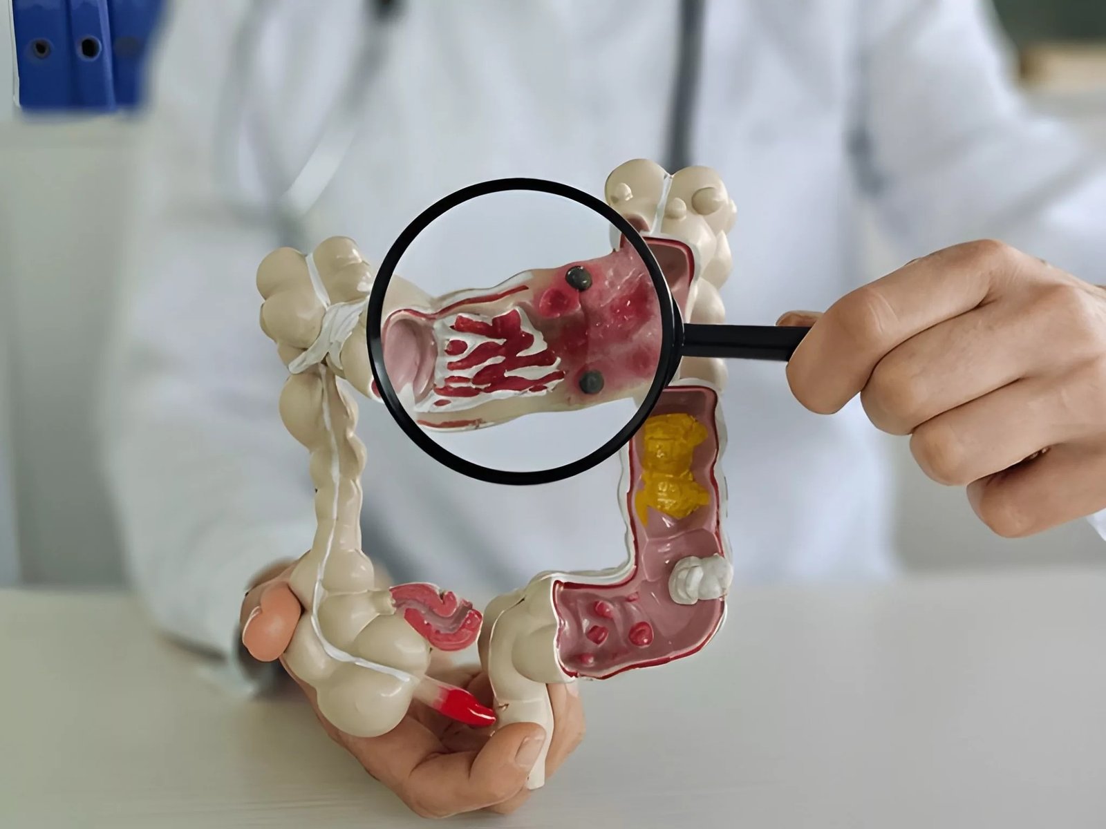
Anastomotic Leak After An Anastomotic Leak is one of the most serious complications that can occur after gastrointestinal surgery. Learning about this condition, its symptoms, and treatment options is critical for any patient undergoing surgery involving bowel or other body channel anastomoses.
What Is an Anastomotic Leak?: Anastomotic Leak After
Anastomotic leaks occur when an anastomotic junction fails to heal properly. This condition can result in the leakage of luminal contents into the peritoneal cavity or surrounding tissues. Shockingly, about 1.5–23% of all anastomosis procedures result in this condition, depending on the procedure, with the highest risk in low rectal anastomoses.[1] [2]
When a part of the intestine is removed during surgery, the surgeon reconnects (anastomoses) the remaining portions of the intestine to restore intestinal continuity. When this connection doesn’t seal, digestive contents fill the abdominal cavity, which may lead to severe inflammation and infection.
Anastomotic Leak Causes: Understanding Risk Factors
Various patient-specific conditions and surgical factors contribute to the occurrence of anastomotic leaks.[3]
Patient-Related Risk Factors:
Medical Conditions
- Wound healing is affected by diabetes mellitus.
- Anemia, inhibition of oxygen flow to tissues.
- Crohn’s disease causes chronic inflammation
- Obesity makes surgery more difficult to perform, and so is the recovery.
- In malnutrition, tissue repair is compromised
Lifestyle Factors
- Smoking decreases blood circulation and oxygen supply.
- The chronic use of steroids suppresses the immune system.
- Old age slows down the healing procedures.
Surgical Risk Factors:
Anatomical Considerations
- Anastomoses of the colon, especially in the rectum and the sigmoid.
- Esophageal-gastric connections
- Anatomical differences are associated with a higher risk for male patients.[4]
Procedural Factors
- Prolonged surgery duration
- Emergency procedures
- The technical problems during the creation of anastomosis.
- Poor blood supply to the anastomotic site.
Anastomotic Leak Symptoms
For a successful treatment, it is essential to detect timely symptoms of anastomotic leak. Symptoms typically appear between post-operative days 3 and 5, though they can occur earlier or later.[5]
Clinically, leaks can be described in terms of timing. Early leaks tend to be caused by technical problems during surgery or poor blood supply to the anastomosis, and late leaks may be related to such factors as obstruction, chronic inflammation, or underlying disease.
Early Symptoms:
Systemic Signs
- Fever (often the first indicator)
- Increased heart rate (tachycardia)
- Low blood pressure (hypotension)
- General feeling of unwellness
Abdominal Symptoms
- Worsening abdominal pain
- Nausea and vomiting
- Abdominal distension
- Changes in bowel function
Advanced Symptoms:
In late leaks, the patients can develop:[6]
- Signs of sepsis (i.e., confusion, hyperpnea, intense weakness)
- Delayed return of bowel function (ileus)
- Unusual drainage of the wound or discharge.
- Reduced urine output
- Pain in the left shoulder (referred pain due to irritation of the abdomen)
When to Seek Immediate Medical Attention?
Contact your healthcare provider immediately if you experience:
- Call your healthcare provider immediately in case of:
- Continued or increasing abdominal pain.
- Fever above 101°F (38.3°C)
- Infection in the area of the surgical site.
- Acute and intense symptoms.
- Any significant changes to your condition.
Anastomotic Leak Diagnosis
Initial Assessment:
Physical Examination
- The doctor puts gentle pressure against your abdomen, inquiring if it aches or is tender, which may suggest a leak.
- Nurses monitor your temperature, blood pressure, pulse, and rate of respiration, looking for the early signs of infection or other problems.
- Surgical wound inspections involve healthcare workers looking at the surgical incision for possible infection, unusual drainage, or difficulty healing the wound.
- Doctors check for signs of peritonitis, which include a tender abdomen, rigid muscles, and other warning signs.
Laboratory Tests:
- Full blood count (FBC) to detect infection: A blood test that counts your white blood cells as they increase when your body is fighting an infection.
- C-reactive protein (CRP) levels: This blood test shows inflammation in your body, and a high level suggests possible infection or tissue damage. CRP is especially helpful during days 3–5 after surgery, when constantly high levels indicate an anastomotic leak.
- Procalcitonin (PCT): It is another biomarker that increases in bacterial infection. High PCT levels during days 3-5 after the operation may aid in the timely leakage detection and decision-making.
- Arterial blood gas analysis: A blood test is performed to measure oxygen and acid levels in your blood to see how well your organs are working.
- Lactate levels assess severely infected organs, which have a low oxygen supply and high acid levels, by measuring lactic acid present in the blood.
- Clotting studies: Blood tests that check how well your blood clots, which are important for preventing bleeding during any procedures.[7]
Imaging Studies:
CT Scan with Contrast
CT with IV and oral/rectal contrast is a gold standard diagnostic tool in the detection of anastomotic leak, but it is not a 100 percent sensitive test because sometimes small or initial leaks are missed, yet the findings should always be considered in the presence of clinical results and lab indicators.
- Intravenous contrast administration: A special dye is injected into your vein so that the internal structures can be better seen during the CT Scan.
- Detecting gas or fluid outside the bowel lumen: In case of improper healing of the surgical connection, there are bubbles of air or liquid that should not be present. A scan helps to detect them.
- Evaluation of contamination in the abdomen: Doctors may view the extent of spread of leaked contents in your abdominal cavity.
- Assessment of abdominal contamination extent: Doctors can see how much leaked material has spread throughout your abdominal cavity.
- Identification of fluid collections or abscesses: The scan will identify the pockets of infected fluid formed by the leak.[8]
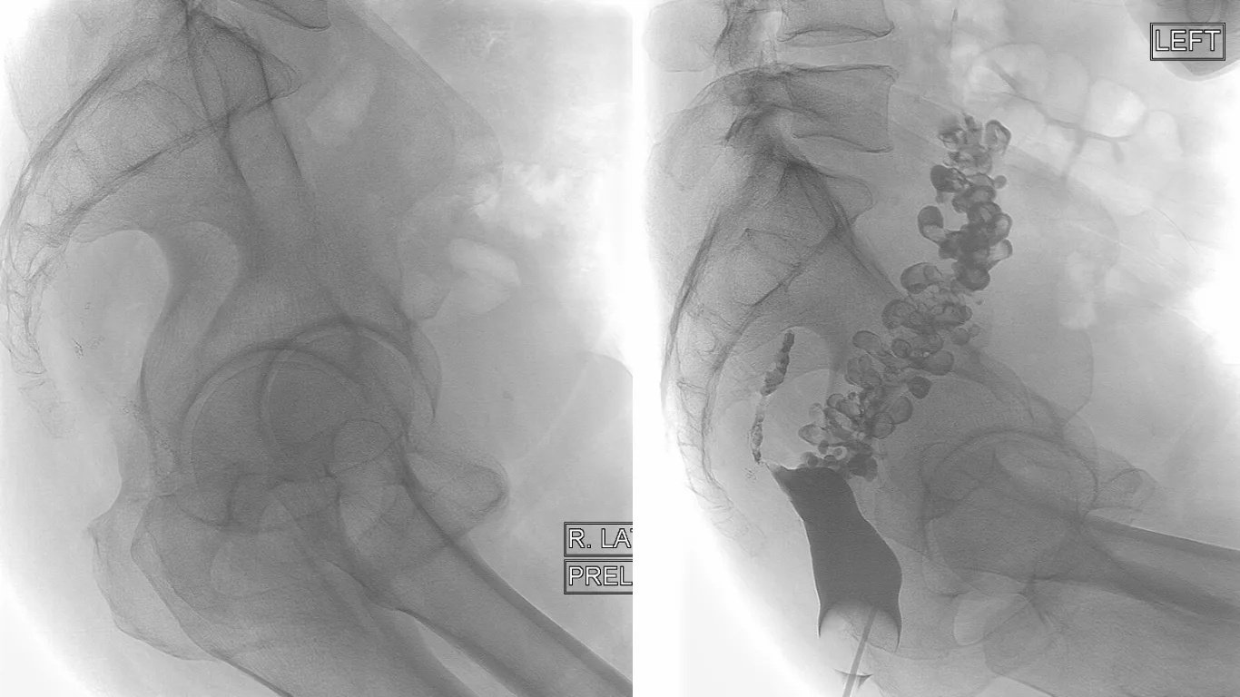
Case courtesy of Henry Knipe, Radiopaedia.org, rID: 29076. Licensed under CC BY-NC-SA 3.0.
Additional Contrast Studies:
- Oral contrast studies for upper GI leaks: For the diagnosis of upper GI leaks, you have to drink a special liquid that shows up the area of leakage on X-rays.
- Contrast enemas for colorectal anastomoses: A contrast liquid is inserted through your rectum to check for leaks in the lower bowel connections.
- Water-soluble contrast agents for safety: Special safe dyes are used that won’t harm you if they leak out during the test.
- Drain fluid testing: In certain cases, especially in upper GI or esophageal leaks, testing of fluid for amylase from surgical drains is helpful.
Screening During Surgery
- Air leak testing with saline immersion: In such tests, surgeons use salt water to fill the area and blow air into the connection to see if bubbles appear, showing a leak.
- Methylene blue instillation: A blue dye is put into the surgical connection to see if any colored liquid leaks out.
- Direct visualization of the anastomosis: The surgeon carefully looks at the surgical site to make sure it is secure.
Treatment Approaches
Immediate Management – Resuscitation & Stabilization:
- Intravenous fluid replacement: IV fluids are given to prevent dehydration and maintain proper blood pressure when you can’t drink.
- Broad-spectrum antibiotic therapy: Strong antibiotics are started immediately to fight any infection from the leaked bowel contents.
- Pain management: Medications are given to control your pain and keep you comfortable during treatment.
- Nil by mouth (NBM) status: You’re not allowed to eat or drink anything to let your bowel rest and heal.
- Urinary catheter insertion for monitoring: A tube is placed in your bladder to accurately measure urine output and monitor kidney function.[9]
Stable Patients (Non-surgical/Conservative Management):
- Extended antibiotic courses: You’ll take antibiotics for weeks or months to completely clear any infection from the leak.
- Bowel rest and nutritional support: Your digestive system gets a break from processing food while you receive nutrition another way.
- Total parenteral nutrition (TPN) if needed: Special liquid nutrition is given directly into your bloodstream through a large IV when you can’t eat.
- Percutaneous drainage of fluid collections: Doctors insert a thin tube through your skin to drain infected fluid without major surgery.
- Close monitoring and serial imaging: You’ll have regular check-ups and repeat scans to make sure the leak is healing properly.
Unstable Patients (Urgent Surgical Management):
- Laparotomy with abdominal washout: Surgeons open your belly and wash out all the infected material with sterile fluid.
- Anastomotic repair or revision: The surgical connection is fixed or completely redone to stop the leak.
- Defunctioning proximal stoma creation: A temporary bag is attached to divert waste away from the leak area to help it heal.
- Complete anastomotic takedown with ostomy: The leaking connection is completely removed, and a stoma is created. This may be temporary or permanent, depending on the patient’s condition and healing.[10]

A woman showing the colostomy in the abdomen
Advanced Treatment Techniques:
Endoluminal Vacuum Therapy
- Endo-SPONGE® systems for rectal leaks: A Special vacuum sponge is placed inside the rectum to help seal leaks from the inside.
- Negative pressure wound therapy: Gentle suction is applied to help draw the leak closed and promote healing.
- Gradual leak closure promotion: The vacuum system slowly encourages the body’s natural healing to close the leak over time.
Minimally Invasive Approaches
- Laparoscopic drainage procedures: Small keyhole surgery is used to drain infected areas without large incisions.
- Endoscopic stent placement: A small tube is inserted through natural body openings to help support the healing area.
- Percutaneous intervention techniques: Various procedures done through the skin to treat the leak without major surgery.
Complications
Immediate Infection-Related Complications:
- Peritonitis (abdominal cavity infection): The lining inside your belly becomes infected from leaked bowel contents, causing severe pain and illness.
- Sepsis and septic shock: The infection spreads throughout your body, causing dangerous drops in blood pressure and organ problems.
- Abscess formation: Pockets of pus develop where infected fluid gets trapped, requiring drainage or antibiotics.
- Multi-organ dysfunction: Severe infection causes your kidneys, lungs, heart, and other organs to stop working properly.[11]
Surgical Complications:
- Wound dehiscence: Your surgical incision opens up or doesn’t heal properly, potentially requiring additional surgery.
- Bleeding complications: Excessive bleeding occurs from the surgical site or is related to blood-clotting problems.
- Respiratory complications: These are mostly the result of prolonged bed rest, pain, or the effects of severe infection.
- Thromboembolic events: As a result of prolonged immobility and inflammation, blood clots form in your legs or lungs.[12]
Long-Term Complications:
Gastrointestinal
- Adhesion formation
- Bowel obstruction
- Chronic abdominal pain
- Functional bowel disorders
Anastomotic Leak Recovery
Anastomotic leak recovery is a slow process that needs great attention, medical guidance, and comprehensive care coordination.[13]
Hospital Recovery Phase:
Initial Recovery (1-2 weeks)
- Intensive monitoring and treatment
- Gradual improvement of your symptoms
- Control of infections is achieved
- Rationalization of nutritional therapy.
Extended Stay Considerations
For complicated cases, hospital stays may go beyond 4 weeks and include:
- Monitoring infection levels
- Sustaining enough oral or other nutritional sources
- Wound healing
- Completion of education for the patient and family
Preparing to Go Home:
Signs You’re Ready to Leave the Hospital
Your doctors will make sure several things are in place before you head home:
- Your body is stable: Your temperature, blood pressure, and heart rate are normal, and your blood tests show you’re on the right track.
- Infection is under control: Blood markers show your body has successfully fought off the infection.
- You can eat properly: Either you’re eating enough regular food, or you have a safe plan for getting nutrition at home.
- Your injuries are healing properly: Your surgical areas are closing and not showing any infection.
- You and your family are fully aware of what to do.
Home Care Preparation
- Training to manage Ostomy (where necessary)
- Wound care instructions
- Knowledge of activity limitations.
- Follow-up appointments.
- Symptom alert.
Long-Term Recovery:
What to Expect Month by Month
First Month (Weeks 1-4):
You spend the first four weeks healing. You will be concerned about taking drugs when prescribed, eating, and letting your body heal itself. At this stage, do not think that you will be able to feel normal yet.
Months 2-3: You may be in a position to take short walks, perform minor household chores, and start enjoying your favorite activities. Energy will be restored slowly.
Months 4-6: Life begins to become more normal. You can go back to work (with some accommodations when necessary), rejoin most of your activities, and feel like you are recovering your old self.
Six Months and Beyond: Your doctor will check your full recovery. At this stage, many individuals believe they are totally recovered, but it varies with each individual.
Factors Affecting Recovery:
- Young age and good health
- Detection and management of leakages at an early stage.
- Proper nutrition.
- Medical adherence.
- Strong support system
Recovery Challenges:
- Multiple comorbidities
- Delayed diagnosis
- Complex leak locations
- Nutritional deficiencies
- Limited social support
Everyone heals differently. Your recovery may not progress like that of another person, and that is absolutely normal. Others recover easily after some time, and some recover slowly. It is important to keep moving in the right direction, even though it might be slow.
You should trust your medical team, be patient with yourself, and have small successes along the way. Making it through an anastomotic leak is a significant effort, and healing is taking you closer to feeling normal again.
Prevention Strategies
Although not every anastomotic leak can be prevented, some steps can help minimize the risk:[14]
Pre-Operative Optimization:
Medical Management
- Blood sugar control in diabetic patients
- Smoking cessation programs
- Nutritional supplementation
- Anemia correction
- Medication review and adjustment
Risk Assessment
- Comprehensive health evaluation
- Surgical risk stratification
- Alternative procedure consideration
- Informed consent discussions
Intraoperative Measures:
Surgical Techniques
- Optimal anastomotic technique selection
- Adequate blood supply is ensured
- Tension-free anastomosis creation
- Intraoperative leak testing
- Indocyanine green (ICG) fluorescence intraoperative perfusion is an increasingly popular technique to establish the adequacy of blood flow and minimize the possibility of leakage.[15]
- Protective ostomy consideration
Post-Operative Care:
Monitoring Protocols:
- Regular vital sign assessment
- Laboratory value tracking
- Early symptom recognition
- Patient education reinforcement
Living with Ostomy After Anastomotic Leak
Many patients require temporary or permanent ostomy creation as part of their treatment:[16]
Temporary Ostomy Benefits:
- Allows anastomotic healing
- Reduces contamination risk
- Improves infection control
- Facilitates the recovery process
Ostomy Reversal Considerations:
- Adequate healing confirmation
- Patient health optimization
- Imaging study clearance
- Multidisciplinary team evaluation[17]
Conclusion
Anastomotic leak is a serious but manageable surgical complication. You can improve its outcome if you detect symptoms at an early phase, prompt medical intervention, and use comprehensive treatment approaches. With appropriate medical care and support, most patients return to normal activities.[18]
Knowledge of the symptoms, treatment choices, and recovery journey will enable the patient and their families to play a key role in making care decisions. The prognosis of anastomotic leak is improving with the increased development of surgical techniques, diagnostic methods, and treatment choices.
In case you have anastomotic leak or have gone through this complication and are now recovering, collaborate with your care team, take their advice seriously, and do not hesitate to ask them questions or raise concerns. It is your involvement in care that will be critical to good outcomes.
References
[1] McDermott F D, Heeney A, Kelly M E, Steele R J, Carlson G L, Winter D C. Systematic review of preoperative, intraoperative and postoperative risk factors for colorectal anastomotic leaks. Br J Surg. 2015;102(05):462–479. doi: 10.1002/bjs.9697.
[2] International Study Group of Rectal Cancer.International consensus on reporting anastomotic leaks after colorectal surgery.Dis Colon Rectum. 2025;68(8):1010–1018
[3] Matthiessen P, Hallböök O, Rutegård J, Sjödahl R. Risk factors for anastomotic leakage after anterior resection of the rectum.Colorectal Dis.2004;6(6):462-469.
[4] Matthiessen P, Hallböök O, Rutegård J, Sjödahl R. Risk factors for anastomotic leakage after anterior resection of the rectum.Colorectal Dis.2004;6(6):462-469.
[5] Bruce J, Krukowski ZH, Al-Khairy G, Russell EM, Park KG. Systematic review of the definition and measurement of anastomotic leak after gastrointestinal surgery.Br J Surg.2001;88(9):1157-1168.
[6] Hyman N, Manchester TL, Osler T, Burns B, Cataldo PA. Anastomotic leaks after intestinal anastomosis: It’s later than you think.Ann Surg.2007;245(2):254-258.
[7] Bellows CF, Webber LS, Albo D, Awad S, Berger DH. Early predictors of anastomotic leaks after colectomy.Tech Coloproctol.2009;13(1):41-47.
[8] Karliczek A, Benaron DA, Zeebregts CJ, Wiggers T, van Dam GM. Intraoperative assessment of microperfusion with visible light spectroscopy for prediction of anastomotic leakage.Br J Surg.2010;97(7):1094-1101.
[9] Rahbari NN, Weitz J, Hohenberger W, Heald RJ, Moran B, Ulrich A, Holm T, et al. Definition and grading of anastomotic leakage following anterior resection of the rectum: a proposal by the International Study Group of Rectal Cancer.Surgery.2010;147(3):339-351.
[10] Ashraf SQ, Burns EM, Jani A, Altman S, Young JD, Faiz O. The economic impact of anastomotic leakage after anterior resections in English NHS hospitals: A retrospective analysis.Colorectal Dis.2013;15(11):1409-1416.
[11] Alves A, Panis Y, Pocard M, Regimbeau JM, Valleur P. Management of anastomotic leakage after nondiverted large bowel resection.J Am Coll Surg.1999;189(6):554-559.
[12] Vignali A, Gianotti L, Braga M, Radaelli G, Malvezzi L, Di Carlo V. Altered microperfusion at the rectal stump is predictive for rectal anastomotic leak.Dis Colon Rectum.2000;43(1):76-82.
[13] Hyman N, Manchester TL, Osler T, Burns B, Cataldo PA. Anastomotic leaks after intestinal anastomosis: It’s later than you think.Ann Surg.2007;245(2):254-258.
[14] Sciuto A, Merola G, De Palma GD, Sodo M, Pirozzi F, Bracale UM, Bracale U. Predictive factors for anastomotic leakage after laparoscopic colorectal surgery.World J Gastroenterol.2018;24(21):2247-2260.
[15] Montedori A, Cirocchi R, Farinella E, Sciannameo F, Abraha I. Covering ileo- or colostomy in anterior resection for rectal carcinoma.Cochrane Database Syst Rev.2010;5:CD006878.
[16] Carlsson E, Persson E, Löfgren A, Nyman J, Gellerstedt M, Jeppsson B. Ostomy patients’ perceptions of quality of care before and after the introduction of the enhanced recovery after surgery (ERAS) programme.Scand J Caring Sci.2012;26(4):791-797.
[17] Nugent KP, Daniels P, Stewart B, Patankar R, Johnson CD. Quality of life in stoma patients.Dis Colon Rectum.1999;42(12):1569-1574.
[18] Rahbari NN, Weitz J, Hohenberger W, et al. Definition and grading of anastomotic leakage following anterior resection of the rectum: a proposal by the International Study Group of Rectal Cancer.Surgery.2010;147(3):339-351.