

Angiokeratoma Those Black When you find small, black, or dark red bumps on your skin, it can be alarming, especially when they are on your genitals, such as the scrotum or labia majora. You probably have angiokeratoma, a benign vascular skin lesion that is more common than you might think. But it’s often misunderstood, misdiagnosed, and unnecessarily feared.
What Is Angiokeratoma?: Angiokeratoma Those Black
Angiokeratoma is a harmless skin condition caused by dilation (ectasia) of tiny blood vessels (capillaries) under the skin. The skin above the capillaries becomes thicker as the capillaries increase in size. It produces a pin-sized bump which is red when fresh, purple or even black when old.[1]
It is only by the name that you can see: angio is vessels, kerato refers to thickened skin (hyperkeratosis), toma is growth. And so literally, it is a thickened growth of blood vessels.
These lesions are normally 1 to 5 millimeters in diameter, or a light pin-head to a small pea. They can be either single or multiple. They are either smooth or rough-warted. And although they sound scary, they are benign and will not become malignant in the long run.
Angiokeratomas are harmless capillary ectasias of the superficial dermis and are usually asymptomatic, as they are represented by blue-red papules of hyperkeratosis, which can occur anywhere in the body.
The Different Types of Angiokeratoma
Angiokeratomas are not all similar. The clinicians recognize the various types based on their location, appearance, and potential causes.
Angiokeratoma of Fordyce
This is the most widespread one. It was named Fordyce angiokeratoma after dermatologist John Addison Fordyce, who described it in the scrotal skin of an elderly patient (60 years) in 1896. Angiokeratoma of Fordyce is mainly seen in the scrotum of men, but may occur in the labia majora in women. It is infrequently manifested on the penis shaft, inner thighs, or lower abdomen.[2]
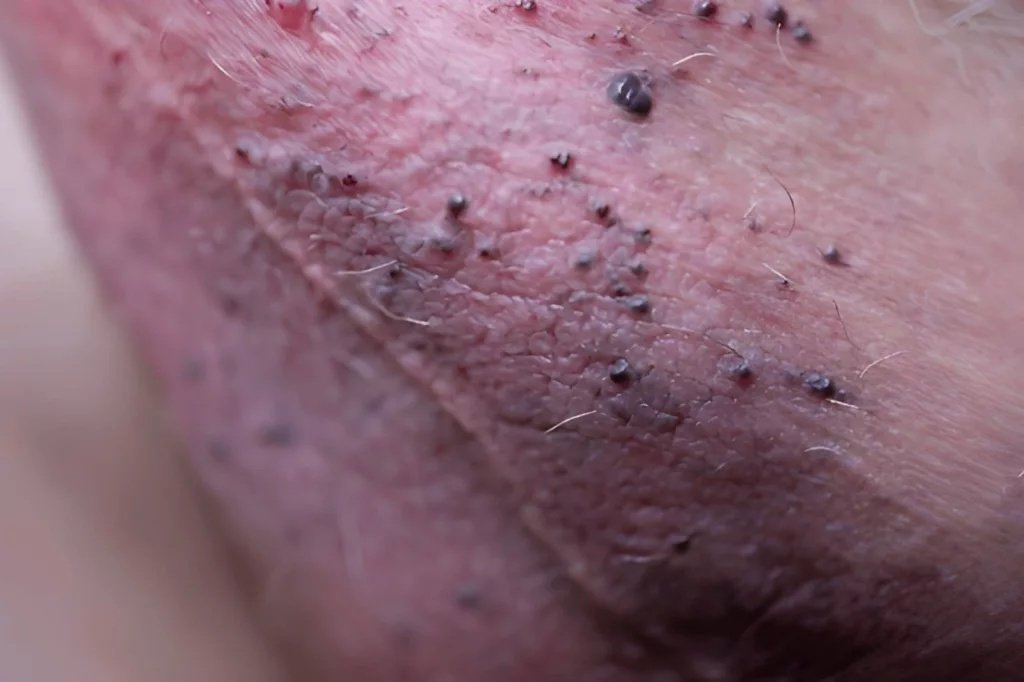
Scrotal angiokeratomas presenting as small, dark, keratotic papules on the skin
Angiokeratomas are known to occur in the general population with an approximate frequency of 0.16/1000 people and 14 percent of the angiokeratomas are the type of Fordyce which is more common in males and with age.
Angiokeratoma of Mibelli
This rarer form is more likely to occur in adolescent girls and is commonly found in the webbing of the fingers and toes, and on the knees and elbows. The lesions bleed easily and can be mistaken for warts.[3]
Angiokeratoma Circumscriptum
It is a congenital type, i.e., it can be present at birth or manifested during early childhood. Normally, it occurs in the form of flat, discolored spots that slowly turn raised and scaly. It is most commonly found on the buttocks or thighs.
Solitary Angiokeratoma
One dark, bluish-black papule that usually occurs in the lower extremities. It is completely harmless and does not require any treatment unless it is bleeding or makes a person uncomfortable.
Angiokeratoma Corporis Diffusum
This is the worst type; it is not because of the threat the lesions pose, but because they are a sign of a metabolic deficiency. The U.S. National Library of Medicine indicates that Angiokeratoma corporis diffusum is a sign of Fabry disease, which is a rare genetic disorder occurring in about 1 in 40,000 to 60,000 men. This type of lesion is common and normally appears in groups on the lower trunk and upper thighs.[4]
What Causes Angiokeratoma?
The high pressure in the local veins and capillaries causes most angiokeratomas. The small blood vessels close to the skin surface enlarge when the pressure rises in the veins. Over time, the skin on such swollen vessels becomes thick, and hyperkeratosis is formed.[5]
This process can be triggered or worsened by a number of reasons:
- Venous insufficiency or localized venous hypertension
- Varicocele (enlarged scrotal veins)
- Inguinal hernia (a condition that raises the pressure of the abdomen and the veins).
- The hemorrhoids in women that increase the pelvic venous pressure.
- Pregnancy that significantly increases the blood volume and pelvic pressure.
- Angiokeratomas of the vulva are associated with oral contraceptive use in women.
- The changes in hormones at puberty or menopause.
- Fabry disease (GLA gene mutations causing enzyme deficiency)
Vulvar lesions may have a link with vulvar varicosities, oral contraceptive pills, oral hemorrhoids, hysterectomy, or high venous pressure during pregnancy. In scrotal lesions, in particular, their increased incidence in older men is due to local increases in venous pressure caused by thrombophlebitis scrotum and inguinal hernia.
In most of the isolated cases, there is never a single underlying cause. The lesions merely occur as an aging process in the blood vessels around the skin.
Angiokeratoma: Symptoms and Appearance
What Angiokeratoma of the Scrotum Looks Like?
Black spots on the scrotum are often the first thing that brings a patient to a doctor. These are red, purple, or dark blue, non-blanchable, elevated spots. Fordyce angiokeratomas are well-circumscribed, domed papules of 2-5 mm and are mostly found on the scrotum. They are usually bilateral in nature.[6]
What does Angiokeratoma of the Labia Majora look like?
The same is the case with black spots on the labia majora. They manifest in the form of dark red to black papules, which can cluster together. In women, the distribution of Fordyce angiokeratoma is over the labia majora. Such spots are frequently similar to genital warts or melanoma, and that is why it is important to make a diagnosis.
Other Common Symptoms
The majority of angiokeratomas do not have any symptoms. But some individuals do suffer:
- Itch or slight burning in the area of the lesion.
- Bleeding after minor trauma, friction, or sexual activity
- Pain in case a blood clot occurs in the lesion.
- Pain in the physical movement of sensitive parts.
Angiokeratomas may have blood clots. These clots are not harmful, unlike those that affect the circulation of blood. They do not predispose to stroke or heart attack, but they may be painful.
Angiokeratoma Diagnosis
The diagnosis is mostly clinical. A dermatologist or urologist examines the lesions and takes into account the age, localization and the overall health of the patient. Several tools help confirm the diagnosis:
Dermoscopy enables a clinician to see the lesions in the skin with the help of magnification without incising the skin. It can reveal the characteristic lacunae (vascular spaces) of angiokeratoma. This is especially useful for ruling out melanoma, which can look similar to an untrained eye.[7]
Skin biopsy is the gold standard for uncertain cases. A small tissue sample is analyzed under a microscope. Histologically, angiokeratomas show dilated blood vessels in the upper dermis with overlying epidermal thickening.

Histological features of angiokeratoma showing large dilated blood vessels in the superficial dermis with overlying hyperkeratosis (H&E stain). Image credit:CC BY-SA 3.0
In case Fabry disease is suspected, a blood test must be done to check the alpha-galactosidase A enzyme activity, and the diagnosis will be verified with the help of genetic testing of the GLA gene in male patients and in women who become carriers.
Angiokeratoma vs. Other Skin Conditions: A Quick Comparison
| Feature | Angiokeratoma | Genital Warts | Melanoma | CherryAngioma |
|---|---|---|---|---|
| Cause | Dilated capillaries | HPV virus | Malignant cells | Benign vessel growth |
| Color | Red, purple, black | Flesh-colored | Varied (often dark) | Bright red |
| Texture | Rough, warty | Soft, cauliflower-like | Flat or raised | Smooth |
| Location | Scrotum, vulva, legs | Genitals, anus | Anywhere | Trunk, arms |
| Contagious? | No | Yes | No | No |
| Cancer risk? | No | Low (some HPV strains) | Yes | No |
| Bleeds easily? | Yes | Rarely | Sometimes | Rarely |
How to Stop Angiokeratoma Bleeding?
The most dangerous symptom that angiokeratoma may present is bleeding. It may occur without any predetermined reasons or following minor friction or trauma. It is essential to know how to prevent the bleeding of angiokeratoma in the shortest possible time and safely.[8]
Immediate steps:
- Firmly and directly press the bleeding area with a clean cloth or gauze. The preliminary management of bleeding from an angiokeratoma is to put direct pressure on the site.
- Checking to maintain the pressure for at least 10 to 15 minutes without raising it.
- Apply an ice wrapped in a cloth to the area. Cold leads to vasoconstriction that aids in slowing and preventing blood flow.
- Do not rub or scratch the area.
- See a doctor in case of bleeding not stop in 30 minutes or if it happens frequently.
These measures treat the acute episode. They do not treat the underlying lesion. To ensure a permanent solution, a medical professional provides definitive treatment.
After the management of bleeding, check for the raised intra-abdominal pressure as the possible cause, in particular, when there are any systemic manifestations. Also, examine the intra-abdominal mass clinically to rule out hernias and urinary tract tumors.
Treatment Options for Angiokeratoma
Most angiokeratomas need no treatment at all. The leading causes of morbidity are blood loss, anxiety, and overtreatment because of physician misdiagnosis. However, treatment is a viable option in case lesions bleed regularly, hurt, or cause psychological discomfort.
Laser Therapy
This is one of the most effective and least invasive options. In 2006, a study comparing the efficacy of pulsed-dye laser in scrotal angiokeratoma treatment of 12 patients showed that all patients responded well to the treatment, with transient bruising and minimal procedural bleeding being the only side effects. Multiple sessions may be needed for larger or multiple lesions.[9]
Cryotherapy
Liquid nitrogen freezes and destroys the lesion. It is fast, office-based, and is usually well-tolerated. Electrocoagulation and cryotherapyare the most common interventions in the treatment of fordyce angiokeratoma, and are equally effective and safe, but vary in patient discomfort and cosmetic results.[10]
Electrocautery / Electrofulguration
Electric current can burn and seal the abnormal vessels. It is effective on small lesions and is a part of the outpatient surgery under local anesthesia.
Surgical Excision
Big lesions that do not respond to other therapies might need excision. It is a good technique that has a minimal risk of scarring.
Topical Rapamycin
This is an emerging option. A case report published in JAAD Case Reports described a patient who reported sustained resolution of pain and a significant improvement in the size and number of scrotal angiokeratomas after seven months of twice-daily topical application of rapamycin 0.25% cream. This treatment works by inhibiting vessel growth factors. It is still being studied but shows real promise.[11]
What Not to Do
Avoid picking, scratching, or attempting to remove lesions at home. This risks infection and can trigger significant bleeding. Never assume a new dark spot on sensitive skin is an angiokeratoma without a proper diagnosis.
When Should You See a Doctor?
Not every angiokeratoma demands an urgent clinic visit, but some situations absolutely do. See a doctor if:[12]
- A lesion bleeds repeatedly or does not stop bleeding within 30 minutes
- You notice rapid growth or a sudden change in color
- New lesions appear across a wide area of the body simultaneously
- You have burning pain in your hands and feet alongside skin lesions (possible Fabry disease)
- You are uncertain whether what you see is an angiokeratoma or something else
Early and accurate diagnosis prevents unnecessary anxiety and rules out conditions that do require treatment.
Angiokeratoma and Mental Health
The psychological effect of this condition is one of the sides that receives almost no attention. Spots on the genitals carry social stigma. Most individuals think that they have a sexually transmitted disease, and they do not seek assistance due to shame or embarrassment.
Angiokeratoma is not a sexually transmitted disease or infection. Angiokeratoma lesions of the scrotum or the vulva can appear as genital warts, but are essentially different.[13]
Knowing this matters. Self-consciousness and anxiety are actual and legitimate reactions. A simple conversation with a doctor, about a confirmed diagnosis and that this is a normal, benign condition, can ease much of the mental load.
Conclusion:
Angiokeratoma is not a life sentence or an indication of a sexually transmitted disease. It is a vascular skin disorder characterized by enlarged capillaries towards the surface of the skin. Even the appearance of the spots can be frightening, particularly when they present in the form of black spots on the scrotum or on the labia majora, yet they are harmless in the overwhelming majority of cases.
Real control of angiokeratoma occurs when you know what causes it, the types of angiokeratoma, and how to prevent angiokeratoma bleeding when it happens. Be it you or a loved one who has angiokeratoma of Fordyce, a single spot on the leg, or general spots related to Fabry disease, there are treatment options and supportive management options to follow.
The greatest step? A thorough diagnosis should be done by a dermatologist. From there, the path forward is far less frightening than the unknown.
References
[1] Schiller PI, Itin PH. Angiokeratomas: an overview.Dermatology. 1996;193(4):275–282. doi:10.1159/000246270
[2] Imperial R, Helwig EB. Angiokeratoma of the scrotum (Fordyce type).Journal of Urology. 1967;98(3):379–387. doi:10.1016/S0022-5347(17)62879-X
[3] Ghosh SK, Bandyopadhyay D, Ghosh A. Angiokeratoma of Mibelli: successful treatment with the 532-nm frequency-doubled Nd:YAG laser.Journal of Cosmetic and Laser Therapy. 2010;12(6):289–292. doi:10.3109/14764172.2010.520107
[4] Brady RO, Gal AE, Bradley RM, Martensson E, Warshaw AL, Laster L. Enzymatic defect in Fabry’s disease: ceramidetrihexosidase deficiency.New England Journal of Medicine. 1967;276(21):1163–1167. doi:10.1056/NEJM196705252762101
[5] Erkek E, Basar MM, Bagci Y, Karaduman A, Bukulmez G. Fordyce angiokeratomas as clues to local venous hypertension.Archives of Dermatology. 2005;141(10):1325–1326. doi:10.1001/archderm.141.10.1325
[6] Sion-Vardy N, Manor E, Puterman M, Bodner L. Angiokeratoma of the oral cavity.Medicina Oral, Patología Oral y Cirugía Bucal. 2008;13(1):E39–E42.
[7] Patrizi A, Neri I, Bianchi F, Passarini B, Varotti C. Dermoscopy of angiokeratoma of Fordyce.Acta Dermato-Venereologica. 2004;84(2):179–180. doi:10.1080/00015550310017498
[8] Bechara FG, Jansen T, Altmeyer P, Hoffmann K. Angiokeratoma of Fordyce: treatment with Nd:YAG laser.Acta Dermato-Venereologica. 2004;84(5):392–393. doi:10.1080/00015550410026460
[9] Lapins J, Emtestam L, Marcusson JA. Angiokeratomas in Fabry’s disease and Fordyce’s disease: successful treatment with copper vapour laser.Acta Dermato-Venereologica. 1993;73(2):133–135.
[10] Kaur S, Bhalla M, Bhatt DL. Comparative study of electrocautery versus cryotherapy in the treatment of Fordyce angiokeratomas.Indian Journal of Dermatology, Venereology and Leprology. 2003;69(3):188–190.
[11] Fackler N, Mounir M, Bhate K. Scrotal angiokeratomas treated with topical rapamycin 0.25% cream: a case report with sustained resolution.JAAD Case Reports. 2020;6(8):768–770. doi:10.1016/j.jdcr.2020.06.022
[12] Germain DP. Fabry disease.Orphanet Journal of Rare Diseases. 2010;5:30. doi:10.1186/1750-1172-5-30
[13] Sampogna F, Abeni D, Gieler U, et al. Mapping the burden of skin disease: the role of psychological impact.British Journal of Dermatology. 2018;179(4):816–825. doi:10.1111/bjd.16624