

Fluorosis Symptoms Effective Fluorosis is a condition characterized by excess deposition of fluorides in the hard tissue structures, like bone and teeth. Dental fluorosis (DF) refers to excessive fluoride deposition in the teeth, while skeletal fluorosis is the term given to too much fluoride in the bones and joints. Long periods of too much fluoride consumption are the main cause of this disorder.
In a study, the prevalence of dental fluorosis in Pakistan was found to be 42.1% with the highest number of patients lying in the 11-20 years age bracket.[1] Skeletal fluorosis is highly prevalent in regions of Africa and Asia, with some areas being declared fluorosis-endemic areas.[2]
Dental fluorosis presents with discolored teeth, and skeletal fluorosis manifests as bone deformities with associated pain and stiffness. Doctors treat it with symptomatic management.
Fluorosis Types: Fluorosis Symptoms Effective
There are different types of fluorosis, which are discussed below:
Dental Fluorosis:
As evident from the name, this type affects your teeth and is mostly diagnosed in young patients. The main reason for dental problems is overexposure to fluorides during the tooth development, i.e., the first 8 years of life. Apart from discoloration, DF doesn’t cause any symptoms or significant damage to the tooth. Therefore, most patients seek treatment for aesthetic concerns only. It is further classified into:
- Questionable: Your tooth’s enamel shows a few white flecks or spots that are not easily noticeable.
- Very mild: Small and scattered spots are seen that cover less than 25% of the tooth surface.
- Mild: Discolored areas cover more than 25% but less than 50% of your tooth surface.
- Moderate: Yellow-brown spots/specks cover more than 50% of the visible tooth.
- Severe: This is characterized by large regions of white, yellow, or brown discoloration. You may also see roughened enamel surfaces in patients with severe fluorosis. Such an extent has significant adverse effects on patients’ psychological and social well-being, and oral health-related quality of life. [3]
Skeletal Fluorosis:
This type impacts your bones and joints. In this particular variant, fluoride deposits in the bones, leading to different health problems. Moreover, it tends to an increase in bone density, i.e., osteocondensation, which makes the bone brittle, thereby increasing the chances of fracture/breakage. In addition to osteocondensation, patients undergo ossification of soft tissues like intra-osseous membranes and ligaments, which means your body’s soft tissues turn into bone.[4]Therefore, you can experience joint pain, arthritis, and osteoporosis. Like dental fluorosis, the development of skeletal fluorosis is attributed to prolonged ingestion of too much fluoride.
Osteocondensation and consequent ossification of ligaments and intramembraneous membranes.
Non-Skeletal Fluorosis:
In the early stages of skeletal fluorosis, patients develop symptoms in regions other than the skeletal structures. Patients encounter problems in the gastrointestinal tract, common manifestations of which include belly pain, diarrhea, bloating, and loss of appetite. Later on, this progresses to skeletal fluorosis.
Fluorosis Symptoms
Symptoms of the disorder vary depending on the type and the severity of exposure. The greater the exposure, the more aggravated the symptoms. Individuals suffering from fluorosis notice the following symptoms:
Teeth Discoloration:
The most evident and widely discussed manifestation of fluorosis is teeth discoloration. Thus, many patients with severe condition observe a mottled appearance of their teeth, attributed to the numerous dark spots on the outer tooth surfaces. Researchers initially noted the typical tooth stains in the residents of Colorado Springs. Therefore, it was named Colorado Brown Stains.
Conventionally, it was believed that discoloration of the teeth was the only evident feature of dental fluorosis. Present-day studies also confirm the fact that there is no link between dental fluorosis and increased risk of dental caries.[5]
Presentations of excessive fluoride deposition in the tooth structure vary from small white specks to light brown or even dark brown large spots. Moderate DF is the most common type seen in young patients.[6]
Cases with severe condition often claim to have pits within their teeth that have different coloration. According to studies, the canines, premolars, and second permanent molars have a higher incidence of dental fluorosis as compared to other teeth in the oral cavity.[7]
Gastric Symptoms:
In the non-skeletal form, many patients develop a variety of gastric issues. The common problems associated with this type include abdominal pain, bloating, and diarrhea. Studies suggest that non-skeletal fluorosis can present with a wide array of symptoms that may mimic seronegative arthritis.[8]Patients often complain of:
- Abdominal pain
- Nausea
- Loss of appetite
When investigating the impact of dental and non-skeletal fluorosis, researchers found that the non-skeletal type of the disease is linked to a variety of symptoms. The most common non-skeletal manifestations in individuals exposed to excessive fluorides were dyspepsia, i.e., inability to digest food properly, followed by fatigue and muscle weakness.[9] You may also experience nausea, vomiting, and constipation as consequences of non-skeletal fluorosis.
Musculoskeletal Manifestations:
Muscle Wasting & Pain
In addition to gastric upset, patients encounter muscular problems. One of the most prevalent complications of overdeposition of fluorides is muscle wasting. The induction of this degenerative phenomenon under the influence of fluorides leads to the development of different symptoms. Individuals report stiffness of neck and back muscles, which is accompanied by pain. In serious cases, sufferers are even unable to carry out the basic daily household chores.[10]
Osteosclerosis
The leaching of fluorides into your bones is linked to different skeletal complications. You may experience joint stiffness, sporadic pain, and chronic joint pain, attributed to osteosclerosis, i.e., abnormally increased bone density due to reduced bone resorption and calcification of soft tissues like ligaments. Muscle wasting and skeletal deformities are also not uncommon in this disorder.[11]
Bone/Spinal Deformities
Investigative analysis has revealed that skeletal fluorosis leaves a permanent impact on bones, joints, and even your spine. Radiographical analyses show that chronic fluoride exposure is linked to different deformities, including kyphosis (abnormal bending of the spine at the waist level), and neurological complications like paraplegia (paralysis of the lower limbs) and quadriplegia (paralysis of upper and lower limbs).[12]
Bone Fractures
The process of osteoarthritis makes your bones brittle and prone to easy breaking. Therefore, many patients suffer from long bone fractures. In one clinical case, a patient developed skeletal fluorosis due to underlying hyperparathyroidism. The 51-year-old patient presented with chronic musculoskeletal pain and an increased risk of recurrent fractures. Moreover, the radiographs revealed generalized osteosclerosis.[13]

Skeletal fluorosis patients experience repeated fractures in bones.
Neuropsychological Symptoms:
The consequences of excess fluoride uptake in the body are widespread.
Myelopathy
It is a condition characterized by spinal cord decompression that causes pain, numbness, and muscle weakness etc. There are multiple accounts in which the patients reported myelopathy as a result of fluorosis. Cervical compressive myelopathy was seen in a case report of skeletal fluorosis, which presented with muscle weakness and pain.[14]
Similarly, two female patients developed thoracic myelopathy secondary to fluorosis-induced spinal stenosis. One of the patients had altered sensations in the limbs, while the other patient presented with paraplegia and loss of bladder control.[15]
Radiculopathy
The fluorosis-induced ossification of soft tissues leads to numerous complications, including spinal cord compression. The changes in the soft tissues cause impingement of neighboring nerves, which eventually leads to radiculopathy, i.e., pinching of nerves at the root level. Therefore, it may be a presenting feature of skeletal fluorosis.[16]
Cervical myelopathy and radiculopathy can make things worse for patients. One such case affected with skeletal fluorosis, a 40-year-old woman, reported chronic low back pain, numbness in her legs, and an inability to walk.[17]
Cognitive Issues
Sadly, this condition can also have negative impacts on your brain. Thus, several studies have noted mild cognitive impairments in individuals from areas endemic to fluorosis. A study conducted in India concluded that there is a direct association between fluorosis and impaired cognition in school-going children.[18]
The same effect can be seen in older adults. Another study found that there is a potential for cognitive function impairment in older people drinking high-fluoride water.[19] The neurological aftermath of this condition may also lead to sleeping disorders.
Fluorosis Causes
This condition arises when an individual is exposed to high amounts of dietary fluorides for a long period. The most common source of the disorder is fluoride-rich drinking water supplies in areas of Africa and Asia.
Drinking Water:
DF arises when a child in the tooth development age constantly drinks fluoride-rich water or eats foods with high fluoride. The extra fluoride ions replace the hydroxyl ions in the tooth structure, leading to the formation of calcium fluoroapatite crystals (tooth minerals) instead of the normal calcium hydroxyapatite. This structure, i.e., calcium fluorapatite, is robust and more resistant to caries. However, it is disliked by people due to its poor aesthetics.[20]
According to the World Health Organization (WHO), the optimal recommended fluoride level in drinking water is 0.7 parts per million (ppm) or 0.7 milligrams per liter (mg/L). Persistent consumption of water having fluoride concentrations above 1.5 ppm can cause dental fluorosis. However, skeletal fluorosis occurs when this value reaches 5 to 10 ppm.
Similarly, a study found groundwater fluoride concentrations >1.5 mg/L in Pakistan, which puts around 13 million people at the risk of fluorosis.[21]. In a region of Lahore city (along Multan road), researchers found a highly toxic zone of water supply that carries a fluoride concentration of 8-21.3 mg/L.[22]
Toothpaste:
Rarely, but swallowing large amounts of fluoridated toothpaste can also increase your chances of fluorosis. According to a study published in 2024, dental fluorosis was much more common in children with a history of fluoride toothpaste ingestion.[23]
Risk Factors:
Clinicians identify certain risk factors associated with fluorosis. The following cases increase your propensity to fall prey to the disorder:
- Residing in a fluorosis endemic region where the groundwater has more than optimal fluoride levels
- Regular consumption of drinking water that has more than 0.7 mg/L of fluoride
- Regular ingestion of fluoride toothpastes for long periods
- Infants who are given formula milk made with fluoridated water are also at high risk
Fluorosis Diagnosis
The diagnosis of DF is made on physical examination of the teeth by the dentist. Often, examination of your white/brown teeth specks is followed by a history taking which comprises questions regarding the area of young age (to identify a connection to the fluorosis endemic region). Your health provider might also take dental X-rays to rule out enamel disorders like enamel hypoplasia.
Dentists often use dental X-rays to differentiate between dental fluorosis and enamel hypoplasia.
Diagnosing non-skeletal and skeletal fluorosis is very difficult due to the overlap with a wide array of health conditions. To rule out other diseases, your doctor may order a set of different diagnostic tests, including:
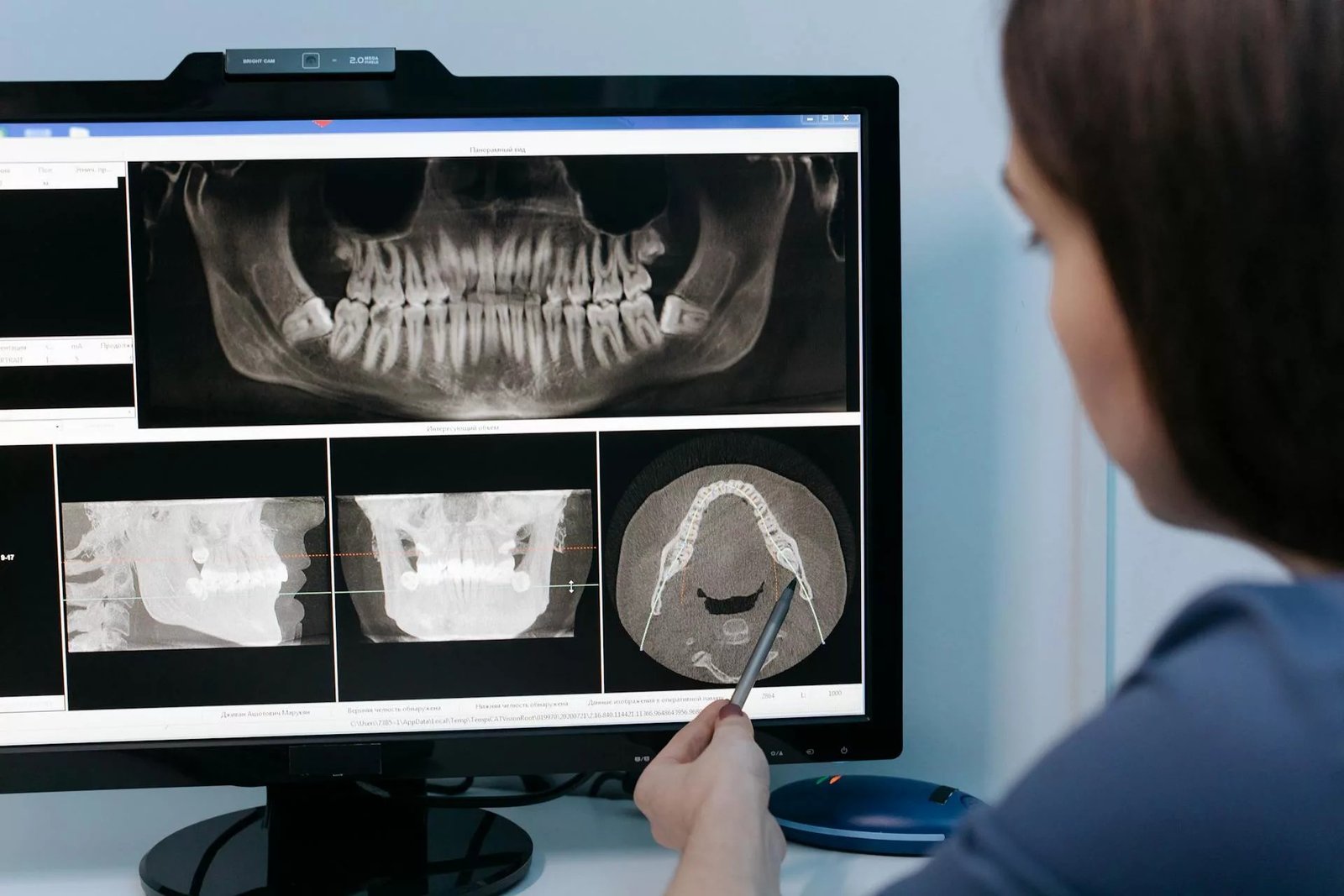
Imaging Tests:
Different imaging tests, such as Computed tomography (CT) scans, X-rays, provide a picture of your hard structures. However, an important test in skeletal fluorosis diagnosis is the Dual-energy X-ray Absorptiometry (DEXA) scan. This painless test uses two different X-ray beams to measure mineral bone density (BMD). This particular test is useful in diagnosing fluorosis-associated bone complications like osetosclerosis.
Blood Test:
Doctors perform plasma fluoride levels to confirm a diagnosis of fluorosis. There is no definitive scale to diagnose this condition from blood levels. However, in most patients with fluorosis, there is a raised level, i.e., greater than 4 µmol/L.
Urinalysis:
In this case, urinalysis helps check the level of fluorides in your pee. Studies show that high levels of fluorides in body fluids, i.e., blood and urine, provide ample data in the diagnosis of fluorosis.[24]
Differential Diagnosis:
Enamel Hypoplasia Vs Fluorosis
Both these dental conditions are characterized by changes in the structure of enamel. However, enamel hypolasia is a disorder in which there is incomplete mineralization/formation of the enamel layer. This results in thin and weak enamel, which is prone to breakage and caries/decay. On the other hand, fluorosis is an enamel formation defect that appears as white or brown spots in the tooth, but there is no weakening of the enamel.
Different conditions fall under the differential diagnosis of skeletal fluorosis, including:
- Joint ankylosis
- Spondyloarthropathies
- Diffuse idiopathic skeletal hyperostosis
Fluorosis Treatment
Dental Fluorosis Treatment:
Apart from aesthetic concerns, DF does not pose any functional or masticatory problems. Therefore, dentists advise aesthetic treatments for moderate to severe cases. Your dentist may advise the following cosmetic procedures:
Teeth Whitening
Teeth bleaching to change your enamel color is a highly prevalent dental procedure for fluorosis patients. Research suggests that it is one of the most promising treatments for dental fluorosis, along with resin infiltration and microabrasion.[25]

Dental whitening/bleaching helps change the color of teeth with fluoride stains.
In this procedure, the dentist applies a bleaching gel (containing acid) to your teeth for a specific period. The acid bleaches the enamel and lightens its color, thereby improving your appearance. This strategy works best for mild fluorosis.
Microabrasion
Enamel microabrasion involves removing a small layer of enamel from your teeth to eliminate fluorosis stains. It has proven to be a safe and effective approach that is minimally invasive. Moreover, as it involves removing the stained enamel, the results of enamel microabrasion for mild-to-moderate fluorosis are long-lasting.[26]
Dental Bonding/Resin Infiltration
Several dentists use resin bonding to cover the fluorosed tooth surfaces. Tooth-colored composite resins provide a near-natural look while hiding the underlying defect.
Dental Veneers
Minimal procedures like dental veneers offer good results in managing DF. Custom-made veneers are made over minimally prepared teeth. Made out of porcelain, zirconia, or resin, veneers cover only the outer surface of your teeth. Veneers are not as strong and long-lasting as dental crowns. Doctors take mouth impressions for veneer preparation.
Dental Crowns
Another long-lasting treatment option for the condition is a dental crown. Made from materials similar to veneers, dental crowns cover the entire structure of your tooth. Thus, they are more robust.
To make a custom-made crown, your dentist will prepare (cut the tooth), take an impression of your mouth, and fix the crown with dental cement (after it is prepared by a dental lab).
Non-Skeletal & Skeletal Fluorosis Treatment:
The first step in the management of this type is to eliminate the source of the disorder. Using water purification plans like reverse osmosis and distillation helps lower the fluoride content. After management, doctors manage the conditions with anabolic steroids to alleviate osteoporotic symptoms. NSAIDs help lower pain and inflammation. Calcium and vitamin D supplementation can slow down the process of fluorosis-induced bone hardening. Hence, they may also be advised.
How To Prevent Fluorosis?
The only way to prevent this condition is to know about your groundwater fluoride levels and make amends to ensure optimal levels in your drinking water.
Final Word
Fluorosis is a condition characterized by overdeposition of fluorides in your hard tissues, like teeth and bones. Dental fluorosis is more common than non-skeletal and skeletal fluorosis and manifests as white to brown specks/spots on tooth surfaces. On the other hand, skeletal fluorosis involves osteosclerosis and hardening of bone, which makes them brittle. Therefore, patients experience increased chances of bone breakage, myelopathies/radiculopathies, muscle pain/stiffness, and abdominal symptoms.
The main cause of this condition is prolonged ingestion of heavily fluoridated water (above 1.5 mg/L) and toothpaste. Doctors treat DF with cosmetic dental procedures like veneers, crowns, microabrasions, resin bonding, and teeth bleaching. Skeletal fluorosis treatment involves calcium and vitamin D supplementation to reduce bone hardening, while NSAIDs and steroids alleviate symptoms.
References
[1] Khan, F., Ajmal, A., Shawana, M., Khurshid, A. A., & Ahmed, J. (2022). Prevalence of Dental Fluorosis amongst patients attending the tertiary care hospital, Peshawar, Pakistan: Prevalence of Dental Fluorosis amongst Patients.Pakistan BioMedical Journal, 55-59.
[2] Srivastava, S., & Flora, S. J. S. (2020). Fluoride in drinking water and skeletal fluorosis: a review of the global impact.Current environmental health reports,7(2), 140-146.
[3] Montanher, P. L., Velasco, S. M., Montanher, R. C. P., Souza, T. M., Mamani, M. P., Bastos, J. R. M., … & Bastos, R. S. (2024). Impact of dental fluorosis on the oral health-related quality of life: a systematic review.Clinical Oral Investigations,28(11), 599.
[4] Sellami, M., Riahi, H., Maatallah, K., Ferjani, H., Bouaziz, M. C., & Ladeb, M. F. (2020). Skeletal fluorosis: don’t miss the diagnosis!. Skeletal radiology, 49(3), 345-357.
[5] Duarte, M. B. S., Carvalho, V. R., Hilgert, L. A., Ribeiro, A. P. D., Leal, S. C., & Takeshita, E. M. (2021). Is there an association between dental caries, fluorosis, and molar-incisor hypomineralization?.Journal of Applied Oral Science,29, e20200890.
[6] Idowu, E. A., Ibiyemi, O., Taiwo, O. O., & Afolabi, A. O. (2022). Presentation and management of dental fluorosis in a resource-limited facility in North-Central, Nigeria.Nigerian Dental Journal,30(2), 31-45.
[7] Idowu, E. A., Ibiyemi, O., Taiwo, O. O., & Afolabi, A. O. (2022). Presentation and management of dental fluorosis in a resource-limited facility in North-Central, Nigeria.Nigerian Dental Journal,30(2), 31-45.
[8] Gupta, R., Kumar, A. N., Bandhu, S., & Gupta, S. (2007). Skeletal fluorosis mimicking seronegative arthritis.Scandinavian journal of rheumatology,36(2), 154-155.
[9] Shruthi, M. N., & Anil, N. S. (2018). A comparative study of dental fluorosis and non-skeletal manifestations of fluorosis in areas with different water fluoride concentrations in rural Kolar.Journal of family medicine and primary care,7(6), 1222-1228.
[10] Mulualem, D., Hailu, D., Tessema, M., & Whiting, S. J. (2022). Association of dietary calcium intake with dental, skeletal and non-skeletal fluorosis among women in the Ethiopian Rift Valley.International Journal of Environmental Research and Public Health,19(4), 2119.
[11] Srivastava, S., & Flora, S. J. S. (2020). Fluoride in drinking water and skeletal fluorosis: a review of the global impact.Current environmental health reports,7(2), 140-146.
[12] Choubisa, S. L. (2022). Radiological findings more important and reliable in the diagnosis of skeletal fluorosis.Austin Med Sci,7(2), 1-4.
[13] Cook, F. J., Seagrove-Guffey, M., Mumm, S., Veis, D. J., McAlister, W. H., Bijanki, V. N., … & Whyte, M. P. (2021). Non-endemic skeletal fluorosis: Causes and associated secondary hyperparathyroidism (case report and literature review).Bone,145, 115839.
[14] Singhai, A., Mishra, V. N., & Ingle, V. (2022). A case report of skeletal fluorosis leading to cervical compressive myelopathy and a review of literature.MGM Journal of Medical Sciences,9(3), 435-438.
[15] Shah, D., Dhawale, A., Chaudhary, K., & Achalare, A. (2021). Skeletal Fluorosis With Thoracic Myelopathy: A Report of 2 Cases.International Journal of Spine Surgery,14(s4), S89-S95.
[16] Gassara, Z., Feki, A., Fourati, H., Akrout, R., & Baklouti, S. (2021). Spinal cord compression as the presenting feature of skeletal fluorosis.Authorea Preprints.
[17] Ambwani, V., Dongre, N., Singh, V., & Paliwal, V. K. (2021). Fluorosis causing spinal cord compression.Practical Neurology,21(1), 69-70.
[18] Prabhakar, A., Abdulkhayarkutty, K., Cheruvallil, S. V., & Sudhakaran, P. (2021). Effect of endemic fluorosis on cognitive function of school children in Alappuzha District, Kerala: A cross sectional study.Annals of Indian Academy of Neurology,24(5), 715-720.
[19] Ren, C., Zhang, P., Yao, X. Y., Li, H. H., Chen, R., Zhang, C. Y., & Geng, D. Q. (2021). The cognitive impairment and risk factors of the older people living in high fluorosis areas: DKK1 need attention.BMC Public Health,21(1), 2237.
[20] Simmer, J. P., Hardy, N. C., Chinoy, A. F., Bartlett, J. D., & Hu, J. C. (2020). How fluoride protects dental enamel from demineralization.Journal of International Society of Preventive and Community Dentistry,10(2), 134-141.
[21] Ling, Y., Podgorski, J., Sadiq, M., Rasheed, H., Eqani, S. A. M. A. S., & Berg, M. (2022). Monitoring and prediction of high fluoride concentrations in groundwater in Pakistan. Science of the Total Environment, 839, 156058
[22] Yasar, A., Javed, T., Kausar, F., Shamshad, J., Hayat Khan, M. U., & Iqbal, R. (2021). Ground water toxicity due to fluoride contamination in Southwestern Lahore, Punjab, Pakistan.Water Supply,21(6), 3126-3140.
[23] Siraj, F., Alam, M. S., Khan, I. A., & Arbab, K. N. (2024). Usage of Fluoridated toothpaste in children and Fluorosis of permanent teeth.Isra Medical Journal,16(1).
[24] Mondal, N. K. (2021). Diagnosis of fluorosis by analysis of fluoride content in body fluids using ion selective electrode method. InTranslational Urinomics(pp. 121-127). Cham: Springer International Publishing.
[25] Di Giovanni, T., Eliades, T., & Papageorgiou, S. N. (2018). Interventions for dental fluorosis: A systematic review.Journal of Esthetic and Restorative Dentistry,30(6), 502-508.
[26] Goel, A., Arya, A., Arora, A., Grewal, M. S., & Verma, S. (2021). Microabrasion-a conservative approach for mild to moderate fluorosis–a case report.Journal Of Evolution Of Medical And Dental Sciences,10, 30.