

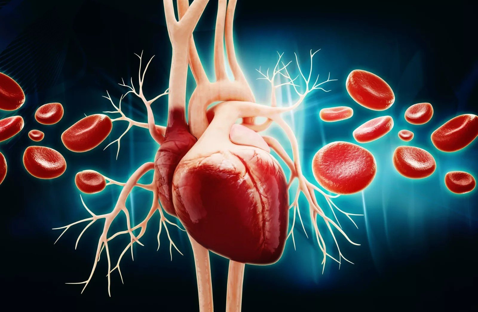
Hypovolemic Shock Blood Hypovolemic shock is a life-threatening condition that occurs when there is a significant loss of blood or fluid volume, preventing the heart from maintaining adequate cardiac output to meet the body’s needs. This reduction in circulating volume leads to decreased tissue perfusion and oxygen delivery, which can quickly progress to cellular hypoxia, organ dysfunction, and ultimately death if not treated promptly.Hemorrhagic shock. New England Journal of Medicine, 2018. 378(4): p. 370-379.” style=”position:relative;color:#309b65;cursor:help;border-bottom:1px dotted #309b65;font-weight:bold”>[1] Significant blood loss from trauma, internal bleeding, severe dehydration, and childbirth complications are the common causes of this condition. Patients typically present with mild to severe symptoms such as rapid heart rate, sweating, anxiety, and low urine output, and as shock progresses, hypotension, altered mental status, and weak pulses may develop. Immediate, effective treatment is essential to improve the condition and prevent complications. Treatment starts with restoring blood volume and can ultimately include surgical intervention to control active bleeding.
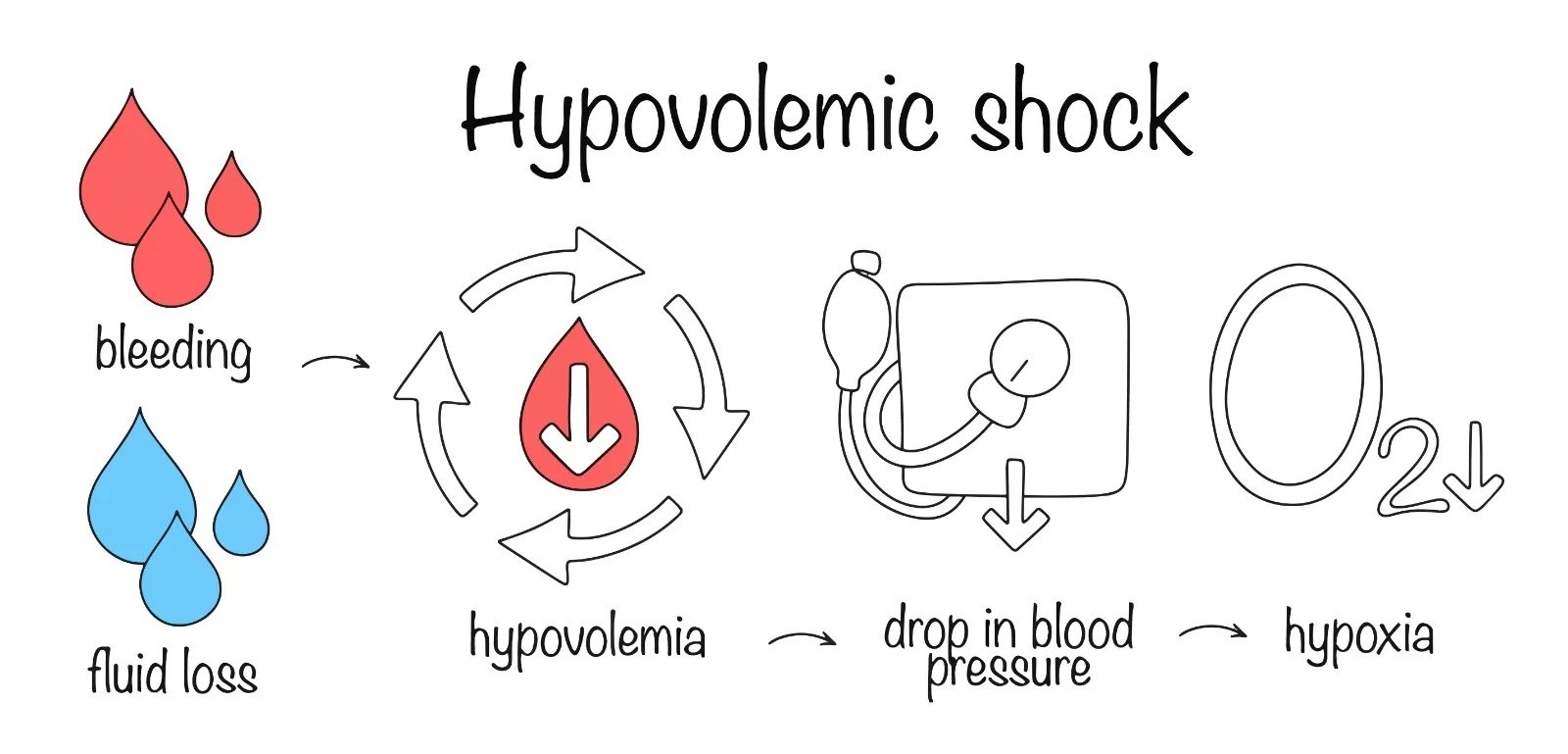
Causes of Hypovolemic Shock: Hypovolemic Shock Blood
Hypovolemic shock results from both non-hemorrhagic (fluid loss) and hemorrhagic (blood loss) causes. The causes of hemorrhagic shock include:
- Trauma
- Gastrointestinal bleeding
- Postoperative or intraoperative bleeding
- Postpartum hemorrhage
- Ruptured aneurysms
- Gynecologic bleeding (including uterine and vaginal bleeding)
On the other hand, the non-hemorrhagic hypovolemic shock arises from significant loss of body fluids that lowers the adequate circulating volume. Renal causes of non-hemorrhagic shock include excessive water and sodium loss that can occur with the use of diuretics, salt-wasting nephropathies, and osmotic diuresis from hyperglycemia.
Gastrointestinal causes involve diarrhea, severe vomiting, and excessive losses through fistulas or stomas.
Skin is the key site of fluid loss. Individuals who exercise in hot environments lose a significant volume of fluid. Patients with compromised skin barriers or burns may lose up to two liters of fluid per hour.[2]
Third-spacing or fluid sequestration occurs when fluid shifts from the intravascular space into the interstitial or “third” space. This shifting results in intravascular volume depletion and potential hypovolemic shocks. It occurs in intestinal obstruction, burns,pancreatitis, obstruction of a major vein, as postoperative complications, or in any other pathological condition that results in a massive inflammatory response.[3]
Pathophysiology of Hypovolemic Shock
Hypovolemia lowers the cardiac output, impairing tissue perfusion and oxygen delivery. All these events lead to tissue hypoxia, disrupting cellular function. Initially, compensatory mechanisms such as tachycardia, vasoconstriction, and activation of the renin-angiotensin-aldosterone system attempt to preserve circulation. As shock progresses, inadequate oxygen delivery leads to tissue hypoxia, acidosis, and impairment of endothelial-dependent vasodilation. Inflammatory and anti-inflammatory mediators are activated, further contributing to systemic dysfunction. If left uncorrected, the continuing nutrient and oxygen deprivation results in progressive cellular damage, endothelial dysfunction, and systemic acidosis.
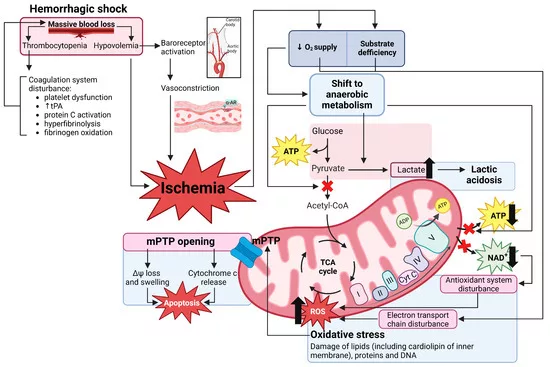
Pathogenesis of hemorrhagic shock. The massive blood loss leads to hypovolemia and thrombocytopenia, resulting in peripheral vasoconstriction and coagulopathy, respectively. Image Courtesy: Hemorrhagic Shock and Mitochondria: Pathophysiology and Therapeutic Approaches by Andrianova et al. 2025, doi.org/10.3390/ijms26051843, available via https://www.mdpi.com/1422-0067/26/5/1843, CC BY4.0.
Stages of Hypovolemic Shock
Hypovolemic shock progresses over a series of stages. These stages include:
Stage I:
Stage I includes losing up to 15% of the total blood volume (approximately 750 mL). In this stage, the heart rate is normal or minimally elevated, and there is no change in respiratory rate, pulse pressure, and blood pressure.[4]
Stage II:
In this stage, the volume loss ranges from 15 to 30% (750 to 1500 mL). The pulse pressure begins to narrow, and the systolic pressure remains normal or slightly decreased. However, the respiratory rate and heart rate become elevated (20 to 24 RR, and 100 to 120 BPM).[5]
Stage III:
Stage III involves the volume loss from 30 to 40% of the total blood volume (1500 to 2000 mL). Respiratory and heart rate become significantly elevated (above 120 BPM). There is a drop in blood pressure, changes in the mental status of the patient, and a decrease in urine output.[6]
Stage IV:
In stage IV, you can lose more than 40% of your blood volume. Tachycardia becomes highly pronounced (above 120 BPM), and hypotension with narrow pulse pressure becomes less than 25 mmHg.[7]
Symptoms of Hypovolemic Shock
Patients can present with various symptoms and signs based on the underlying causes. Potential causes of hypovolemia are gastrointestinal, hemorrhage, skin losses, renal, and third-space sequestration. The initial and progressive symptoms include:
- Tachycardia
- Tachypnea
- Clammy and pale skin
- Fatigued or feeling weak
- Confusion or anxiety
- Thirst
- Sweating
- Hypotension
- Oliguria
- Fainting
- Dizziness
Cause-Specific (Hemorrhagic) symptoms include:
- External or internal bleeding (scalp, peritoneal, thoracic, or retroperitoneal spaces)
- Muscle or subcutaneous swelling
- Bruising due to internal bleeding
- Hematemesis (bloody vomiting)
- Black or bloody stool (melena, hematochezia)
Symptoms due to Gastrointestinal Losses include
- Nausea
- Vomiting
- Diarrhea
- Abdominal pain
- Fever
- High ostomy output
Patients with skin losses, such as burns or exudative lesions of the skin, present pain and symptoms related to that specific skin condition.
Patients with renal losses can present the following symptoms:
- Headache
- Dizziness
- Lethargy
- Fatigue
- Dehydration
- Arrhythmia
- Hypotension
- Muscle weakness[8]
- Constipation
- Polyuria
- Kidney stones
- Electrolyte disturbances
The typical symptoms of hypovolemic shock depend on the electrolyte imbalances, acid-base disorders, and volume depletion. As shock advances, patients may develop an altered mental status due to inadequate cerebral, coronary, and mesenteric perfusion.
Diagnosis of Hypovolemic Shock
Doctors first look at the physical symptoms and take a detailed medical history, and then order further tests if required.
History & Physical Examination:
The physical presentation of hypovolemia varies significantly in children and adults. Older adults exhibit nonspecific symptoms that can make the diagnosis more challenging. Physical findings that suggest volume depletion are decreased skin turgor, dry mucous membrane, clammy skin, cold, tachycardia, cyanosis, low jugular venous pressure, and prolonged capillary refill.[9] Additionally, a history of trauma, recent surgery, or overt bleeding can be present in people with hemorrhagic shock.
Laboratory Tests:
The doctors recommend these initial laboratory tests: complete blood count (CBC), basic metabolic panel/blood chemistry (electrolytes, glucose), blood lactate, coagulation studies (PT/INR, aPTT), renal function (creatinine, BUN), blood type & crossmatch when bleeding is suspected, and arterial/venous blood gas if acid–base status is needed.
Abnormal laboratory findings in vomiting, diuretics, and gastrointestinal losses include:
- Hypokalemia, hyponatremia, and metabolic acidosis or alkalosis
- Elevated creatinine and blood urea nitrogen (BUN) are usually due to dehydration.
- Hemoconcentration with elevated hematocrit; serum albumin may appear elevated, but can also be reduced in protein-losing states
Abnormal laboratory findings in prolonged hypoperfusion or acute kidney injury include:
- Hyperkalemia, metabolic acidosis, and elevated urine specific gravity (concentrated urine).
- Low urine sodium, along with concentrated urine or low urine output, can suggest that the body is low on fluids. However, these findings can also be caused by other conditions like diuretic use or kidney disease, so they should be interpreted together with the patient’s symptoms and other test results.[10]
Imaging Tests:
The commonly recommended tests to confirm the diagnosis of hypovolemic shock include:
- A CT Scan is used to visualize the internal injuries or sources of fluid loss or bleeding.
- Focused Assessment with Sonography for Trauma (FAST) ultrasound is used for the same purpose as a CT scan.
- Echocardiography and endoscopy for the evaluation of cardiac function and gastrointestinal bleeding, respectively. If the imaging studies find evidence of fluid loss or organ injury, there will be a high clinical suspicion of the disorder.
Hemodynamic Monitoring:
Clinicians measure the hemodynamic profiles through pulmonary artery catheters. The measured parameters include cardiac output, mixed venous oxygen saturation, and systemic vascular resistance. A low mixed venous oxygen saturation indicates tissue hypoxia, while an abnormally high value may reflect poor cardiac output or impaired oxygen extraction.
Management & Treatment of Hypovolemic Shock
The initial step in managing this condition is to differentiate between hemorrhagic and non-hemorrhagic causes. Immediate recovery and control of the bleeding source are essential for hemorrhagic shock. It can improve survival and lower blood product transfusions. The healthcare providers achieve control over the bleeding source via direct pressure, fracture stabilization, or surgical interventions.
Prehospital Care:
The prehospital care team works to prevent further injury. Immediate transport of the sick patient is the most critical aspect of prehospital care, along with the immediate initiation of treatment. Direct pressure on the external bleeding vessels can prevent loss of blood. To avoid further injury, they immobilize the cervical spine of the patient and move him on the stretcher. Immobilization of the fracture can further minimize the blood loss and neurovascular injury. Positive pressure ventilation diminishes venous return and cardiac output and can worsen the shock state. Ventilation and oxygenation are necessary, but excessive pressure ventilation can cause death to a patient with hypovolemic shock.
Emergency Department Care:
There are three goals in the emergency care of a patient with hypovolemic shock. These goals include maximization of the oxygen delivery, control of further blood loss, and fluid resuscitation.
Management of Non-Hemorrhagic Hypovolemic Shock:
Providers ensure adequate oxygenation and ventilation, restore circulating volume, and optimize oxygen saturation. Fluid resuscitation is started with warm isotonic crystalloids (30 mL/kg), typically given in 500 mL boluses for adults.[11]
Vasopressors
Medications such as norepinephrine, dopamine, dobutamine, or epinephrine should only be started after adequate fluid resuscitation. Starting vasopressors too early can worsen tissue hypoperfusion due to vasoconstriction.[12]
In hemorrhagic shock, excessive crystalloids should be avoided. Instead, early use of blood products in balanced transfusion protocols (packed RBCs, plasma, platelets) is preferred to avoid dilutional coagulopathy.
Prognosis
Prognosis depends on the cause and severity of the hypovolemic shock. The prognosis only worsens when patients progress to multiorgan failure. Patients with preexisting comorbidities can also experience poorer prognostic outcomes.
Complications
The most common complication of hypovolemic shock is circulatory failure. Additional complications can occur either due to hypovolemia or as a result of the treatment. The potential complications are:
- Coagulopathy
- Abdominal compartment syndrome
- Hypothermia
- Acute respiratory distress syndrome
- Renal failure
- Stroke
- Myocardial infarction
- Transfusion-related infections or reactions
- Cognitive dysfunction
- Limb amputation
- Condition-specific complications such as Sheehan syndrome (postpartum hemorrhage), Asherman syndrome (uterine scarring after obstetric interventions), or psychological sequelae such as post-traumatic stress disorder)
Hypovolemic Shock versus Hemorrhagic Shock
Hemorrhagic shock is a subset of hypovolemic shock. The loss of whole blood specifically causes it, while hypovolemic shock is a significant reduction in the circulating blood or plasma. Some of the differences between the two are summarized below in Table 1.
Table 1: Difference between hypovolemic and hemorrhagic Shock
| Features | Hypovolemic Shock | Hemorrhagic Shock |
|---|---|---|
| Causes | DiarrheaVomiting, etc. | Gastrointestinal bleedingRuptures, etc. |
| Type of fluid loss | Blood/plasma or gastrointestinal fluids | Only blood |
| Treatment focus | Replacement of fluids or blood and treatment of the underlying cause. | Replace blood and control bleeding. |
Final Thoughts
Hypovolemic shock is a severe and life-threatening condition. Early recognition and immediate interventions are crucial for the reduction of morbidity and mortality. Treatment focuses on rapidly restoring blood volume and stabilizing vital functions via intravenous fluid resuscitation, transfusion of blood if required, and medications to support the cardiac output and blood pressure. Sometimes the patient needs to control active bleeding through surgical interventions. Effective management requires a coordinated, interprofessional approach for prehospital and in-hospital treatment of the condition.
References
[1] Cannon, J.W., Hemorrhagic shock. New England Journal of Medicine, 2018. 378(4): p. 370-379.
[2] Taghavi, S., A. Nassar, and R. Askari, Hypovolemic shock. 2018.
[3] Hooper, N. and T.J. Armstrong, Hemorrhagic shock, in StatPearls [internet]. 2022, StatPearls Publishing.
[4] Hooper, N. and T.J. Armstrong, Hemorrhagic shock, in StatPearls [internet]. 2022, StatPearls Publishing.
[5] Hooper, N. and T.J. Armstrong, Hemorrhagic shock, in StatPearls [internet]. 2022, StatPearls Publishing.
[6] Hooper, N. and T.J. Armstrong, Hemorrhagic shock, in StatPearls [internet]. 2022, StatPearls Publishing.
[7] Hooper, N. and T.J. Armstrong, Hemorrhagic shock, in StatPearls [internet]. 2022, StatPearls Publishing.
[8] Garg, Y., et al., Primary aldosteronism diagnosis in the intensive care unit: resistant alkalosis and hypokalemia during severe sepsis with hyperlactatemia: a case report. Journal of Medical Case Reports, 2025. 19(1): p. 192.
[9] Jegerlehner, S., et al., Association of central capillary refill time with mortality in adult trauma patients: a secondary analysis of the crash-2 randomised controlled trial data. Scandinavian Journal of Trauma, Resuscitation and Emergency Medicine, 2025. 33(1): p. 82.
[10] Hato, T. and R. Ng, Diagnostic value of urine sodium concentration in hyponatremia due to syndrome of inappropriate antidiuretic hormone secretion versus hypovolemia. Hawaii medical journal, 2010. 69(11): p. 264.
[11] Investigators, P., A randomized trial of protocol-based care for early septic shock. New England Journal of Medicine, 2014. 370(18): p. 1683-1693.
[12] Shagana, J., et al., Hypovolemic shock-A review. Drug Invention Today, 2018. 10(7): p. 1102-05.