
, which may...)

Klebsiella Infection Risks Klebsiella infection is a bacterial infection caused by the Klebsiella species (primarily, K. pneumoniae), which may cause severe disorders such as pneumonia, urinary tract infection (UTI), bloodstream infection, and wound infection. The most pathogenic species is K. pneumoniae. It is a gram-negative, encapsulated, and non-motile bacterium that was first described by Carl Friedlander in 1882. It is an important cause of hospital-acquired and ventilator-associated pneumonia, particularly in critically ill patients. Surveillance studies report that K. pneumoniae accounts for a significant proportion of hospital-acquired pneumonia worldwide, though prevalence varies by region and setting.[1] There are two other Klebsiella species that cause human illness. These are K. oxytoca and K. granulomatis.[2]
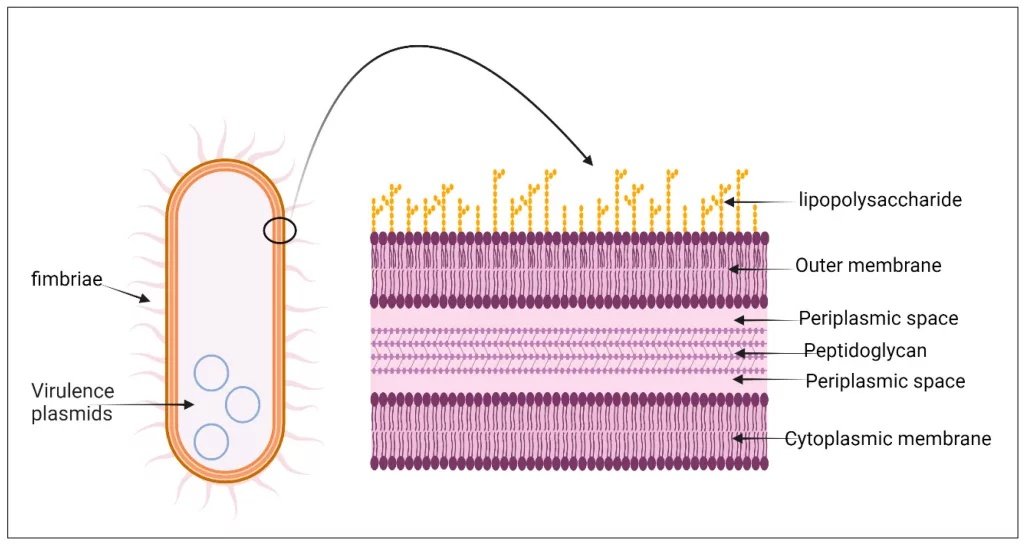
The cell wall structure ofK. pneumoniae. Like most other gram-negative bacteria, it is composed of an outer membrane that contains lipopolysaccharide, periplasmic spaces, a thin layer of peptidoglycan, and a cytoplasmic membrane. Image Courtesy: Hypervirulent Klebsiella pneumoniae: Insights into Virulence, Antibiotic Resistance, and Fight Strategies Against a Superbug et al, 2025, doi.org/10.3390/ph18050724, available via: https://www.mdpi.com/1424-8247/18/5/724, CC BY 4.0.
Causes of Klebsiella Infection: Klebsiella Infection Risks
Klebsiella infections occur when the bacteria, which are normally commensal organisms in the human gastrointestinal tract, obtain an opportunity to cause disease. The spread of these germs in hospitals is either by touch or by contaminated equipment. They primarily target individuals with compromised immunity, such as diabetic and ventilated patients.[3] Infection happens primarily in healthcare facilities through direct contact. Translocation of endogenous gut flora also plays an important role. Certain risk factors increase host susceptibility, allowing normally harmless colonizing bacteria to become pathogenic.
Transmission between People
The hands are considered the most common way through which Klebsiella passes from one person to another. The infection transmits when medical personnel come in contact with one patient and then the other, without cleaning their hands properly. Klebsiella species can survive in moist environments such as sinks, drains, feces, and respiratory secretions, contributing to environmental contamination. Patient rooms may become hotspots during outbreaks. Medical equipment such as breathing tubes, urinary catheters, or IV lines provides direct entry points into normally sterile body sites. Klebsiella is not transmitted via airborne spread. In the community, infection may occur due to contaminated water or soil, though healthcare-associated transmission remains the most common source.
Endogenous Sources
These are the bacteria that commonly hang in the silence of your intestines or throat; however, when host defenses are disrupted by antibiotics, surgery, chemotherapy, or critical illness, bacterial translocation into the bloodstream may occur. Patients with diabetes are particularly vulnerable due to impaired neutrophil function and altered immune responses, rather than glucose directly promoting bacterial growth.
Who is at Risk?
Natural protective mechanisms of your body weaken due to old age, disease, or treatment. This renders Klebsiella even more dangerous. The typical causes of the infection are as follows:
- Individuals who have a weak immune system because of cancer medications, HIV, or steroids are prone to infection.[4]
- The longer you spend in the hospital, the more chances you have of picking it up, particularly in ICUs.
- Diabetes, too, is a risk factor since it interferes with the cells that fight infections.
- Alcohol addicts are the most susceptible; the alcohol weakens the lungs, impairing airway protective reflexes and pulmonary immunity.
- Tubes and devices, such as pneumonia ventilators and UTI catheters, may be the most frequent cause of infection.
- Neonates, older adults, and postoperative patients are also at higher risk.
- Effective prevention relies on hand hygiene, device stewardship, and active surveillance programs.
Symptoms of Klebsiella Infection
Klebsiella infections, mainly from K. pneumoniae, present symptoms varying by site. The symptoms can be more severe in immunocompromised individuals, such as diabetics or alcohol use disorder.
Patient’s experience pneumonia symptoms, including:
- High fever
- Chills
- Productive cough with bloody sputum[5]
Other symptoms include:
- Chest pain
- Shortness of breath
- Signs of lobar consolidation on examination, including crackles
UTIs typically present with:
- Dysuria
- Frequent or urgent urination
- Suprapubic pain
- Hematuria
- Flank pain
- Nausea
- Vomiting
Bacteremia or sepsis may occur with symptoms of:
- Sudden fever
- Chills
- Tachycardia
- Hypotension
- Confusion
- Abdominal pain
- Rapid breathing
- Skin rash may occur in severe systemic infection
Other symptoms include:
- Meningitis (fever, headache, neck stiffness, photophobia, and seizures)
- Wound and skin infections (redness, swelling, pus, pain)
- Liver abscess, particularly with hypervirulent strains
Klebsiella Infection Diagnosis
Diagnosis of Klebsiella infections assumes clinical examination using history and physical examination. Confirmation goes further to clinicians, laboratory, and imaging techniques.
Physical Examination and History Taking
Patients state the following risk factors, such as hospitalization, the use of antibiotics, diabetes, alcoholism, or ventilator support. Physical findings may include fever, tachycardia, tachypnea, pulmonary crackles or consolidation, flank tenderness in pyelonephritis, and abdominal tenderness or jaundice in liver abscess.
Laboratory Tests
Lab tests that aid in diagnosing Klebsiella infection are:
Gram Staining
This preliminary quick test on direct specimens such as sputum, urine, pus, or blood smears identifies Gram-negative, plump rods with a prominent capsule. They can either look like a safety pin or a bipolar stain because of the shrinkage of capsules.[6] However, A “safety-pin” appearance is not a consistent or reliable feature of Klebsiella species and should not be considered diagnostic.
Culture Methods
The specimens (blood, sputum, urine, CSF, wounds) are cultured on selective media. Mucoid, lactose-fermenting pink colonies are visible in MacConkey agar. Blood agar results in butyric acid odor, grey-white colonies in the form of domes.[7]
Antimicrobial Susceptibility Tests (AST)
Disk diffusion or MIC testing guides antibiotic selection, particularly in multidrug-resistant infections.
Imaging Studies
- Chest X-ray pneumonia presents with dense lobar consolidation (common in the upper lobes), fissures that are bulging, cavitation, abscess, or pleural effusion.
- Hypoechoic liver abscesses (hypervirulent strains), splenomegaly, or peritonitis are detected by Abdominal CT or ultrasound. Edema of pyelonephritis is confirmed by the use of a flank ultrasound. Imaging also assists in abscess drainage.
Molecular Tests
Polymerase chain reaction (PCR) or (Matrix-Assisted Laser Desorption/Ionization Time-of-Flight) MALDI-TOF mass spectrometry rapidly identifies species and resistance genes directly from cultures.
Klebsiella Infections Management and Treatment
Klebsiella (mainly K. pneumoniae) infections require management that focuses on control of the source and targeted antibiotics depending on susceptibility and supportive care, in addition to modifications in multidrug-resistant (MDR).
Initial Supportive Care
The medical practitionerwill stabilize sepsis or shock patients using IV fluids, oxygen, and vasopressors. They resort to mechanical ventilation for severe pneumonia. Prompt removal of infected or unnecessary devices is critical. It may reduce bacterial load. ICU monitoring is required for severe cases.
Antibiotic Therapy
Empiric IV therapy may include:[8]
- Piperacillin-tazobactam or cefepime for suspected susceptible strains.
- Vancomycin may be included only for empiric MRSA coverage and has no activity against the Klebsiella species.
- Ceftriaxone or levofloxacin for low-risk community-acquired infections when susceptibility is confirmed.
Treatment of Carbapenem-Resistant Enterobacteriaceae (CRE)
The following are the options for CRE:
- As initial therapy against susceptible severe infections, ceftazidime-avibactam, meropenem-vaborbactam, or imipenem-relebactam.
- Cefiderocol for cases of pyelonephritis.
Hypervirulent Klebsiella Infections
Management includes prompt drainage of liver abscesses and aggressive antibiotic therapy. Combination regimens such as meropenem plus rifampicin have been reported in refractory cases, but are not standard first-line therapy and should be guided by specialists.[9]
Prognosis of Klebsiella Infection
The prognosis for Klebsiella infections varies widely depending on the infection site, patient health, and bacterial resistance. Timely treatment often leads to recovery in milder cases. Severe forms, such as bloodstream infections or pneumonia, carry higher risks. Advanced age, ICU admission, severe sepsis, and immunosuppression are associated with poorer outcomes. Pneumonia and UTIs in healthy outpatients resolve with antibiotics like ceftriaxone, and liver abscesses from hyper-virulent strains respond to drainage plus antibiotics, with good recovery.
Differential Diagnosis
The differential diagnosis of Klebsiella infections depends on the affected site, such as pneumonia, UTI, or bacteremia. They can be distinguished from similar presentations using clinical history, imaging, and microbiology.
Pneumonia
Klebsiella pneumonia mimics community-acquired cases from Streptococcus pneumoniae or Staphylococcus aureus. It also mimics hospital-acquired infections caused by Pseudomonas aeruginosa, Acinetobacter, or Legionella. Non-infectious options include lung malignancy, ARDS, abscess, or empyema.
UTIS
Different from Escherichia coli, Proteus, or Enterococcus UTIs, symptoms include dysuria and flank pain.
Bacteremia
Bloodstream infections resemble those from E. coli, other Enterobacteriaceae, or gram-positives like Staphylococcus, and sepsis signs like fever and hypotension prompt broad differentials, including viral illnesses or endocarditis.
Klebsiella Infections Prevention
The aim of preventing the development of Klebsiella infection should focus on the lack of transmission of the infection in the health care institutions, where nosocomial spread is most common. You should minimize host susceptibility through hygiene, stewardship, and surveillance.
- Medical personnel should carry out hand hygiene either prior to or after contact with a patient with alcohol based sanitizers.
- Disinfect high-touch areas (bedrails, monitors, doorknobs, etc.) with sporicidal agents every day to decrease the risk of transmission.
- Isolation or cohorting of colonized patients.
- Interact with them using gowns and gloves.
- Avoid using unnecessary broad-spectrum agents and carbapenems.
Final Remakes
Klebsiella infections pose a major clinical challenge due to their virulence, ability to acquire antimicrobial resistance, and propensity for nosocomial spread. High-risk infections include severe pneumonia with bloody sputum, complicated UTIs, and disseminated infections caused by hypervirulent strains. Accurate diagnosis, targeted antimicrobial therapy, source control, and strict infection control practices remain essential to reduce morbidity and prevent outbreaks.
References
[1] Ashurst, J. V., & Dawson, A. (2018). Klebsiella pneumonia.
[2] Sahly, H., & Podschun, R. (1997). Clinical, bacteriological, and serological aspects of Klebsiella infections and their spondylarthropathic sequelae.Clinical Diagnostic Laboratory Immunology,4(4), 393-399.
[3] Ergul, A. B., Cetin, S., Altintop, Y. A., Bozdemir, S. E., Ozcan, A., Altug, U., … & Torun, Y. A. (2017). Evaluation of microorganisms causing ventilator-associated pneumonia in a pediatric intensive care unit.The Eurasian Journal of Medicine,49(2), 87.
[4] Sahly, H., Podschun, R., & Ullmann, U. (2002). Klebsiella infections in the immunocompromised host.The biology and pathology of innate immunity mechanisms, 237-249.
[5] , S., Yu, S., Peng, M., Qin, J., Xu, C., Qian, J., … & Zhou, P. (2021). Clinical features and development of Sepsis in Klebsiella pneumoniae infected liver abscess patients: a retrospective analysis of 135 cases.BMC Infectious Diseases,21(1), 597.
[6] Osman, E. A., El-Amin, N., Adrees, E. A., Al-Hassan, L., & Mukhtar, M. (2020). Comparing conventional, biochemical and genotypic methods for accurate identification of Klebsiella pneumoniae in Sudan.Access microbiology,2(3), e000096.
[7] Para, R. A., Fomda, B. A., Jan, R. A., Shah, S., & Koul, P. A. (2018). Microbial etiology in hospitalized North Indian adults with community-acquired pneumonia.Lung India,35(2), 108-115.
[8] Tamma PD, Aitken SL, Bonomo RA, Mathers AJ, van Duin D, Clancy CJ. Infectious Diseases Society of America guidance on the treatment of antimicrobial-resistant gram-negative infections.Clin Infect Dis. 2024;78(6):e1–e24. doi:10.1093/cid/ciad516.
[9] Chen, J., Zhang, H., & Liao, X. (2023). Hypervirulent klebsiella pneumoniae.Infection and drug resistance, 5243-5249.