

Larsen Syndrome Rare Larsen syndrome is one of the rare genetic disorders that impacts the skeletal and connective tissues of your body. The inherited disorder mainly causes facial disfiguration, joint and limb malformations, and spinal deformities. While joint deformities contribute to significant debilitation, severe spinal deformities can even interfere with normal breathing (by putting pressure on the breathing apparatus). Patients suffering from this congenital skeletal disorder are often plagued with other non-bony problems, likehearing loss, cardiac issues, and renal problems. You won’t find the disorder very commonly, and according to the National Organization for Rare Disorders, the syndrome has a frequency of 1 in 100,000.
A genetic mutation in a development-related gene (most commonly FLNB) leads to problems. Correction of bone deformities usually requires surgical intervention. Therefore, doctors fix your joints and spine with different types of surgeries. Non-interventional therapies like physical therapy, orthotic support, and bracing can help improve quality of life. Chiropractic manipulation is generally avoided due to the risk of cervical instability, but supervised physiotherapy can help improve function. Early treatment helps prevent aggravation of the disease and protects you from complications.
Larsen Syndrome Symptoms: Larsen Syndrome Rare
The most commonly encountered symptoms of the disorder include:
Facial Abnormalities
Most individuals have a flat nose and a deficient middle face, i.e., midface hypoplasia. Cephalometric analyses have revealed midface hypolasia and mandibular retrognathism, which lead to a flattened appearance of the patient.
The forehead is large and prominent, which is known as frontal bossing. Additionally, most patients possess a wide set of eyes. Ocular hypertelorism is a part of many genetic disorders. Doctors refer to the facial appearance as “dish-like” due to the prominent forehead, depressed nasal bridge, and hypertelorism.
Dental Problems
Young patients are also plagued with multiple dental complications like microdontia (small teeth), supernumerary teeth, and malocclusion. Cleft palate is a common pathology that requires extensive treatment. Usually, orthodontists, general and oral surgeons collaborate to fix appearance.
Joint And Limb Issues
Typical features of the disease include spatulate thumbs (blunt and square-shaped). You may also find extra bones in the wrists and hands of such patients. Improper fusion of bones (ossification) results in dwarf (short) limbs. Other common symptoms include:
Dislocations:
Most patients are born with dislocations of joints like the hips, knees, and elbows. In a rare case, an infant was born with bilateral dislocated hips and knees, along with severe clubfeet. These dislocations can cause pain, and if left untreated, they can lead to degenerative joint disease.
Contractures:
There may also be joint contractures, like hip flexion contracture, in which the hip joint is stuck in a fixed position. This interferes with daily activities like walking. Clinical studies hypothesized that hip flexion contracture has an impact on the alignment of the spine (sagittal spine), which further complicates things. There is a wide range of joint symptoms. While contractures are common, you may also encounter patients with hypermobility. Such individuals have an unusually large range of joint movement.
Foot deformities:
Feet abnormalities are salient features of this particular disorder. Symptoms can range from clubbing (talipes equinovarus) to z-feet deformity. Skewfoot or “z”-shaped/”c”-shaped foot is a congenital anomaly in which the toes point inward while the heel points outward.
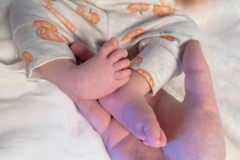
The image shows an infant with an inward-tilted clubfoot due to Larsen syndrome.
In clubfoot deformity, the foot is twisted inward and downward. Larsen syndrome is a noteworthy contributor to pediatric foot alignment deformities (especially clubfoot). There may also be other associated skeletal problems in this disorder.
Spinal Deformities
Abnormal Curvature of the Spine:
In the case of Larsen syndrome, many people encounter abnormal curvatures of the spine, which are believed to arise from hypoplasia and abnormal segmentations of the vertebrae. These spinal abnormalities are directly linked to complications like muscle weakness and even breathing compromises.
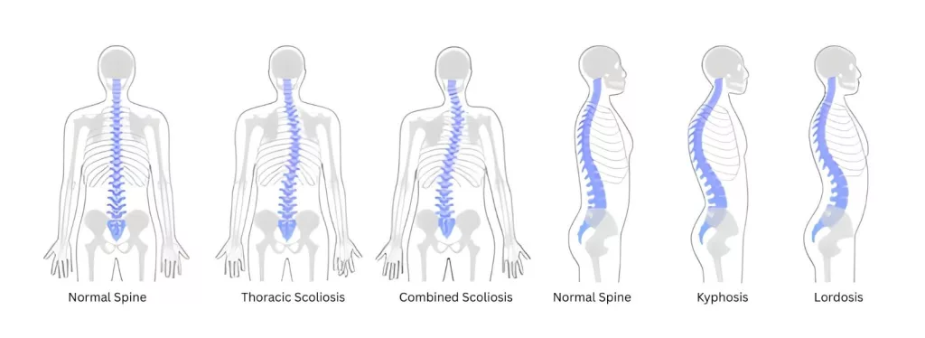
The illustration shows the different types of spinal curvatures, i.e., kyphosis, lordosis, and scoliosis, compared to a normal spine curvature.
Scoliosis is a side-to-side abnormal curvature of the spine. Patients are tilted towards one side due to the abnormally curved spine. On the other hand, kyphosis is the forward bending or rounding of the upper back. Numerous patients develop Larsen syndrome-induced early-onset scoliosis, which needs surgical intervention. Early treatment has proven to be immensely beneficial.
You may find both conditions present at the same time. Thoracolumbar kyphoscoliosis is not uncommon, and subluxations with abnormal curvatures can even prove to be fatal (due to breathing hindrance). A 5-year-old girl with severe kyphoscoliosis was referred for spinal surgery due to severe respiratory dysfunction. The respiratory function drastically improved after surgery. Hence, doctors recognize severe thoracic scoliosis as a cause for respiratory dysfunction. People may also experience nerve impingement symptoms like back pain, muscle weakness (of the limbs), and fatigue.
Other Issues
Hearing Problems:
Loss of hearing is a pretty common phenomenon in such patients. Malformed ear bones (auditory ossicles) contribute to hearing difficulties. Conductive hearing loss is the most frequent type and is attributed to ossicular malformations and middle ear infections. Cleft palate and eustachian tube dysfunction (hollow tubes that connect the mouth, nose, and ears) also contribute to the disability.
Respiratory problems:
Abnormal spine curves can cause shortness of breath (dyspnea) or episodic pauses in breathing (apnea). Due to compromised respiratory health, patients may also fall prey to repeated respiratory infections. Patients may also present with tracheomalacia/tracheobronchomalacia (weakness of the windpipe/bronchial cartilage, which leads to airway constriction).
Heart and kidney disorders:
Such individuals are also prone to developing renal and cardiac abnormalities. Children may suffer from life-threatening heart defects like valvular insufficiency (when one or more valves don’t close properly), aneurysms of the ductus arteriosus (abnormal dilation of the vessel that connects the aorta and pulmonary artery in fetal life), and aortic dilation (enlargement of the aorta).
Larsen Syndrome Causes
The genetic disorder is an outcome of a mutation in the FLNB gene. Most cases occur due to autosomal dominant FLNB mutations, many of which arise de novo. You can inherit a mutated gene from your parents or acquire the mutation later in life. This gene instructs the production of a protein called filamin B. This protein is crucial in the development of your skeleton during the intra-uterine life. It binds to another protein, actin, and promotes bone growth.
In the womb, actin-binding proteins like filamin B transduce signals for cell growth. It is expressed in the chondrocytes (cells that produce collagen) present in the growth plate. The loss of filamin B results in shortening of the long bones and an overall delay in ossification.
Larsen Syndrome Inheritance Pattern:
The disease is usually inherited in an autosomal dominant pattern, which means that only one copy of the altered gene is sufficient to cause Larsen syndrome. However, there have been cases in which the genes are passed in an autosomal recessive pattern. A mutated B4GALT7 gene is the causative gene when the disease is caused by recessive inheritance. Generally, the disease in this pattern is more severe. This variant of the disease is known as Larsen of La Reunion Island syndrome (LRS). In rare instances, there is a mutation in the CHST3 gene.
Larsen Syndrome Diagnosis
Physical Examination and History:
Your healthcare provider will physically examine your joints, spine, and craniofacial regions. Curvatures of the spine, paired with joint and spinal deformities, point toward an underlying genetic disorder. Your doctor may also ask about the history of Larsen syndrome in your family. To confirm the diagnosis, doctors further order genetic and imaging tests.
Imaging Tests:
Joint dislocation is a unique feature of the disease that can be diagnosed in a fetal ultrasound. Larsen syndrome is prenatally diagnosed on ultrasounds of fetuses having congenital knee dislocation.
X-rays have been used conventionally in visualizing bone and joint deformities. However, more advanced imaging scans CT scans and MRI scans, are used to diagnose spinal and joint issues. These imaging tests help appreciate different parameters of the skeletal structures.
EOS imaging is a type of imaging best suited for syndromes like Larsen syndrome. This technology creates 2-dimensional and 3-dimensional X-ray images of the skeletal system, which do a great job at diagnosing multiple issues like dysplasia, kyphosis/scoliosis at the same time. The horizontal and vertical X-ray beams ensure maximum area visualized with minimal exposure to radiation.
Genetic testing:
Most syndromes have a genetic component; thus, genetic testing is frequently used to identify the gene mutations leading to the disorder. In the case of Larsen syndrome suspicion, technicians search for mutations in the FLNB gene.
Diseases like Otopalatodigital syndrome, boomerang dysplasia fall in the differential diagnosis of Larsen syndrome.
Larsen Syndrome Treatment
In severe, potentially life-threatening cases, doctors first target the serious complications like respiratory issues, cardiac, and renal complications.
Surgery
Joint surgery
Reduction of dislocated joints:
Surgeons perform open reduction (surgical) of the dislocated joints in infants. Most commonly reduced joints are the hips and knees. Individuals having knee dislocations due to syndromic disorders can not be treated with closed reduction and need surgical procedures like open reduction and arthrotomy. After reduction, orthopedic surgeons stabilize the joint with the help of stiff casts and splints.
Osteotomy:
Different types of bone cutting procedures (osteotomies) are performed to realign, reshape, and/or relengthen the abnormal bones. These procedures are usually paired with joint repair surgeries for better results.
Clubfoot Correction:
The vast majority of surgeons correct the inward twisted foot by lengthening the Achilles tendon of the foot in a procedure called tenotomy. It is a minimally invasive surgical procedure that has proven to be reliable (but has its side effects).
Spinal Surgery
Spinal fusion (Arthrodesis):
In this procedure, a surgeon joins two or more vertebrae to stabilize the spine and improve function. This intervention is performed in early life to prevent potential neurological defects. The ideal age for a fusion surgery is 18 months in patients with mild deformity. It is the mode of correction for pediatric kyphosis and scoliosis. The surgeons fix the corrected spinal bones in place with the help of rods and screws.
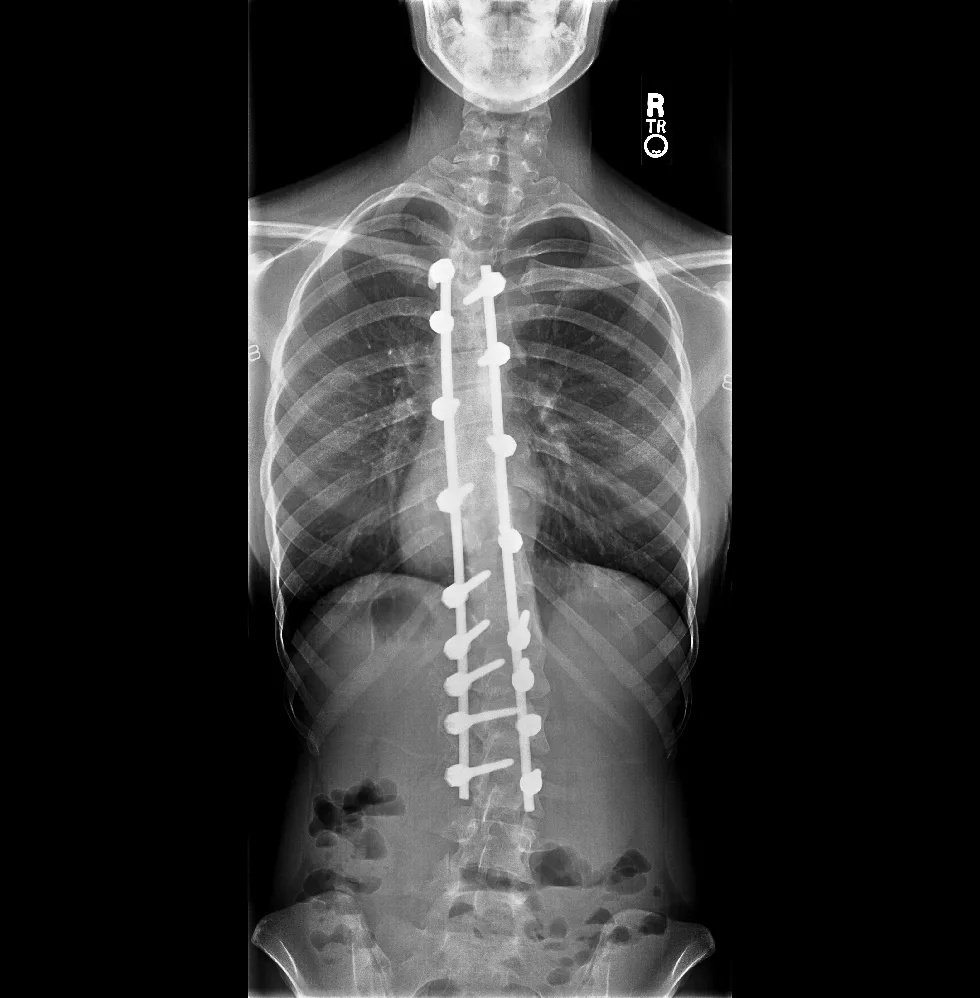
The image shows the post-operative X-ray image of a scoliosis patient. The spine is fixed in place with rods and screws.
Decompression
To relieve pressure exerted on the nerves by spine curvature, doctors perform decompression surgeries. In a lot of cases, a decompression is done with a spinal fusion surgery.
Non-Surgical Treatment
Several non-surgical approaches can help alleviate symptoms and improve patients’ quality. Some of the strategies are used solely, while others are employed in conjunction with surgical treatments.
Bracing and Support:
In some mild scoliosis cases of scoliosis, doctors might recommend a brace to growing children for spine alignment. Cervical bracing may be advised in cases where decompression is not performed. This is done to prevent the progression of cervical myelopathy (a neurologic condition due to cord compression).
Physical Therapy:
Mild spinal deformities can be corrected with physiotherapy. These non-invasive therapies are known to improve the function, strength, and range of joint motion in patients. Kinetotherapy is a type of physical therapy that uses specific movements/exercises to correct joint deformities and spinal abnormalities.
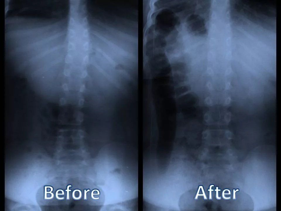
The X-ray image shows the before and after of a child’s spine (with Larsen syndrome ) being treated with chiropractic care.
Recent studies reveal that kinetotherapy can significantly help patients acquire motor skills aligned with their age-appropriate neuromotor development.
Ponseti Treatment:
In this non-surgical procedure, healthcare providers gently stretch the foot and brace it with a plaster cast to correct clubfeet. The casts are changed weekly (or after several weeks), depending on the severity of the disease. Clinicians can achieve effective results in syndromic clubfoot, but it requires a good number of corrective casts.
Hearing Aids:
As hearing loss is a highly common issue in this particular syndrome, doctors advise hearing aids to patients.
Larsen Syndrome Life Expectancy:
With good medical care, Larsen syndrome life expectancy is good, and most patients live to adulthood. Patients can live a normal life with early and aggressive treatment. However, prognosis varies depending on spinal severity, respiratory compromise, and cardiac complications.
Wrapping Up
Larsen syndrome is a rare, genetic disorder that affects your bones, joints, spine, and heart and kidneys. Patients have a dish-like facial appearance due to widely spaced eyes and a depressed midface. Infants are plagued with joint deformities (congenital dislocations, joint contractures, clubfeet, etc.) and spine anomalies (infantile kyphosis, scoliosis), which can potentially lead to serious breathing issues.
Mutations in the FLNB gene (responsible for the filamin B protein) lead to this disorder. It is transmitted majorly in an autosomal dominant pattern, but may also move in an autosomal recessive pattern, in which case it is called Larsen of La Reunion Island syndrome. Family history and imaging tests like ultrasound (during pregnancy), X-rays, MRI scans, CT scans, and EOS scans can help diagnose the disease.
Doctors fix the hard tissue issues with surgery. Joint reductions and fixations, osteotomies, and tenotomies help correct bone dysfunction. However, decompression and spinal fusion procedures treat kyphoscoliosis. Physical therapy (kinetotherapy) and Ponseti treatment (for clubfoot) can offer some help too.
References
[1] Yasunaga, M., Ishikawa, H., Yanagita, K., & Tamaoki, S. (2021). An orthodontic perspective on Larsen syndrome.BMC Oral Health,21(1), 111.
[2] Kaissi, A. A., Gubin, A., Ryabykh, S., Dougales, V., Al Kaissi, H., Kircher, S. G., & Grill, F. (2025). The Orthopedic Strategy for Patients with Larsen Syndrome.Surgical Techniques Development,14(2), 10.
[3] Matar, H. E., & Garg, N. K. (2017). Management of joint dislocations of the lower limb in Larsen syndrome: practical approach.The Annals of The Royal College of Surgeons of England,99(1), e8-e10.
[4] Nair, K. R., Sreedharan, S., Gopinath, N. P., Surendran, S., Patinharayil, G., Fazil, V. M., … & CU, J. (2025). A case report of Larsen syndrome–A rare genetic disorder affecting the musculoskeletal system.Journal of Orthopaedic Reports,4(2), 100421.
[5] ousafeiris, V., Dreyer, M. A., & Thomas, A. (2023). Pediatric foot alignment deformities. InStatPearls [Internet]. StatPearls Publishing.
[6] Siafaka, A., Angelis, S., Piagkou, M., Apostolopoulos, A., Troupis, T., & Filippou, D. (2023). Larsen syndrome and associated spinal deformities.Cureus,15(7).
[7] Furuya, M., Takeoka, Y., Yurube, T., Ito, M., Suzuki, T., Kakutani, K., & Uno, K. (2024). Two-Staged Surgery for Kyphoscoliosis in Larsen Syndrome with A 30-Year Follow-Up: A Case Report.Spine Surgery and Related Research,8(3), 338-341.
[8] Sousa Marques, L. H., Martins, D. V., Juares, G. L., Lorenzetti, F. T. M., & da Costa Monsanto, R. (2017). Otologic manifestations of Larsen syndrome.International Journal of Pediatric Otorhinolaryngology,101, 223-229.
[9] Lin, C. J., Purohit, M., & Averill, L. W. (2025). Larsen Syndrome. InAnesthesia Care of Patients with Skeletal Dysplasia(pp. 95-108). Cham: Springer Nature Switzerland.
[10] Hu, J., Lu, J., Lian, G., Ferland, R. J., Dettenhofer, M., & Sheen, V. L. (2014). Formin 1 and filamin B physically interact to coordinate chondrocyte proliferation and differentiation in the growth plate.Human molecular genetics,23(17), 4663-4673.
[11] Alessandri, J. L., Celse, T., Spodenkiewicz, M., Calaya, A., Dumont, C., Jacquemont, M. L., … & Dospeux, J. (2024). Prenatal and neonatal phenotype of Larsen of La Reunion Island syndrome (B4GALT7-linkeropathy).European Journal of Medical Genetics,69, 104940.
[12] Cavoretto, P. I., Castoldi, M., Corbella, G., Forte, A., Moharamzadeh, D., Emedoli, D., … & De Pellegrin, M. (2023). Prenatal diagnosis and postnatal outcome of fetal congenital knee dislocation: systematic review of literature.Ultrasound in Obstetrics & Gynecology,62(6), 778-787.
[13] Sakaura, H., Matsuoka, T., Iwasaki, M., Yonenobu, K., & Yoshikawa, H. (2007). Surgical treatment of cervical kyphosis in Larsen syndrome: report of 3 cases and review of the literature.Spine,32(1), E39-E44.
[14] Kaissi, A. A., Ganger, R., Klaushofer, K., & Grill, F. (2011). The management of knee dislocation in a child with Larsen syndrome.Clinics,66, 1295-1299.
[15] Kayondo, T. K., El-Adwar, K. L., Abdullah, E. A., & Mosa, M. M. (2024). Reliability of percutaneous Achilles tenotomy in the treatment of idiopathic clubfoot.Journal of Pediatric Orthopaedics,44(6), e566-e569.
[16] Icleanu, I. (2025). The Efficacy of Kinetotherapy in Larsen Syndrome in Children.SEA: Practical Application of Science,13(37).
[17] Meseguer, E. F., Sánchez, S. R., Almero, L. P., Bataller, A. R., & Rey, M. M. (2021). Syndromic clubfoot beyond arthrogryposis and myelomeningocele: orthopedic treatment with Ponseti method.Revista Española de Cirugía Ortopédica y Traumatología (English Edition),65(3), 180-185.