
 or neoplastic meningitis is a cancer involving the meninges of the...)

Leptomeningeal Carcinomatosis Diagnosis Leptomeningeal carcinomatosis (LMC) or neoplastic meningitis is a cancer involving the meninges of the brain and spinal cord. LMC occurs because of the spread of solid tumors, like blood cancers and even brain tumors, from their primary site to the cerebrospinal fluid (CSF) and meninges.
Symptoms of the LMC are very diverse and severe, depending on the part of the brain and spinal cord it infiltrates. For the definite and timely diagnosis of leptomeningeal carcinomatosis, it is important to clearly understand the pathophysiology and causes of it.
Pathophysiology of Leptomeningeal Carcinomatosis: Leptomeningeal Carcinomatosis Diagnosis
There are three layers around our brain and spinal cord. Of these three, the inner two layers, arachnoid mater and pia mater, are collectively referred to as the leptomeninges. The CSF flows in the space present between the leptomeninges, called the subarachnoid space.
In leptomeningeal carcinomatosis, cancer cells migrate from other parts of the body, such as the breast or lungs, and invade the leptomeninges of the brain. It usually occurs in the advanced stages of cancer, causing a wide range of neurological symptoms.
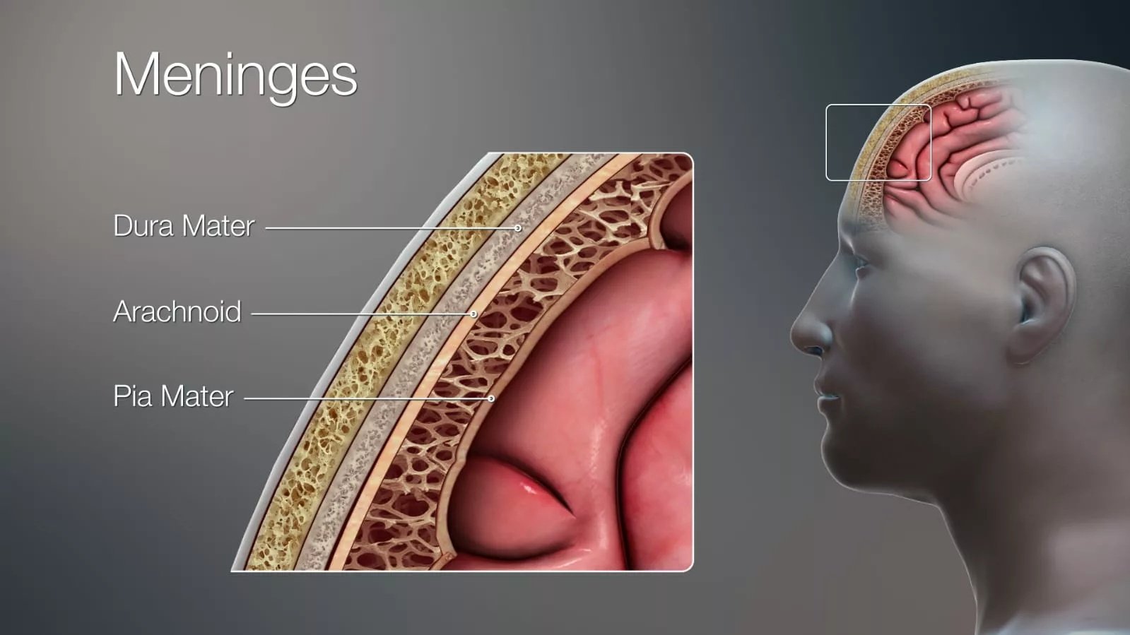
Image courtesy: 3D Medical Illustration Meninges by Ajokley, licensed under Creative Commons Attribution 4.0 International (CC BY 4.0) and Creative Commons Attribution‑ShareAlike 4.0 International (CC BY‑SA 4.0). Source: Wikimedia Commons – 3D Medical Illustration Meninges Details
How do Cancer Cells Reach the Leptomeninges?
Cancer cells take different pathways to spread out from their primary site and reach the leptomeninges:
- Through Bloodstream: The tumors that grow aggressively, such as those in the breast, lungs, and melanoma, enter the bloodstream in their advanced stages and circulate throughout the body. Some of these cells reach the choroid plexus, which is a CSF-producing brain structure. After invading the choroid plexus, these cancer cells infiltrate the leptomeninges and enter the CSF.[1]
- Through Direct Extension: Tumors growing close to the meninges, like brain tissue tumors, penetrate the nearby coverings, which may lead to leptomeningeal carcinomatosis.
- Through Perineural or Perivascular: Cancer cells can ride along the nerves (peri-neural) or the blood vessels (peri-vascular), accompanying these cells to the meninges. Although this route is not common, it is observed in the nerves involving cancers, such as head and neck tumors.[2]
Although the lymphatic system is also the main route of cancer spread in the rest of the body, the brain and spinal cord have no conventional lymphatic vessels. So, the cancerous cells do not spread through this route in the leptomeninges.
What happens after the Cancer Cells enter the CSF?
After penetrating the leptomeninges, cancer cells enter the CSF and spread throughout the brain and spinal cord. Research explains that LMC has two main forms:
- Free-floating tumor cells, which spread across the brain and spinal cord.
- Plaques and nodules that adhere to the brain and spinal cord surfaces and ultimately become so large as to block the CSF flow and increase the intracranial pressure. These two patterns can coexist.[3]
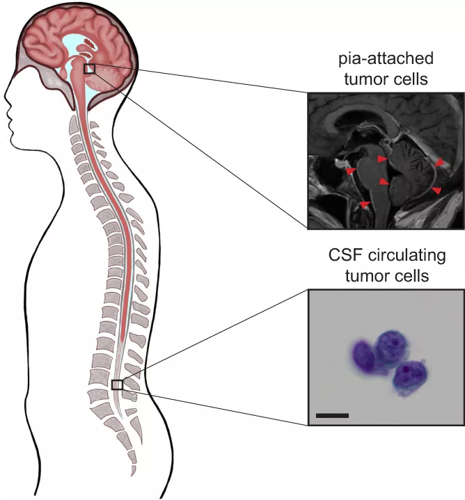
Image courtesy: Image by Ibrahim et al., licensed under Creative Commons Attribution 3.0 Unported (CC BY 3.0). Source: PMC – Leptomeningeal metastatic cells adopt two phenotypic states
Because the CSF is an immunoprivileged site with reduced immune surveillance, tumor cells are more likely to survive and spread within it.
Causes of Leptomeningeal Carcinomatosis
Different cancers in the body cause the LMC.
- Most commonly, the solid tumors, like breast, lungs, and melanoma, spread to the CNS.
- Gut tumors and CNS tumors like medulloblastoma and lymphoma cause LMC, especially in children.
- In children and young adults, hematological cancers like acute lymphoblastic leukemia, non-Hodgkin lymphoma are among the most common causes of LMC.
How common is Leptomeningeal Carcinomatosis?
While LMC is rare in the general population, estimates suggest up to 110,000 cases occur per year in the U.S. alone. However, the exact data is even more than that because of the undiagnosis of many cases.[4]
- LMC is observed in approximately 5% to 15% of patients with cancer.[5]
- About 5% to 8% of cases of solid cancers, like lung, melanoma, and breast cancers, cause LMC. [6]
- Approximately 5-15% of the hematological (blood) cancers, like leukemia and lymphoma, lead to leptomeningeal disease in their advanced stage.[7]
In autopsies of patients having neurological symptoms, have even higher rates of LMC. Some studies of autopsies claimed 18-20% of the patients with CNS-related symptoms had LMC, indicating underdiagnosis during life.[8]
Why Cases of LMC are Increasing?
The cases of the LMC are increasing day by day because of the modern cancer therapies and advanced imaging techniques.
- Improved cancer survival: Advanced cancer treatment options have improved the patient’s survival, which provides more time for the tumor to spread to the brain and spinal cord.
- Better imaging techniques: MRI and other neuro-imaging detect even the minor and diffuse LMC that used to be missed before.
Symptoms of Leptomeningeal Carcinomatosis
Because LMC affects different areas of the CNS, symptoms vary based on whether the brain, cranial nerves, or spinal cord is involved.
Cerebral Symptoms:
When the tumor cells move from the CSF and seep down the brain, the following symptoms may occur:
- Headache: It is a common early symptom you may feel. Inflammation or disturbed CSF flow causes the meningeal irritation, leading to headache.
- Nausea & Vomiting: These symptoms usually come with the headache because of increased intracranial pressure.
- Altered Mental Status: When the tumor grows in the brain and starts disrupting the neural activity, it leads to confusion, memory loss, and difficulty in concentration.
- Seizures: When the tumor involves the cortex of the brain, it results in seizures.[9]
Cranial Nerve Symptoms:
After infiltrating the brain tissues, LMC starts invading the cranial nerves, causing different symptoms depending on the specific nerve involved.[10]
- Diplopia (Double Vision)
- Facial weakness and numbness
- Hearing loss
- Swallowing difficulty, hoarseness
- Tongue weakness
As the cancer progresses, these symptoms worsen.
Spinal Cord Symptoms:
As CSF also flows to the spinal cord, it transmits the cancerous cells there, too.
- Back pain: Sharp and shooting pain in the back and neck.
- Leg Weakness: Weakness and numbness in the leg due to damage to the nerves that supply your legs.
- Bowel or Bladder Dysfunction: Incontinence ( loss of control over the bladder) or retention due to sphincter dysfunction.
These are the early symptoms that ultimately lead to loss of control over muscles.
How is Leptomeningeal Carcinomatosis diagnosed?
Once symptoms of leptomeningeal carcinomatosis appear, its diagnosis becomes important, which depends on the combination of clinical suspicion, neuroimaging, and CSF analysis.
Magnetic Resonance Imaging (MRI):
MRI of both the brain and the spine is the first-line imaging study. It indicates whether the LMC is linear, nodular, or curvilinear. It has very high sensitivity and specificity for the leptomeningeal disease.[11]
CSF Cytology:
Finding malignant cells in the CSF helps to diagnose the leptomeningeal tumor. Your healthcare provider takes a sample of CSF from your brain and spinal cord by lumbar puncture or spinal tap. This sample is then examined under a microscope to identify any cancerous cells.[12]
The initial CSF sample sometimes can’t detect the cancer cells in about half of the cases, even with a high suspicion of LMC. Therefore, the doctors often repeat the testing if the patient presents with strong clinical symptoms of LMC.
Advanced CSF Testing:
When traditional CSF cytology yields negative results despite a strong suspicion of LMC, healthcare professionals typically choose advanced CSF testing, a newer method of diagnosing LMC.
Unlike the old CSF cytology, in which the whole tumor cells are detected, advanced CSF testing involves the finding of circulating tumor DNA (ctDNA) or tumor-derived RNA fragments. These markers are more sensitive to detection and provide a more accurate diagnosis.[13]
CSF Flow:
Normally, the CSF flows freely in the brain and spinal cord to protect and nourish them. But in leptomeningeal neoplasm, the cancerous cells stick to the surfaces of the brain in the CSF pathways and block the normal flow of CSF. To detect this blockage, doctors use a special type of scan.
The healthcare providers inject a small amount of radioactive tracer, such as Indium 111 or Technetium 99m, into the spinal fluid, and then they track its movement using imaging. If the tracer is stuck elsewhere, it signals a blockage in the CSF flow.[14]
This test is useful in cases where your doctor wants to inject the cancer medicines directly into the CSF. If the CSF flow test confirms the blockage, the drug may not adequately reach all areas of the brain and spinal cord.
Meningeal Biopsy:
This method is not common for the LMC diagnosis, but your doctor may consider it in situations where other tests may not provide satisfactory results.
In this method, a biopsy of the meninges may be performed, which is studied to confirm the leptomengeal carcinomatosis.
Differential Diagnosis of Leptomeningeal Carcinomatosis
Although the symptoms we have discussed above point toward the LMC, these are not confined to leptomeningeal. Many other diseases also present with the same symptoms, which makes the diagnosis challenging.
Infectious Meningitis:
Chronic infections like tuberculosis (TB), fungal meningitis have similar symptoms to leptomeningeal carcinomatosis, like headache, changed mental status, cranial nerve palsies, and abnormal CSF findings.
However, these infectious diseases are not aggressive, and the CSF findings show elevated white blood cells (WBCS).
Autoimmune & Inflammatory Disorders:
Autoimmune and inflammatory disorders like neurosarcoidosis, CNS vasculitis, and autoimmune encephalitis present similarly to LMC.
But these disorders also have systemic signs such as fever, rash, and joint pain. Apart from these systemic signs, your doctor can rule out autoimmune and inflammatory diseases based on CSF analysis and specific autoimmune panels.
Sub-Arachnoid Hemorrhage:
Patients with subarachnoid hemorrhage may present with early symptoms similar to LMC, i.e., severe headache, vomiting, and meningeal signs. However, a CT Scan helps distinguish it from LMC by revealing blood in the subarachnoid space.
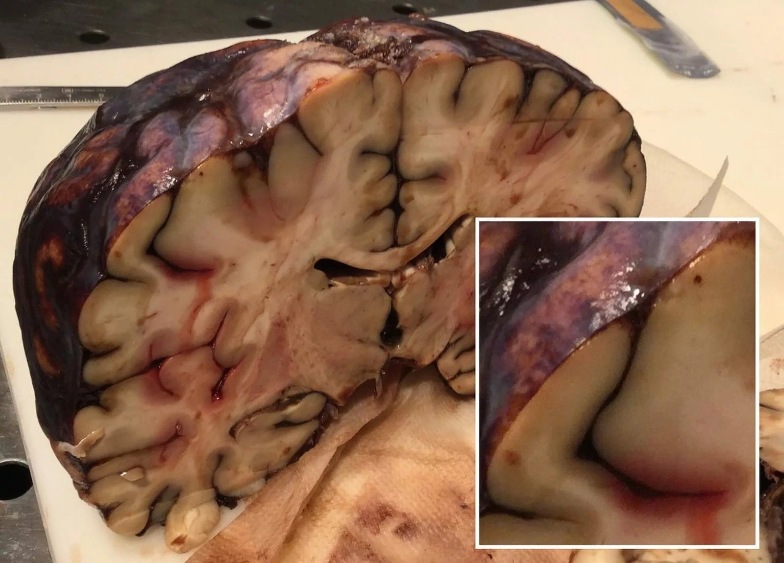
Gross pathology of subarachnoid hemorrhage by Mikael Häggström, M.D., public domain. Source: Wikimedia Commons – Gross pathology of subarachnoid hemorrhage
Multiple Sclerosis:
This disease involves the loss or damage to the myelin sheath, the protective coating around nerve fibers, leading to slower or blocked nerve signals.
Although multiple sclerosis results in vision loss, numbness, and weakness in the muscles that mimic the LMC symptoms, it has specific features on MRI and a distinct CSF profile.
Brain Metastases:
Unlike the leptomeningeal disease, in which the disease spreads through CSF, brain metastases are caused by the cancer spreading in the brain tissues. The well-explained differences between them are given below.
Difference between Brain Metastases & Leptomeningeal Carcinomatosis
| Feature | Brain Metastases | Leptomeningeal Carcinomatosis |
|---|---|---|
| Site of Spread | Brain tissue (parenchyma) | Leptomeninges and CSF |
| Route of Spread | Through the bloodstream | Through CSF or nearby tumor invasion |
| Common Primary Cancers | Lung, breast, melanoma, kidney, colon | Lung, breast, melanoma, leukemia, lymphoma |
| Typical Symptoms | Seizures, weakness, speech issues, headache | Headache, vision changes, cranial nerve palsies, leg weakness, back pain |
| MRI Findings | Mass lesions with swelling | Diffuse enhancement along the brain surface, cranial nerves, or spinal cord |
| CSF Findings | Usually normal unless the meninges are involved | Abnormal showing cancer cells, high protein, low glucose, high pressure |
| Prognosis | Often manageable with treatment | Poorer due to widespread CSF involvement |
| Treatment Options | Surgery, radiosurgery, whole-brain radiation, systemic therapy | Intrathecal chemo, targeted radiation, systemic therapy in select cases |
| Long-Term Control | Possible in some with early and targeted treatment | Rare but mostly symptom relief and short-term control |
These differential diagnoses help the healthcare provider rule out similar presenting diseases to ensure the appropriate treatment.
Management & Treatment Options for Leptomeningeal Carcinomatosis
After the successful diagnosis of leptomeningeal carcinomatosis, your doctor will plan for the treatment and management of it.
Unfortunately, the patient with leptomeningeal carcinomatosis already has advanced cancer somewhere in the body. So the disease has no cure, but it can be managed.
Intrathecal Chemotherapy:
Our brain and spinal cord have a protective barrier, the blood-brain barrier (BBB), which stops the entry of many drugs and chemicals. So to overcome this issue, doctors inject the drugs directly into the CSF, allowing the drugs to bypass the BBB. This is achieved through a lumbar puncture or an implanted device known as an Ommaya reservoir.
An Ommaya reservoir is a small device that a doctor places surgically under the scalp. It connects to the brain’s CSF system through a thin catheter. The common drugs that are induced in the CSF are:
- Methotreaxate
- Cytarabine
- Thiotepa
In some cases, your healthcare provider may use liposomal cytarabine, which has a long-acting formulation to maintain the drug level over time.
Systemic Therapies:
Certain intravenously and orally taken drugs like osimertinib and tucatinib cross the blood-brain barrier and reach the leptomeninges. Nivolumab is under study and has the potential to activate the immune cells in CSF, which then help fight against the cancer cells.[15]
Radiation:
In nodular cases causing CSF blockage, doctors use focal radiation that focuses precisely on the area blocking the CSF or pressing nerves. This method is less effective in diffuse cases of LMC.
Supportive & Palliative Care:
In cases where cancer has progressed severely that the chances of recovery become low, supportive and palliative care helps manage the disease. The main focus of this is not curing the disease but relieving the symptoms, improving quality of life, and helping patients and their families cope emotionally with the disease. These are some supportive and palliative care drugs:
- Corticosteroids, like dexamethasone, reduce the swelling and ease the symptoms such as headache and nausea.
- Antiseizures
- Pain killer & anti-nausea medications
Palliative care specialists offer emotional support, daily care planning, and guidance in end-of-life decisions. They may help patients maintain their dignity during a difficult time.
Why Is LMC So Difficult to Treat?
Leptomeningeal carcinomatosis has no cure and is challenging to treat because cancer cells in it spread rapidly and widely throughout the CSF. It quickly involves the whole brain and spinal cord.
- It also spreads diffusely, which is challenging to target through radiation and surgery.
- Blood-brain barrier restricts the entry of many systemic drugs into the CSF. Even if given these drugs directly into the CSF, they may not reach the target areas because of the blockage of CSF flow.
LMC typically occurs in patients with advanced-stage cancers who may also be coping with systemic treatment side effects.
Prognosis & Life Expectancy of Leptomeningeal Carcinomatosis
Unfortunately, leptomeningeal carcinomatosis has a worse prognosis. Although treatment may improve the quality of life, LMC is not curable.
- Life Expectancy Without Treatment: Most studies claimed half of the patients passed away within 4-6 weeks after the symptoms began if LMC was left untreated.
- Life Expectancy With Treatment: Many patients may live slightly longer with the treatment. A study showed the data that half of the patients died in 2-6 months.[16]
Some patients, those with breast cancer or hematological cancer, respond well to therapy and may survive longer.
How to prevent Leptomeningeal Carcinomatosis?
Currently, it is not guaranteed to completely prevent the LMC as it is a late-stage event. Controlling the primary cancer site and stopping its spread to other places may help in its prevention.
Early, more aggressive treatment for the cancers of the breast and lung lowers the risk of their spreading to the leptomeninges.
How do I take care of myself if I have an LMC diagnosis?
Even though LMC is a serious condition, there are still things you can do to take care of yourself physically, mentally, and emotionally.
- Follow your treatment plan, attend appointments, report any new symptoms right away, and ask questions to your doctor if anything is unclear.
- Rest when you are tired, because LMC itself and its treatment make the patient fatigued. If you have walking difficulties, let your healthcare team know. They may help you with physical therapy or mobility aids.
- Eat small and frequent meals even when you are not hungry. These frequent eating habits keep your energy up.
- Try to keep yourself mentally healthy by talking to a counselor, joining support groups, and having open conversations with your loved ones.
You might not like to think about end-of-life planning, but doing so early gives you control and peace of mind. Talk openly with your family and care team about your goals and wishes.
Conclusion
Leptomeningeal carcinomatosis is caused by the spread of cancerous cells from the breast, lung, or other cancer sites to the brain’s protective layers, the meninges. The cancerous cells enter the CSF and invade the brain and spinal cord, causing several serious neurological symptoms ranging from headache to cranial nerve palsies. Its diagnosis is done through CSF cytology and an MRI study. Although it is not a curable disease, some medications are available to improve the quality and quantity of patients’ lives.
References
[1] Rhun, E. L., Taillibert, S., & Chamberlain, M. C. (2013). Carcinomatous meningitis: Leptomeningeal metastases in solid tumors.Surgical Neurology International,4(Suppl 4), S265. https://doi.org/10.4103/2152-7806.111304
[2] Sullivan, L. M., & Smee, R. (2006). Leptomeningeal carcinomatosis from perineural invasion of a lip squamous cell carcinoma. Australasian radiology, 50(3), 262–266. https://doi.org/10.1111/j.1440-1673.2006.01577.x
[3] Remsik, J., Chi, Y., Tong, X., Sener, U., Derderian, C., Park, A., Saadeh, F., Bale, T., & Boire, A. (2022). Leptomeningeal metastatic cells adopt two phenotypic states. Cancer reports (Hoboken, N.J.), 5(4), e1236. https://doi.org/10.1002/cnr2.1236
[4] LMC. Nayar, G., Ejikeme, T., Chongsathidkiet, P., Elsamadicy, A. A., Blackwell, K. L., Clarke, J. M., Lad, S. P., & Fecci, P. E. (2017). Leptomeningeal disease: Current diagnostic and therapeutic strategies. Oncotarget, 8(42), 73312. https://doi.org/10.18632/oncotarget.20272
[5] Bhambhvani, H. P., Rodrigues, A. J., Umeh-Garcia, M. C., & Gephart, M. H. (2020). Leptomeningeal Carcinomatosis: Molecular Landscape, Current Management, and Emerging Therapies. Neurosurgery Clinics of North America, 31(4), 613. https://doi.org/10.1016/j.nec.2020.06.010
[6] Batool A, Kasi A. Leptomeningeal Carcinomatosis. [Updated 2023 Mar 27]. In: StatPearls [Internet]. Treasure Island (FL): StatPearls Publishing; 2025 Jan-. Available from: https://www.ncbi.nlm.nih.gov/books/NBK499862/
[7] Batool A, Kasi A. Leptomeningeal Carcinomatosis. [Updated 2023 Mar 27]. In: StatPearls [Internet]. Treasure Island (FL): StatPearls Publishing; 2025 Jan-. Available from: https://www.ncbi.nlm.nih.gov/books/NBK499862/
[8] Le Rhun, E., Taillibert, S., & Chamberlain, M. C. (2013). Carcinomatous meningitis: Leptomeningeal metastases in solid tumors. Surgical neurology international, 4(Suppl 4), S265–S288. https://doi.org/10.4103/2152-7806.111304
[9] Zima, L. A., Tulpule, S., Samson, K., & Shonka, N. (2019). Seizure prevalence, contributing factors, and prognostic factors in patients with leptomeningeal disease. Journal of the neurological sciences, 403, 19–23. https://doi.org/10.1016/j.jns.2019.05.032
[10] Trivedi, R. A., Nichols, P., Coley, S., Cadoux-Hudson, T. A., & Donaghy, M. (2000). Leptomeningeal glioblastoma presenting with multiple cranial neuropathies and confusion. Clinical neurology and neurosurgery, 102(4), 223–226. https://doi.org/10.1016/s0303-8467(00)00115-3
[11] Clarke, J. L., Perez, H. R., Jacks, L. M., Panageas, K. S., & Deangelis, L. M. (2010). Leptomeningeal metastases in the MRI era. Neurology, 74(18), 1449–1454. https://doi.org/10.1212/WNL.0b013e3181dc1a69
[12] Rhun, E. L., Taillibert, S., & Chamberlain, M. C. (2013). Carcinomatous meningitis: Leptomeningeal metastases in solid tumors.Surgical Neurology International,4(Suppl 4), S265. https://doi.org/10.4103/2152-7806.111304
[13] Malani, R., Fleisher, M., Kumthekar, P., Lin, X., Omuro, A., Groves, M. D., Lin, N. U., Melisko, M., Lassman, A. B., Jeyapalan, S., Seidman, A., Skakodub, A., Boire, A., DeAngelis, L. M., Rosenblum, M., Raizer, J., & Pentsova, E. (2020). Cerebrospinal fluid circulating tumor cells as a quantifiable measurement of leptomeningeal metastases in patients with HER2-positive cancer. Journal of Neuro-Oncology, 148(3), 599. https://doi.org/10.1007/s11060-020-03555-z
[14] Nayar, G., Ejikeme, T., Chongsathidkiet, P., Elsamadicy, A. A., Blackwell, K. L., Clarke, J. M., Lad, S. P., & Fecci, P. E. (2017). Leptomeningeal disease: Current diagnostic and therapeutic strategies. Oncotarget, 8(42), 73312. https://doi.org/10.18632/oncotarget.20272
[15] Park, S., Lee, M. H., Seong, M., Kim, S. T., Kang, J. H., Cho, B. C., Lee, K. H., Cho, E. K., Sun, J. M., Lee, S. H., Ahn, J. S., Park, K., & Ahn, M. J. (2020). A phase II, multicenter, two-cohort study of 160 mg osimertinib in EGFR T790M-positive non-small-cell lung cancer patients with brain metastases or leptomeningeal disease who progressed on prior EGFR TKI therapy. Annals of oncology: official journal of the European Society for Medical Oncology, 31(10), 1397–1404. https://doi.org/10.1016/j.annonc.2020.06.017
[16] Grossman, S. A., & Krabak, M. J. (1999). Leptomeningeal carcinomatosis. Cancer treatment reviews, 25(2), 103–119. https://doi.org/10.1053/ctrv.1999.0119