
, achromoderma, or depigmentation refers to a skin condition characterized by the...)

Leukoderma White Patches Leukoderma (also spelled leucoderma), achromoderma, or depigmentation refers to a skin condition characterized by the formation of localized white patches on the skin, which is attributed to a total loss of skin pigmentation, i.e., epidermal melanin. Leuko- means white, and derma- means skin. Leukoderma is not a disease itself, but a descriptive term for depigmentation that occurs as a manifestation of various underlying skin or systemic conditions. The most common cause is vitiligo, an autoimmune disorder of the skin that destroys the pigment-producing cells (melanocytes). Although vitiligo is the most frequent cause, the terms vitiligo and leukoderma are not strictly interchangeable, as leukoderma can arise from non-autoimmune causes as well.
Leukoderma can arise anywhere, but is commonly seen on the skin of the hands, feet, and forearms. It can affect people of all ages and skin colors. However, a high prevalence is seen in people aged 10-30 years with darker skin. Different methods can help improve the appearance of the white patch, with camouflage (such as makeup or dyes) being the simplest. Dermatologists manage the condition with light therapy, medical therapies targeted at the underlying cause, and surgical options in selected stable cases.
What Is Leukoderma?: Leukoderma White Patches
Leukoderma is a benign clinical sign in which young patients notice the development of small white dots on their skin. These small dots then coalesce to form tiny patches over the next several months. The onset, progression, and appearance of these patches vary depending on the underlying cause (such as vitiligo, chemical exposure, trauma, or genetic disorders). The patches are well-demarcated lesions having a normal texture. The skin patches are asymptomatic, but the underlying conditions causing the disorder can have presentations. Leukoderma arises on the visible areas, i.e., skin of the face/head, hands, forearms, and feet. Thus, it raises a lot of aesthetic concerns amongst the patients.
The size and appearance of the lesions can vary between individuals, and even between different locations of the same person. In vitiligo-associated leukoderma, lesions may enlarge over time, whereas in other forms (such as post-traumatic or chemical leukoderma), the patches often remain localized and stable.
Types Of Leukoderma
There exist different presentations of the white patches. Clinically, leukoderma is most commonly classified according to the pattern of vitiligo when vitiligo is the underlying cause.
Non-Segmental Leukoderma
The most common (affecting around 90% of the cases) is the non-segmental type. In this type, patches appear symmetrically on both sides of the body. Non-segmental leukoderma is further divided into:
- Focal leukoderma: Depigmentation is limited to one area. This type is commonly seen in children.
- Generalized leukoderma: White patches are widespread and symmetrical (on both sides of the body).
- Universal leukoderma: It is a more aggressive form of generalized leukoderma with depigmentation covering over 80% of the body.
- Mucosal leukoderma: Pigment loss affects the mucous membranes instead of the skin. Mucosal leucoderma is seen in the lining of the mouth and the genitals.
- Acrofacial leukoderma: This type is characterized by a specific pattern of depigmentation spots. It primarily affects your facial region (around the lips, nostrils, eyes, and mouth) and extremities (hands and feet). It can co-exist with other forms of pigmentation pathologies.[1]
Segmental Leukoderma
A rare type of leukoderma is the segmental leukoderma, which is known to affect around 10% cases. Depigmented spots appear only on one side of the body. Generally, the progression of the pigment loss is rapid initially, but stabilizes later on.
Psychological Impact Of Leucoderma
The milky white patches do not cause any discomfort. However, sometimes patients experience some itching or slight inflammation. The primary burden of leukoderma is psychological and social rather than physical, particularly when the depigmentation is visible. It was revealed in a study that many patients from rural areas had no physical problems, but a large majority (75%) showed psychological disturbances from the disease. Individuals were shy and ashamed of social interaction. Thus, they also reported fear and prejudice due to the defective skin appearance.[2]
Most patients seek treatment for cutaneous disorders for aesthetic concerns only. An Indian study concluded that the prevalence of anxiety is higher in individuals with leukoderma than in healthy persons.[3]
What Causes Leukoderma?
As already mentioned, leukoderma can be a presentation of multiple underlying infirmities. Therefore, you can develop white, depigmented patches on the skin as a result of the following conditions:
Autoimmune Disorders
The most common cause of leukoderma is an autoimmune disorder known as vitiligo. In this disease, your immune system mistakenly destroys the pigment-producing cells (melanocytes) present in your skin and mucosa. Due to the obliteration of the melanocytes, there is a shortage of pigment in the skin region that leads to the development of leukoderma. Patients usually notice multiple white patches on each side of the body. Typically, the patches appear on the skin that is frequently exposed to the sun.[4]
Chemical Exposure
Skin exposure to chemicals like phenolic compounds, cosmetics, catechol derivatives, and industrial rubber/pesticides can cause localized depigmentation. This type looks similar to vitiligo and is known as contact leukoderma or chemical leukoderma.
Most people notice pea-sized or confetti-sized macules. Studies show that phenols and aromatic/aliphatic catechol derivatives induce melanocytotoxicity (toxicity of melanocytes), which causes suboptimal performance of the pigment-producing cells. Occupational exposure to chemicals has been shown to play a crucial role in the occurrence of chemical leukoderma.[5]
Even contact with pesticides can induce hypopigmented lesions on the hands. Such lesions don’t cause any pain or discomfort.[6]
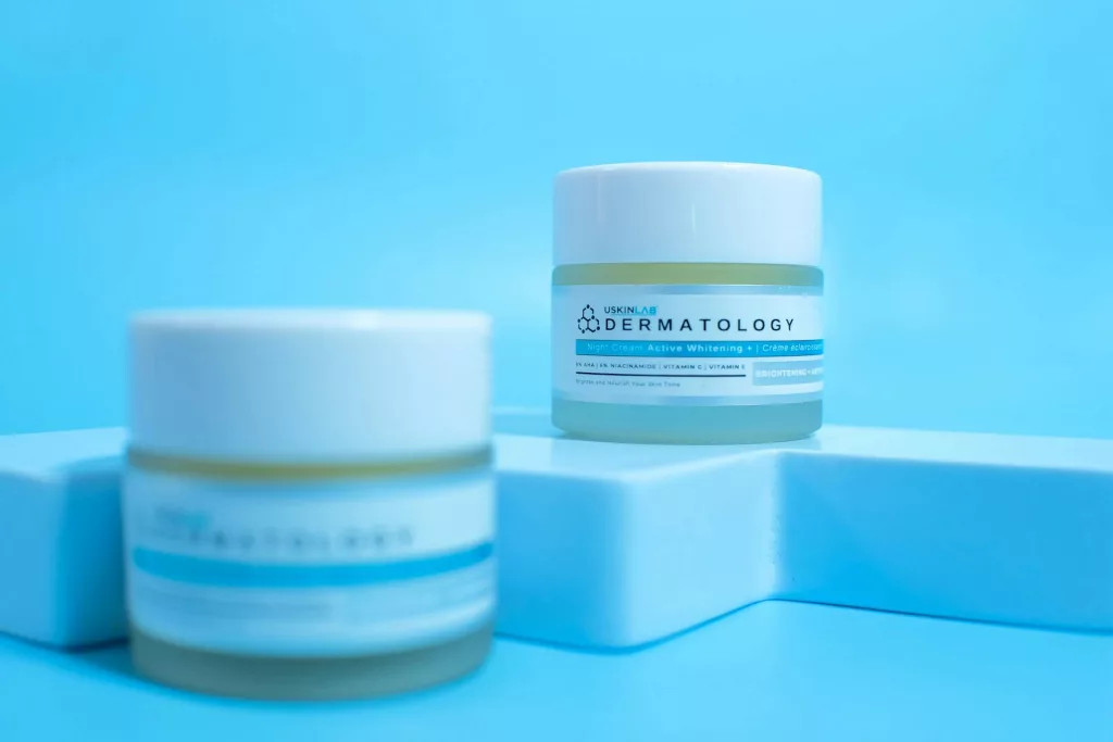
Cosmetic products like skin-lightening creams can cause leukoderma.
The prevalence of this type has increased recently due to the widespread use of cosmetics.[7] Thus, the use of skin-lightening therapies also puts you at a higher risk of chemical leucoderma.[8]
Rare cases have shown eyelid leukoderma arising due to the use of eyelash extensions. Hence, you should be careful when using such cosmetic products.[9]
Injury/Trauma
Some people develop white patches on the skin after an injury to the region. This is most commonly observed in injuries that cause scarring. Wound surface after sunburns and burns tends to undergo depigmentation. Usually, partial and full-thickness burns lead to hypo- or depigmentation like this.[10]
Procedures like laser treatment and chemical peels can cause scarring and indirectly contribute to whitening. Therefore, healthcare workers must be careful while performing these procedures.
Drugs
Certain drugs can also serve as the foundation for skin depigmentation. Drug-induced leukoderma is associated with skin-whitening drugs like rhododendrol.[11]
Moreover, women receiving therapy for breast cancer are at a higher risk of developing depigmentation. This is because anti-cancer drugs like cyclin-dependent kinase 4/6 inhibitors are increasingly linked to vitiligo-like leucoderma. Thus, oncologists must keep an eye on the skin changes as well.[12]
Genetic Issues
You may notice white skin patches in newborns suffering from congenital disorders. Waardenburg syndrome is a rare genetic disorder that affects the color of the hair, skin, and eyes. In this disease, congenital leukoderma is seen on the skin of the face, trunk, and limbs alongwith sensorineural hearing loss.[13] Another rare genetic condition associated with depigmentation is piebaldism, characterized by congenital absence of melanocytes in affected skin and hair.
Health Conditions
Various disorders are linked to achromoderma. Melanoma is a cancerous skin condition affecting melanocytes. It is known to induce drug-induced leukoderma/vitiligo. Non-segmental variant of leucoderma is the most common finding in melanoma cases.[14]
Halo nevus is a benign skin condition that has a unique type of skin lesion. The disorder appears as a depigmented ring (halo) of leukoderma surrounding a red mole. Another malady found to have a link with pigment loss in the skin is Darier disease (keratosis follicular). It is a rare, inherited disorder that mainly causes wart-like papules on the chest, scalp, back, and nails. An early manifestation of the disease is guttate leukoderma, characterized by the formation of small white spots on the skin.[15]
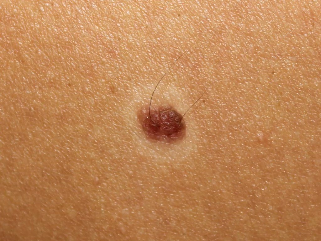
A halo nevus can lead to leukoderma.
Allergic contact dermatitis (ACD) is an autoimmune disorder that causes a red, itchy skin rash accompanied by inflammation and blisters. There have been cases of chemical leukoderma arising after allergic contact dermatitis.[16]
How Is Leukoderma Diagnosed?
In most cases, a dermatologist diagnoses the condition with a physical examination of the lesions. Moreover, health professionals perform several tests to reach a confirmed diagnosis.
Wood Lamp Examination
In this diagnostic technique, the doctor uses a special light, i.e., Ultraviolet (UV) backlight, in a dark room to observe the skin lesion. This test helps distinguish true depigmentation from hypopigmentation but does not determine the exact cause.
Dermoscopy
This is a non-invasive technique in which the doctor uses a special magnifying tool, i.e., a dermatoscope, to clearly visualize the skin lesions. Therefore, it is commonly used to diagnose pathologies of the skin, hair, and nails.
Skin Biopsy
In rare instances, doctors remove a small portion of the lesion and send it to the lab for microscopic evaluation. Skin biopsy is adopted only when doctors want to rule out cancerous conditions.
Leukoderma Vs Vitiligo
Leukoderma is a broad term that refers to the appearance of white lesions on the skin. On the other hand, vitiligo is a condition of white lesions caused by autoimmune destruction of the melanocytes. Leukoderma can arise in response to cosmetic and chemical use, but vitiligo arises only due to a faulty immune system. All vitiligo presents as leukoderma, but not all leukoderma is vitiligo.
Leukoderma Treatment
There is no definitive treatment for leukoderma. However, the main aim of healthcare providers is to fix the underlying condition leading to dermal manifestations. Doctors have noted some success with drugs like Janus Kinase inhibitors (like tofacitinib, ruxolitinib) in the treatment of vitiligo but are not effective for all forms of leukoderma.[17]
There is no science-backed evidence to support domestic strategies. However, patients can feel some improvement by staying hydrated, and taking dietary supplements (containing vitamin C and zinc).
Camouflage
Most people go for non-invasive options like cosmetic camouflage. You can use makeup products and hair dyes to cover the depigmented skin and hair. Camouflaging with cream-based preparations has shown promising results. Self-tanning agents like dihydroxyacetone (DHA) significantly enhance appearance and consequently lead to improved quality of life. Moreover, patients claim that the cream is easy to apply and retains its color stability for an appreciable time.[18]
Light Therapy
Your doctor may advise you to get light therapy for your inactive melanocytes. Narrow-band Ultraviolet B (UV-B) light has proven to be an effective therapy for multiple skin disorders, including eczema, psoriasis, and leukoderma. A 2021 study concluded that ultraviolet light therapy is the most effective treatment for rhododendrol-induced chemical leukoderma.[19]
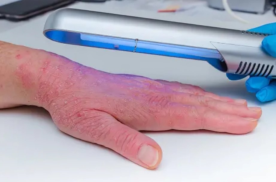
Light therapy (narrow band UV-B) is used to treat multiple skin disorder including leukoderma.
Surgical Management
A permanent fix for visible depigmented skin is surgical intervention. Surgical treatment is considered in stable leukoderma or vitiligo (no progression for at least 6–12 months). Surgeons replace the white, patched skin with a graft taken from a normal/healthy site. Professionals can choose from different types of surgical techniques.
Your doctor may remove the depigmented layer with the help of a high-speed rotating tool in a process called dermabrasion. The abraded skin is then replaced with a thin (split-thickness skin graft). The technique offers consistent and sustained results in repigmentation (over 84 months).[20]
However, other studies show that the most effective technique is the punch minigrafting. In this technique, the doctor creates small holes in white patches. He then takes small circular skin samples, i.e., minigrafts from a healthy site (or a donor) and places the grafts into the holes.[21]
Punch minigrafting improves the appearance of vitiligo/leucoderma by transferring healthy melanocytes (pigment-producing cells) to the depigmented site.
How Can I Prevent Leukoderma?
You can not prevent leukoderma caused by vitiligo. However, you can minimize your risk of chemical/contact type by taking some safety measures. People exposed to industrial phenols, rubber, pesticides, and other melanocytotoxic chemicals must wear gloves. Individuals using skin-lightening products must be cautious not to overapply them. Moreover, patients who have suffered from skin burns must undergo timely checkups with a doctor to effectively manage any changes.
Wrapping Up
Leukoderma is a condition of white (depigmented) patches on the skin, which is the manifestation of some underlying dermal pathology. Vitiligo (an autoimmune disorder) is the major cause of leukoderma across the globe. The disease appears as milky, white patches on sun-exposed skin and is most frequently seen in individuals with darker skin.
Of the two types, i.e., non-segmental and segmental leukoderma, the non-segmental type is more common. It causes anxiety and psychological disturbance in patients, for which they seek treatment. Moreover, apart from the immune system-induced destruction of melanocytes (vitiligo), achromoderma can arise in response to contact with phenolic compounds, rubber, pesticides, and skin-lightening cosmetic products/drugs (like rhododendrol). Some people develop white spots in the skin lesions of burns, nevus, or melanoma.
Dermatologists diagnose the condition with physical examination and diagnostic tests like wood lamp examination and dermoscopy. Doctors usually advise the camouflage of white lesions with dihydroxyacetone cream and cosmetic products. Light therapy (using UV-B light) improves chemical leukoderma. For a more long-lasting solution, surgeons remove the depigmented layer via dermabrasion and replace it with a split-thickness skin graft from a healthy/unaffected site for longer-lasting results. Another surgical technique is punch minigrafting, which has also shown impressive results in revising the appearance of white patches.
References
[1] Schallreuter, K. U., Krüger, C., Rokos, H., Hasse, S., Zothner, C., & Panske, A. (2007). Basic research confirms coexistence of acquired Blaschkolinear Vitiligo and acrofacial Vitiligo.Archives of dermatological research,299(5), 225-230.
[2] Tripathi, K. M., Arya, S., & Singh, V. (2018). Frequency of Occurrence of Different Types of Leucoderma and Vitiligo, Rishi, Dasna, Ghaziabad, India.Int. J. Curr. Microbiol. App. Sci,7(9), 1267-1276.
[3] Saini, S., Yadav, S., & Prasad, M. (2025). Association of Anxiety (Chittodvega) in Leukoderma (Shvitra)–A Cross-Sectional Study.Journal of Ayurveda,19(4), 326-331.
[4] Roy, A. K. (2017). Vitiligo: a white patch that affects the soul.Pigmentary Disorders,4(254), 2376-0427.
[5] Jangra, S., Gulia, H., Singh, J., Dang, A. S., Giri, S. K., Singh, G., … & Kumar, A. (2024). Chemical leukoderma: An insight of pathophysiology and contributing factors.Toxicology and Industrial Health,40(8), 479-495.
[6] Zhao, Q., Nan, L., Mao, C., Wu, Y., & Wu, X. (2024). Chemical Leukoderma Induced by Pesticides.Skin Research and Technology,30(10), e70083.
[7] Bonamonte, D., Vestita, M., Romita, P., Filoni, A., Foti, C., & Angelini, G. (2016). Chemical leukoderma.Dermatitis,27(3), 90-99.
[8] Odedra, S., & Yoo, J. (2021). The risk of chemical leucoderma with skin‐lightening therapies.Clinical & Experimental Dermatology,46(7).
[9] Zhu, J., Luo, L., Guo, Y., & Li, C. (2024). Two Cases of Eyelid Leukoderma Associated With Eyelash Extension.Journal of Cosmetic Dermatology,23(12), 4392.
[10] Maranda, E. L., Wang, M. X., Shareef, S., Tompkins, B. A., Emerson, C., & Badiavas, E. V. (2018). Surgical management of leukoderma after burn: a review.Burns,44(2), 256-262.
[11] Tsutsumi, R., Sugita, K., Abe, Y., Hozumi, Y., Suzuki, T., Yamada, N., … & Yamamoto, O. (2019). Leukoderma induced by rhododendrol is different from leukoderma of vitiligo in pathogenesis: A novel comparative morphological study.Journal of Cutaneous Pathology,46(2), 123-129.
[12] Engler Markowitz, M., Barak Levitt, J. A., Merose, R., Didkovski, E., Snast, I., Oren-Shabtai, M., … & Sherman, S. (2025). Vitiligo-like leucoderma following cyclin-dependent kinase 4 and 6 inhibitor treatment: systematic review of the literature.Clinical and Experimental Dermatology, llaf123.
[13] Milunsky, J. M. (2017). Waardenburg syndrome type I.
[14] Ramondetta, A., Ribero, S., Conti, L., Fava, P., Marra, E., Broganelli, P., … & Quaglino, P. (2020). Clinical and pathological relevance of drug-induced vitiligo in patients treated for metastatic melanoma with anti-PD1 or BRAF/MEK inhibitors.Acta Dermato-Venereologica,100(1), 5585.
[15] Harb, J. N., & Motaparthi, K. (2018). Clinicopathologic findings of guttate leukoderma in Darier disease: a helpful diagnostic feature.JAAD Case Reports,4(3), 262-266.
[16] Lee, E., & Gaspari, A. A. (2025). Chemical Leukoderma after Allergic Contact Dermatitis and Patch Testing.Dermatitis®, 17103568251392912.
[17] Qi, F., Liu, F., & Gao, L. (2021). Janus kinase inhibitors in the treatment of vitiligo: a review.Frontiers in immunology,12, 790125.
[18] Matsunaga, K., Sasaki, M., Okajima, T., Miyaki, M., & Sakaguchi, H. (2020). Improvement in the quality of life of patients with rhododendrol‐induced leukoderma after camouflaging with dihydroxyacetone cream.The Journal of Dermatology,47(7), 801.
[19] Matsunaga, K., Suzuki, K., Ito, A., Tanemura, A., Abe, Y., Suzuki, T., … & Katayama, I. (2021). Rhododendrol‐induced leukoderma update I: clinical findings and treatment.The Journal of dermatology,48(7), 961-968.
[20] Nuntawisuttiwong, N., Yothachai, P., Paringkarn, T., Chaiyabutr, C., Wongpraparut, C., & Silpa-Archa, N. (2024). Sustained Repigmentation in Vitiligo and Leukodermas Using Melanocyte–Keratinocyte Transplantation: 7 Years of Data.Clinical, Cosmetic and Investigational Dermatology, 2447-2457.
[21] Maranda, E. L., Wang, M. X., Shareef, S., Tompkins, B. A., Emerson, C., & Badiavas, E. V. (2018). Surgical management of leukoderma after burn: a review.Burns,44(2), 256-262.