

Leukonychia Your Nails Leukonychia is a common condition of white spots on the nails. “Leuko” means white, and “nychia” means nails. People also call it fortune spots, gift spots, milk spots, or white nails. White streaks or spots can develop on your fingernails or toenails and generally are the outcome of trauma. However, these spots can also develop in response to infections, allergies, and even certain medications. Doctors treat them based on the underlying problem causing the spots. Thus, treatment strategies include antifungal medicines, cessation of drugs, and abstinence from nail products, etc.
Anatomy of the Nail: Leukonychia Your Nails
Before explaining the types of white spots on nails, it is important to know about the anatomy of the nail:
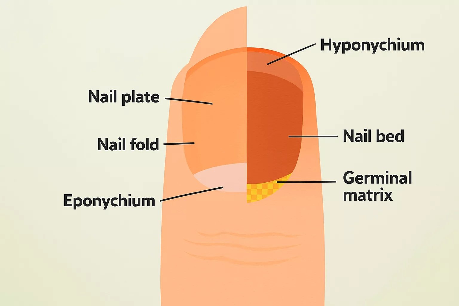
The illustration shows different parts of the nail, including the nail bed, nail plate, and nail matrix
- Nail plate: It is the hard part of the nail that you see. It is made out of keratin.
- Nail bed: It is the skin that is present underneath the nail plate (invisible to the eye).
- Nail matrix: This is the part of the skin (at the base) that connects to the nail plate. It is the point where new keratinized cells grow.
On the sides, you will find the nail folds.
Leukonychia Types
Apparent Leukonychia:
In apparent leukonychia, white spots are present on the nail bed. A distinguishing feature of apparent leukonychia is that the whitening disappears on pressure, unlike true or pseudoleukonychia, where the color change persists.[1] There are different types of presentations of apparent leukonychia:
- Muehrcke lines: Exist as a pair of symmetrical and horizontal lines that run across the nail. These lines mostly indicate some underlying disorder, like chemotherapy. These lines can not be felt, i.e., are non-palpable.
- Lindsay nails: In this type, the nail is divided into two halves with a sharp line. The lower part is white while the upper half is red, brown, or pink. It mostly indicates chronic kidney disease.
- Terry’s nails: In Terry’s nails, you will find whitening in the majority of the nail. However, there is thin distal darkening. The nails appear “washed-out” with just a small strip of brown darkening near the tip.
Pseudoleukonychia:
In this type, white discoloration of the nail plate occurs in response to external factors like cosmetic usage of superficial fungal infections (like onychomycosis). You can notice the spots on the surface of the nail.
True Leukonychia:
In true leukonychia, the white spots originate in the nail matrix (within the new, growing cells of the nail). These cells then increase in size, and the nail grows. Consequently, the white spots appear in the nail plate. True leukonychia is further divided into the following types:
- Leukonychia Partialis: In this type, a part of the nail plate turns white and can be attributed to acquired or hereditary causes. Partial leukonychia is subdivided into three types, based on the appearance of the white spots. In punctate type, you have a single or multiple small white dots on the nail. In striate, there are bands of white discoloration parallel to the nail’s base, while in the longitudinal type, you can see white bands running the length of the nail.

Leukonychia partialis in an African woman.
- Leukonychia Totalis: The totalis type is rare and is characterized by white discoloration of the entire nail. Congenital leukonychia totalis has been clinically reported, which is not linked to any other systemic disorder.[2]
Leukonychia Symptoms
The main presentation of leukonychia is the formation of white discolorations on the nail. The discolorations can vary from one individual to another. Some people report one or two medium-sized specks, while others notice large spots. As mentioned, some part of the nail is covered in white discoloration in leukonychia partialis. However, individuals with leukonychia totalis have a completely white nail plate. Leukonychia can be present in conjunction with other symptoms, including:
Nail Brittleness:
Leukonychia and nail brittleness are known to co-exist in individuals suffering from nutritional deficiencies. Zinc deficiency leads to brittle nails and transverse leukonychia.[3]
Leukonychia Striata (Mee’s lines), aka transverse leukonychia, usually arises due to abruptions in the nail matrix. Mee’s lines are white bands of lines that run transversely on the finger/toe nails.
Onycholysis:
Detachment of the nail plate (onycholysis) from the base can accompany the white spotting on the nails. Significant trauma to the nail can induce simultaneous onycholysis and leukonychia. In a reported clinical case, doctors observed leukonychia and capillary dilatation in proximity to the onychodermal bands. The severe changes in the nail structure arose in response to intense trauma.[4]
Changes in Thickness:
Inflammatory conditions of the nail induce thickening of the nail along with white spotting. Clinicians note that leukonychia and nail plate thinning are seen in several cases of nail lichen nitidus.[5] On the other hand, fungal infections induce nail plate changes such as thickening (hyperkeratosis) and discoloration (leukonychia).[6]
You may also note the formation of ridges and dents with white spot formation. Some iron-deficient individuals also notice spoon-shaping (koilonychia) in association with the discoloration.
Leukonychia Causes
Trauma/Injury:
The most common cause of leukonychia is trauma to the nail. Injury to the nail plate or the matrix can lead to the formation of white spots. Activities that can injure your keratinized finger structures include nail biting and directly hitting/bumping your nails. Studies show that nail biting/forceful nail biting can lead to an array of nail disorders, one of which is leukonychia.[7] Repeated manicures can also increase your risk of getting a nail injury.
Allergy:
Allergic leukonychia can occur if your body recognizes nail products (like nail polishes, nail glosses, polish removers, fake nails, and hardeners) as potential allergens. The chemical compounds in these products can also induce contact dermatitis. In a study about the side effects of gel nail polish, about 8.5% of the participants reported white spots on the nail plates.[8]
Infection:
Another cause of white spotting on the nails is a fungal infection. Infestation by fungi is known to make your nails thick, cracked, or discolored. Fungal leukonychia is a common pathology in immunosuppressed patients. The disorder is frequently associated with Trichophyton species.[9]
Nutritional Deficiencies:
Different types of nutritional deficiencies can have manifestations in your nails. The most notable mineral deficits that can turn your nails white include calcium, iron, and zinc. Iron deficiency is notorious for changing the shape and color of your nails. Similarly, the study by Seshadri and colleagues found that zinc deficiency is linked to transverse leukonychia. Moreover, vitamin deficiency can also contribute to color changes in the nails. A clinical study found true leukonychia in a 26-year-old male who had a long history of nail discoloration. Serum tests revealed low vitamin B12 levels, which were suspected to be the cause of the whitening.[10]
Drugs & Medications:
A wide array of drugs and medications can end up turning your nails white. A 2025 study revealed that exposure to heavy metals like selenium and thallium leads to transverse leukonychia.[11] Similarly, arsenic toxicity can lead to multiple changes in the skin and nails.[12]
Chemotherapeutic drugs are also known to bring about changes in your nail structure. Numerous cancer patients notice white spotting in their nails after starting chemotherapy. Drugs like bleomycin, adriamycin, and vinblastine are associated with leukonychia.[13] Moreover, antibiotics like UTI medicines and sulfonamides can also cause white spots to develop in your nails.
Systemic Diseases:
A list of systemic disorders has been linked to structural and color changes in your nails. Apparent leukonychia is associated with multiple systemic diseases. Experts suggest that Terry’s nails can be an indication of serious health conditions like chronic renal failure, congestive heart failure, and cirrhosis.[14]
Many times, apparent luekonychia is the outcome of systemic disease. The band formation is attributed to abnormal matrix keratinization.[15]
Liver cirrhosis is a common cause of nail plate whitening. Terry’s nails are a common observation in patients with end-stage liver cirrhosis.[16] Diabetes and heart failure cases may also present with milk spots. Autoimmune disorders like psoriasis can also contribute to white nail spotting.
Hereditary Conditions:
The association between hereditary conditions and leukonychia can not be neglected. It has been noted that congenital leukonychia is a part of various hereditary diseases and syndromes.[17] A 7-year-old Thai boy had asymptomatic white spots in the nails and was diagnosed with the rare disorder of congenital leukonychia totalis. [18]
Several hereditary conditions have a direct impact on your nails, including Darier disease, Hailey-Hailey disease, Bauer syndrome, and Bart-Pumphrey syndrome etc.
Leukonychia Diagosis
Your doctor will physically examine the nail and identify the location (nail bed or plate) and type of white dot pattern (striate, punctate, etc.). He will ask you questions about the history of the symptoms. Questions may include any direct trauma to the fingers, underlying comorbidities like diabetes, renal or liver disease, or nutritional deficiencies, etc.
Diagnostic Tests:
Your healthcare provider may also order different diagnostic tests to diagnose the condition responsible for the white discoloration of your nails. The most commonly adopted tests include:
- Mycology: Nail analysis study in which the doctor sends nail clippings for lab analysis.
- Blood test: After drawing a small amount of blood from a vein in your arm, the doctor gets it checked for markers of systemic diseases. It helps them know potentially which disorder is causing leukonychia.
- Potassium hydroxide (KOH) test: This is frequently done to identify the pathogen responsible for skin infection. Here, the doctor removes a small part of the nail (instead of skin) and sends it to the lab. In the lab, technicians dip the nail specimen in KOH solution, which dissolves the nail cells, leaving only the pathogen cells. It aids in diagnosing nail infection-induced leukonychia.
- Biopsy: In rare instances, your doctor may remove a small, fine part of your nail and send it to a lab for microscopic evaluation and lab testing.
Differential Diagnosis:
Terry’s Nails Vs Leukonychia

Terry’s nail is a type of leukonychia in which there is a “washed-out” appearance of the nail with a darker coloration of the margins.
Leukonychia is a generalized term that refers to any type of white spotting or discoloration of the nails. On the other hand, Terry’s nail is a type of apparent leukonychia characterized by a “washed out” look of the nails with a brown/pink strip of nail at the tip.
Leukonychia Treatment
As we know, leukonychia isn’t a disease itself but rather a manifestation of some other disorder. Therefore, the treatment plan depends on the underlying cause of the issue.
Trauma:
In case of trauma-induced white spots grow out and reach a point where you can remove them with a nail cutter/clipper. However, this is a slow process and demands patience. Generally, fingernails take 6-9 months while toenails take 12-18 months for the white specks/spots to grow out.
Infection:
Doctors advise antifungal medications to manage fungal infections of the nails. Topical antifungals can work for mild infections, but for moderate-to-severe and severe infections, doctors prescribe oral antifungals like itraconazole, terbinafine, and fluconazole. Other drugs like albaconazole and posaconazole are also effective in managing onychomycosis.[19]
Nutritional Supplementation:
In case of nutrient deficiencies, nutritional supplementation has been shown to improve the discolorations. The study conducted by Ahmed and colleagues in 2025 found that leukonychia improved with zinc supplementation in deficient patients.
Management of systemic diseases can help, but most of the time, leukonychia arises in severe states, which are difficult to manage. If caused by medicine intake, cessation of specific drugs (with the doctor’s guidance) can help resolve leukonychia.
Leukonychia Prevention
You can adopt the following steps to prevent leukonychia:
- Avoid repeated manicures if your nails are injury-prone.
- Wear protective gear like gloves when playing hard sports or working with tools, etc.
- Keep your nails short and moisturized.
- Minimize use of irritating nail products (gels, polishes, etc).
Wrapping Up
Leukonychia is a condition in which white discolorations appear on the nails (fingernails or toenails). The white spots, specks, or bands can be present on the nail bed (apparent leukonychia), on the nail plate due to some external factor (pseudoleukonychia), or originate from the nail matrix and grow (true leukonychia).
White spotting on the nails usually develops in response to trauma, fungal infections, or underlying systemic disease diabetes, renal disease, and liver failure, etc.). Nutritional deficiencies and certain drugs (arsenic, chemotherapy, and UTI drugs) can also impact the color of your nails. When leukonychia develops secondary to some underlying cause, other symptoms like nail brittleness, changes in nail thickness, and detachment of the nail plate can be seen.
Treatment involves correcting the underlying issue causing the spotting. In trauma cases, patients wait for white spots to grow and then cut them out. Antifungal medications, nutritional supplementation, and cessation of causative drugs/nail products can lead to resolution of leukonychia.
References
[1] Curtis, K. L., & Lipner, S. R. (2025). Punctate Leukonychia. InAtlas of Nail Disorders Across All Skin Colors: A Comprehensive Guide to Navigating Nails(pp. 351-358). Cham: Springer Nature Switzerland.
[2] Pakornphadungsit, K., Suchonwanit, P., Sriphojanart, T., & Chayavichitsilp, P. (2018). Hereditary leukonychia totalis: A case report and review of the literature.Case Reports in Dermatology,10(1), 82-88.
[3] Seshadri, D., & De, D. (2012). Nails in nutritional deficiencies.Indian Journal of Dermatology, Venereology and Leprology,78, 237.
[4] Navarro, L. (2023). Pattern diagnosis of onycholysis.JEADV Clinical Practice,2(2), 213-224.
[5] Tordjman, L., Thomas, J., Tosti, A., & Morrison, B. W. (2025). Inflammatory nail disorders in skin of color: A systematic review of clinical and onychoscopic manifestations.International journal of dermatology,64(6), 1013-1020.
[6] Lee, D. K., & Lipner, S. R. (2022). Optimal diagnosis and management of common nail disorders.Annals of Medicine,54(1), 694-712.
[7] Nandgaye, D. C., Gotefode, S. N., & Moharkar, D. W. The Causes, Diagnosis and Treatment of Nail Disorder.
[8] Putek, J., PRZYBYŁA, T., Szepietowski, J. C., Baran, W., & Batycka-Baran, A. (2020). Side-effects associated with gel nail polish: a self-questionnaire study of 2,118 respondents.Acta Dermato-Venereologica,100(18), 5931.
[9] Saldaña, M., Férez-Blando, K., Domínguez-Cherit, J., Fierro-Arias, L., & Bonifaz, A. (2017). Fungal leukonychia and melanonychia: a review.Current Fungal Infection Reports,11(3), 110-116.
[10] Ahmed, A., Alahmadi, M., Almowald, A., & Almohanna, H. (2025). True leukonychia: case reports and review of the literature.Dermatology Reports.
[11] Cohen, P. R., & Sutton, L. (2025). Forensic onychology of heavy metal exposure: forensic dermatology of the manifestations of heavy metal toxicity in nails.Dermatology Online Journal,31(2).
[12] Rajiv, S. V., George, M., & Nandakumar, G. (2023). Dermatological manifestations of arsenic exposure.Journal of Skin and Sexually Transmitted Diseases,5(1), 14-21.
[13] Samal, K., Mohanty, S., & Satapathy, S. (2021). Chemotherapeutic Drug-Induced Nail Changes: A Prospective Observational Study.Turkish Journal of Dermatology,15(3), 61-65.
[14] Witkowska, A. B., Jasterzbski, T. J., & Schwartz, R. A. (2017). Terry’s nails: a sign of systemic disease.Indian journal of dermatology,62(3), 309-311.
[15] Curtis, K. L., & Lipner, S. R. (2025). Transverse Leukonychia. InAtlas of Nail Disorders Across All Skin Colors: A Comprehensive Guide to Navigating Nails(pp. 359-365). Cham: Springer Nature Switzerland.
[16] Khan, N., & Mervak, J. (2025). White Nails in a Man with Hepatic Cirrhosis. InClinical Cases in Nail Disorders(pp. 107-110). Cham: Springer Nature Switzerland.
[17] Saraniuk, R. V., Polonikov, A. V., & Gosteva, T. A. (2025). Leukonychia associated with hereditary syndromes.Vestnik dermatologii i venerologii.
[18] Pakornphadungsit, K., Suchonwanit, P., Sriphojanart, T., & Chayavichitsilp, P. (2018). Hereditary leukonychia totalis: A case report and review of the literature.Case Reports in Dermatology,10(1), 82-88.
[19] Fávero, M. L., Bonetti, A. F., Domingos, E. L., Tonin, F. S., & Pontarolo, R. (2022). Oral antifungal therapies for toenail onychomycosis: a systematic review with network meta-analysis toenail mycosis: network meta-analysis.Journal of Dermatological Treatment,33(1), 121-130.