

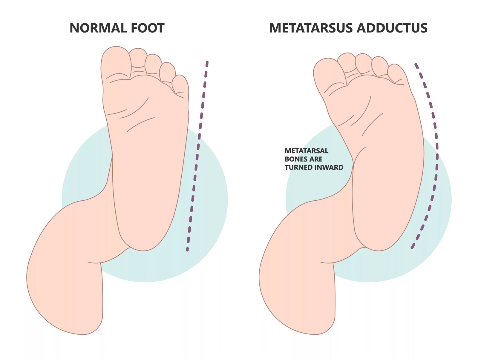
Metatarsus Adductus This Metatarsus adductus is a congenital condition characterized by inward curving of the foot and toes. The deformity is attributed to abnormalities in the bones and tissues of the front part of the foot (forefoot), i.e., metatarsus. It can affect one or both feet. Several other names for the disease include forefoot adduction, metatarsus adductovarus,metatarsus varus, and forefoot varus. Doctors diagnose the foot in infants. It is the most common congenital foot anomaly in infants and is reported to have a prevalence of 1–2% of live births.[1] Although primarily diagnosed in infancy, metatarsus adductus in adults can still cause noticeable foot pain, difficulty finding comfortable footwear, and an increased risk of bunions due to long-standing forefoot deviation.
In most cases, the abnormality resolves without any intervention within the first year of life. However, when needed, doctors advise different foot stretches and exercises. Corrective shoes and casts can also help correct adductus. Surgical intervention is rarely indicated.
Metatarsus Adductus Types: Metatarsus Adductus This
Clinicians use different classification criteria to categorize metatarsus adductus. The most commonly adopted method is to classify the disorder based on the difficulty of correction, i.e., flexibility of the foot. However, you can also find classification based on the severity of the deformity.
Based On Flexibility:
This classification helps healthcare providers decide the treatment plan for the patient. It relies on the flexibility of the deformed foot and is classified as:
Flexible:
This type poses the fewest problems as it resolves on its own (without treatment). Pediatric health professionals can completely straighten the patient’s forefoot using only gentle manipulation. Complete correction can be achieved with ease. The foot has full flexibility.
Semi-flexible:
This type is more difficult to treat. Health professionals cannot achieve complete correction with gentle manipulation. Most pediatric caregivers can partially realign the foot.
Rigid:
The bone and tissue deformity of the forefoot makes it rigid and unresponsive to conventional manipulation techniques. The foot is stiff, and there is no way to straighten it with manual manipulation. Thus, doctors have to resort to intensive treatment strategies like orthotics and even surgery.
Metatarsus Adductus Symptoms
Infants
In infants, parents notice the inward twisting of the foot immediately after birth or during the first year of life. Therefore, most parents report to a pediatrician to fix the deformed foot. Clinicians note adduction of the forefoot. Newborns have a crescent-shaped foot (or feet). Moreover, there is an increased (wider) separation between the big toe and the second toe.
Due to the abnormal pulling of the forefoot, the lateral border of the foot (outer side of the foot) takes a convex shape (instead of straight). This is a hallmark clinical sign. In some cases, you can appreciate a soft-tissue crease on the inner edge of the foot. This crease indicates that the patient is suffering from a more rigid deformity. Despite the deshaping, different motions of the foot (hindfoot movement, etc.) are normal.
Hip Dysplasia:
Analysis of multiple forefoot varus cases has revealed a higher incidence of hip dysplasia in infants. It is an infirmity in which there is improper development of the hip joint, leading to poor fitting of the thigh bone into the acetabulum (bone) of the hip. Patients may develop groin pain, stiffness, and joint instability as a consequence of the disorder. Dysplasia of the hip can occur at birth (congenital) or develop later in life (developmental). The type most commonly associated with forefoot deviation is the developmental hip dysplasia.[2]
Adults
In adults, the C-shaped or banana-shaped foot can cause pain. The twisted podal structures can reduce your foot mobility. Many patients have a reduced range of foot motion. They can’t move the ankle within its full range.
It can also interfere with normal activities. People with metatarsus adductus often have difficulty finding shoes that fit. Moreover, the increased space between the big toe and the second toe adds to footwear problems.
Bunion:
Another issue that arises in such individuals is a bunion. A bunion is a foot deformity characterized by the formation of a bony projection/bump on your big toe. The bump is present on the junction of the big toe and the foot. Some studies conclude that the abnormal attachments of the tendons and muscles to the foot bones in metatarsus adductus put the big toe at a higher risk of valgus deformities (abnormal angling of the bones). Thus, there is a correlation between matatarsus adductus and hallux valgus (bunion).[3]
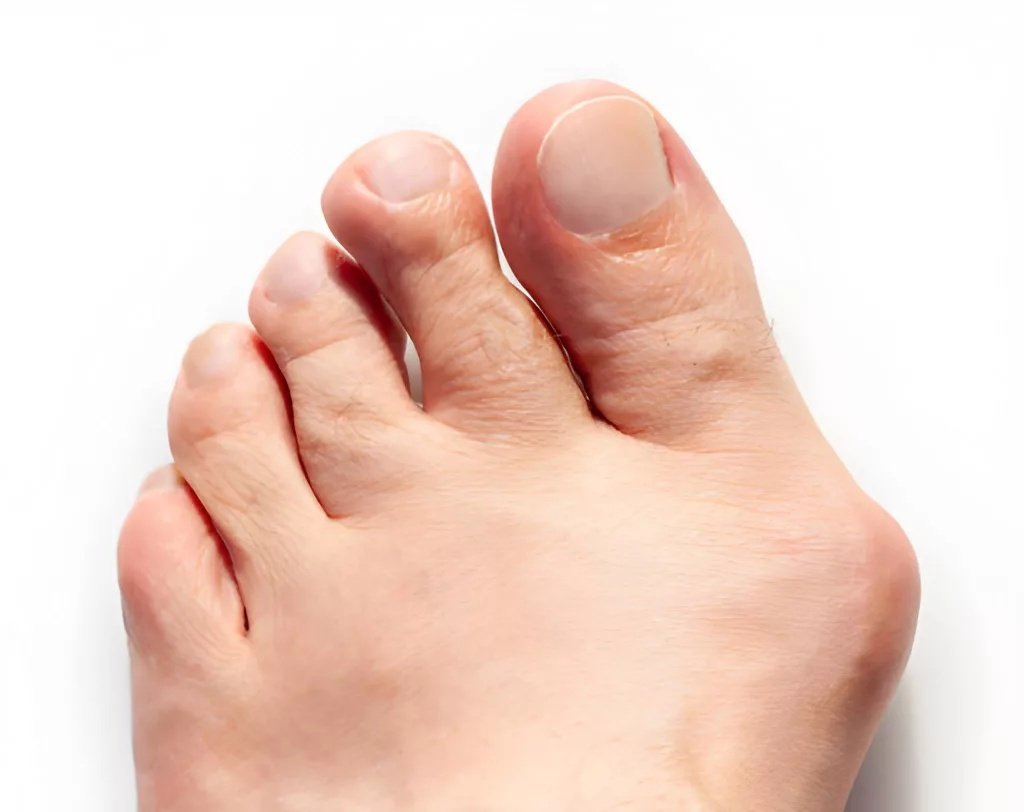
A bunion is frequently associated with metatarsus adductus in adults.
According to one study, the prevalence of metatarsus adductus in patients undergoing surgery for a bunion is found to be 35%.[4]
Metatarsus Varus Causes
The real underlying reason for this twisting of the forefoot is unknown to healthcare providers. However, most healthcare providers agree that it is not strongly genetic, but family history can be a risk factor. Other potential causes of metatarsus adductus include:
Poor Fetal Development:
There is a theory that the compression of the foot during fetal life can contribute to forefoot twisting within the womb. Current reviews suggest that MA arises when there is less room (in the womb) to accommodate the growing fetus. This reduced space leads to molding of the fetus within the uterus, which becomes the basis for musculoskeletal deformities like metatarsus adductus, torticollis, and hip dysplasia.[5]
Fetuses who have their bottoms pointed down in the womb are also prone to developing foot abnormalities. Thus, the Breech position can lead to metatarsus adductus.
Lack of Amniotic Fluid:
Another theory suggests that forefoot varus is the result of a lack of amniotic fluid, a condition known as oligohydramnios. The intrauterine constraints, paired with poor muscle activation in the womb, lead to the foot disorder.
A late pregnancy usually has a higher incidence of complications. Thus, clinicians find late pregnancy as a risk factor for metatarsus adductus.
Metatarsus Adductus Diagnosis
This pedal disorder is diagnosed in infancy. The foot problem can affect one foot, i.e., unilateral, or both feet of an individual, i.e., bilateral. Sometimes, adults notice metatarsus adduction in adulthood. However, the disease doesn’t arise in later life, and it is present since birth. It only aggravates as age passes.
Doctors start by performing a physical examination of the foot. By physically manipulating the foot and analyzing the range of motion, doctors can classify the adduction (rigid, flexible, etc.). It is easy to diagnose the problem in infants. However, there is a resemblance to other congenital foot disorders, and it is the clinician’s job to differentiate.
Your doctor may also ask for your family history of the disease, as a genetic component is considered a potential cause. Your healthcare provider may also look for signs of intoeing (pigeon-toed feet) in your toddler’s feet.
The next step is to classify the disease on the basis of severity, which can help in devising the treatment plan for it.[6]
Classification Based On Disease Severity: Bleck Manual Assessment
This classification is based on the changes in the heel bisector line (HBL). It is an imaginary line that starts from the mid of heel and extends to the forefoot. In normal, healthy newborns, the line typically lies between the second and the third toe web spaces. However, in affected individuals, the line extends beyond the 2nd/3rd toe space. The extent of changes in the HBL determines the class of the disease.
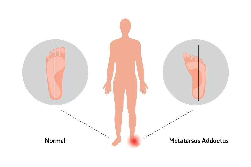
Metatarsus adductus in relation to the Heel Bisector Line (HBL).
It is regarded as an important assessment tool for forefoot adduction.[7]
Mild:
There is a slight deformity, and the HBL extends through the webspace of the 3rd toe.
Moderate:
In the moderate category, the HBL passes through the webspace between the 3rd and the 4th toes.
Severe:
In severely deformed feet, the line is seen extending between the 4th and 5th toe webspace. This indicates the great extent of the foot’s inward tilting.
Imaging Tests
Radiographic imaging plays a pivotal role in the diagnosis of musculoskeletal disorders. However, in the case of metatarsus adductovarus, doctors reach a confirmed diagnosis with physical examination alone. Still, your pediatrician may order a foot X-ray for further evaluation.
Foot X-rays are preferred in adult metatarsus cases. This is because X-ray angles (metatarsus adductus angle) have proven to be a reliable way of measuring the extent of deformity. Moreover, in adults, this helps in accurate pre-operative planning.[8]
Differential Diagnosis
Several congenital foot deformities have presentations similar to forefoot adduction. These disorders require different treatment strategies. Thus, it is crucial to differentiate the disease from other abnormalities like tarsal coalition and clubfoot.
Metatarsus Adductus Vs Clubfoot
Forefoot varus is a self-limiting disorder characterized by a normal heel and inward curving of the forefoot. On the other hand, talipes equinovarus or clubfoot is a severe deformity in which the foot turns inward (with hindfoot involvement) due to shortening of the tendons. Unlike metatarsus adductus, which mostly resolves on its own, clubfoot needs bracing and even surgery.
Metatarsus Adductus Treatment
Doctors generally don’t advise any treatment option because most patients (around 90%) do not need any treatment, and the condition resolves by the first birthday of the child. Moreover, another 5% cases are corrected when the child reaches the walking stage, i.e., 1 to 4 years.
In case of rigid adduction or severe deformity, you will need to consult a pediatric orthopedist. Orthopedists specialize in treating deformities of the skeletal system, and pediatric orthopedists know how to fix abnormal bones, ligaments, and tendons of children. These professionals opt for non-surgical or surgical treatment options, depending on the severity of your child’s disease:
Non-Surgical Management
Your healthcare provider will try to correct the foot twisting with the help of wearables. The most commonly adopted conservative treatment option for metatarsus adductus is serial stretching/serial casting. It involves providing the patient with a series of customized casts that stretch the muscles/joints, bringing them to the normal position. You have to change the casts every week to adjust the foot according to the new position. There is some evidence that non-surgical treatments work for semi-rigid cases.[9]
External medical devices (orthoses) like Denis Brown bar, Wheaton brace, counter rotation system splint, and Fillauer bar can be effective in correcting the foot disorder. You can even get specially modified shoes like Bebax shoes and Ipos antiadductus shoes for metatarsus adductus. Health providers advise you to use a combination of gentle exercise and medical support for the best outcomes. A study concluded that manipulation (exercise) with support devices like rigid straps, Wheaton brace, and reverse last shoe can effectively correct the foot deflection. However, there may be some side effects of the treatment that the clinician needs to look out for. It was also noted that the Wheaton brace and the Bebax shoe have fewer complications than other conservative management options.[10]
Surgical Management
If the disorder is not fixed by 4–6 years of age, doctors advise surgery. Tarsal metatarsal capsulotomy, or Heyman-Herndon tarsometatarsal capsulotomy, is a surgical procedure for metatarsus adductus in which the surgeon cuts the joint capsule in the midfoot and releases the metatarsals (foot bones). It is a surgical strategy adopted for forefoot adductus. This is, at times, paired with procedures like multiple metatarsal osteotomies (cutting and repositioning of multiple foot bones) to correct persistent metatarsus adductus in young children.[11] Generally, metatarsus adductus prognosis is great.
Final Word
Metatarsus adductus (MA) is a congenital foot disorder in which there is inward curving of the forefoot (front part of the foot). Babies have abnormal bones and soft tissues that pull the foot into this position. It is believed that compression during pregnancy, lack of amniotic fluid, and abnormal fetal position in the womb contribute to MA. Many believe that genetics also have a role in MA. Individuals with forefoot adduction are at a higher risk of hip dysplasia and bunions.
In 90% of the cases, the condition corrects itself within the first year of childhood, and for another 5% it resolves during 1-4 years of age. For non-resolving cases, clinicians adopt conservative strategies like splinting, casting, bracing, corrective shoes, and gentle stretching. However, for rare, severe, and rigid cases, doctors go for surgical intervention.
References
[1] Marshall, N., Ward, E., & Williams, C. M. (2018). The identification and appraisal of assessment tools used to evaluate metatarsus adductus: a systematic review of their measurement properties.Journal of foot and ankle research,11(1), 25.
[2] Hutchinson, B. (2010). Pediatric metatarsus adductus and skewfoot deformity.Clinics in Podiatric Medicine and Surgery,27(1), 93-104.
[3] Chen, L., Wang, C., Wang, X., Huang, J., Zhang, C., Zhang, Y., & Ma, X. (2014). A reappraisal of the relationship between metatarsus adductus and hallux valgus.Chinese Medical Journal,127(11), 2067-2072.
[4] Aiyer, A. A., Shariff, R., Ying, L., Shub, J., & Myerson, M. S. (2014). Prevalence of metatarsus adductus in patients undergoing hallux valgus surgery.Foot & ankle international,35(12), 1292-1297.
[5] Freedman, J. D., Eidelman, M., Apt, E., & Kotlarsky, P. (2024). Review of current concepts in metatarsus adductus.Pediatric annals,53(4), e152-e156.
[6] Eamsobhana, P., Rojjananukulpong, K., Ariyawatkul, T., Chotigavanichaya, C., & Kaewpornsawan, K. (2017). Does the parental stretching programs improve metatarsus adductus in newborns?.Journal of Orthopaedic Surgery,25(1), 2309499017690320.
[7] Marshall, N., Ward, E., & Williams, C. M. (2018). The identification and appraisal of assessment tools used to evaluate metatarsus adductus: a systematic review of their measurement properties.Journal of foot and ankle research,11(1), 25.
[8] Marshall, N., Ward, E., & Williams, C. M. (2018). The identification and appraisal of assessment tools used to evaluate metatarsus adductus: a systematic review of their measurement properties.Journal of foot and ankle research,11(1), 25.
[9] Williams, C. M., James, A. M., & Tran, T. (2013). Metatarsus adductus: Development of a non‐surgical treatment pathway.Journal of paediatrics and child health,49(9), E428-E433.
[10] Karimi, M., Kavyani, M., & Tahmasebi, R. (2022). Conservative treatment for metatarsus adductus, a systematic review of literature.The Journal of Foot and Ankle Surgery,61(4), 914-919.
[11] Feng, L., & Sussman, M. (2016). Combined medial cuneiform osteotomy and multiple metatarsal osteotomies for correction of persistent metatarsus adductus in children.Journal of Pediatric Orthopaedics,36(7), 730-735.