


solar lentigines treatment Solar lentigines (singular solar lentigo), also known as liver spots, age spots, sun spots, or senile freckles, are a benign condition characterized by the development of darkened patches of skin. The condition is highly prevalent in older individuals; therefore, it is commonly referred to as senile freckles. Solar lentigines usually develop in response to excessive sun exposure. In one study, around 27% of the population suffered from solar lentigo. The study concludes that of all pigmentary disorders of the skin, solar lentigines have the least impact on the quality of life (QoL), and there are no stigmas attached to it.
Doctors use different types of non-interventional strategies to manage the spots. Apart from aesthetic concerns, these spots do not pose any significant health threat.
What Are Solar Lentigines?
Solar lentigines are harmless, dark spots on the skin that you may have noticed on the skin of the elderly. A single lesion is called a solar lentigo, but as they occur in larger numbers, the condition is called solar lentigines. These old-age spots are generally widespread, being more prominent on the skin that receives more sunlight. The spots are a result of overproduction of the black pigment, i.e., melanin. There is increased melanin production and retention within the epidermis, driven by hyperactive melanocytes rather than true melanocyte proliferation. The number of melanocytes is usually normal or only mildly increased altered keratinocyte–melanocyte signaling and epidermal changes lead to pigment accumulation.
Solar Lentigines Symptoms
Patients notice the flat and small patches on the skin that are non-itchy and painless. Close observation reveals that the patch is well-circumscribed. The shape of these spots can vary from oval to round. However, they may even be irregularly shaped. Moreover, the color of the lentigines can vary between individuals. While most patients notice brown-black skin patches, the shade can vary from tan to dark brown/black.
Usually, the size of these spots is small, but they may range in size from a few millimeters to several centimeters in diameter. Most of the time, lentigos are smooth and flat, but sometimes, they may feel scaly.
Location Of Spots
The spots mostly exist in groups on solar-exposed sites. Therefore, the most common sites of solar lentigines are:
Face:
Your facial skin gets exposed to sunlight more than any other part of the body. Therefore, one of the most common sites for solar lentigines is the face. There have been reported cases of solar lentigo on the cheeks and sides of the face. You can develop them on the temples too. It was revealed in a study that overexposure of the facial skin to sunlight induces abnormalities in the keratinocytic function that contribute to solar lentigines of the face.
Most patients are concerned about the black spots on their skin and seek treatment solely due to aesthetic issues.
Hands:
The back of your hands is prone to developing different skin disorders, including lentigines. Due to the high prevalence of the lesions on the hand, many treatment researchers use hand lentigos to analyze the effect of a specific treatment strategy. In one study, a topical dermocosmetic product was used to fix hand solar lentigines, which proved to be effective.
Arms And Shoulders
Ultraviolet radiation (UVR) exposure of the shoulders and arms increases your likelihood of falling prey to the development of unesthetic spots. A study concluded that the number of solar lentigines on the arms/shoulders is positively associated with daily hours spent outdoors between noon and 3 pm on holiday, days at the beach, and the reported number of sunburns. There is a higher percentage of patients who visit the beach frequently because they expose their skin to sunlight for longer periods and are also prone to sunburns.
Upper Back And Chest
The spots follow the sun-exposed skin areas. Thus, you might also develop solar lentigines on your upper back and chest. These parts are also prone to sunburns. Due to the same source, solar lentigines and sunburns are linked to each other and may co-exist in a person. Solar lentigines can develop in the same areas where you previously had a sunburn.

Solar lentigines can be seen on the upper back of an elderly woman.
In a study, researchers found multiple solar lentigos (on the upper back and shoulders) in an adult patient. The study concluded that the solar lentigos are potential clinical markers of past severe sunburn.
Sun damage can show itself at a greater magnitude on the delicate skin present on the neck, chest, and upper shoulders. This area, known as the decollete area, starts from the chin down and extends to above the chest/breasts. Low necklines expose this intricate skin (which has fewer oil glands and thinner skin layers) to solar damage.
Conditions Associated With Solar Lentigines
Solar damage can manifest in the form of various skin disorders. Therefore, solar lentigines frequently coexist with other manifestations of photoaging, including wrinkling, telangiectasia, and solar elastosis, reflecting long-term ultraviolet-induced structural damage to the skin. In some cases, liver spots arise after a sun damage condition has developed, while in other cases, they set the foundation for another deformity.
Sunburns
Several times, solar lentigos appear on skin affected by sunburns. While lentigines themselves are benign, their presence reflects cumulative UV damage, which is associated with an increased risk of skin cancers, including melanoma, so doctors take solar lentigines growing in the sunburnt skin very seriously.
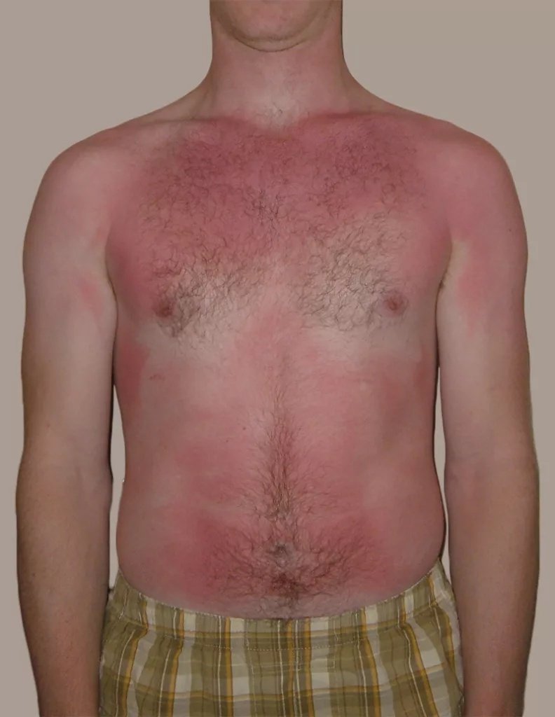
Sunburn are interlinked with solar lentigines
It was concluded in a study that multiple solar lentigines on the upper back and shoulders were associated with intense sunburns in the past. Therefore, these black spots served as indicators for the increased risk of cutaneous melanoma.
Seborrheic Keratosis
Another skin disorder, i.e., seborrheic keratosis (SK), can develop within the senile freckles. The development of this non-cancerous condition changes the lentigines from a flat surface to raised, scaly bumps. There is localized thickening of the skin, accompanied by changes in the texture. Seborrheic keratosis manifests as a waxy, warty growth. Studies show that these exophytic (outward) growths have a characteristic “stuck-on” appearance and can arise from a solar lentigo. Reticular SK frequently arises from lentigines.
The co-occurrence of benign tumors is termed a collision tumor. There have been case reports of seborrheic keratosis co-occurring with solar lentigo and syringoma (a benign tumor of sweat glands).
Lichenoid Keratosis
Liver spots can also undergo inflammation and change into lichenoid keratosis. This condition is characterized by the formation of red, itchy, and scaly lesions within the spots of lentigo. Clinicians have noted inflammation in these lichen-planus-like keratoses. According to a study, solar lentigo is found in about 68% of cases diagnosed with lichenoid keratosis. The inflammation disappears without the need for an intervention. However, the healing process is slow and time-consuming.
What Are The Causes Of Solar Lentigines?
For years, clinicians have believed that sunlight (UV) exposure is the only cause of lentigo. However, recently we have seen researchers focusing on the role of environmental factors in the disease.
Ultra-Violet Radiation
The most common cause of solar lentigines is UV radiation exposure. Therefore, we see lentigines appear on skin parts that are frequently exposed to UVA and UVB type radiation. Excessive exposure of the skin to the sun triggers the body’s protective mechanisms. The sunlight triggers your pigment-producing skin cells, i.e., melanocytes, to produce melanin (the dark pigment), which defends the skin against UV radiation-induced damage. Over time, the deposited melanin clumps together, forming a spot that eventually leads to the formation of lentigines. Thus, hyperpigmentation causes solar lentigines.
Environmental Factors
While the main contributor to solar lentigines is undoubtedly UV exposure, healthcare providers now use the term environment-induced lentigo to explain the unavoidable role of environmental toxicants in the disease onset. Researchers argue that specific receptors in the body, i.e., aryl hydrocarbon receptors, play a critical role in triggering different skin cells (melanocytes, keratinocytes, etc.). Environmental toxicants and UV light trigger these receptors.
In a unique case, a patient developed lentigines on skin areas that are not exposed to the sun. The female patient noticed lentigo lesions on the thighs and lower legs. Patient history revealed frequent occupational radon exposure in a spa (via radon-containing water). This led researchers to suspect radon as a non-solar cause of solar lentigines.
Risk Groups For Sun Spots
The following factors are risk factors for solar lentigines:
Old
Solar lentigines are fairly commonly seen in older adults. The disease is called senile freckles because it is most frequently seen in adults over 50 years of age. Therefore, oldies are more prone to solar lentigo. Sun spots are also key signs of photoaging, i.e., premature aging of the skin due to UV exposure.
Fair Skin
People with fair skin are prone to increased sun damage due to the lack of protective melanin. Thus, you may develop lentigines if you have a fairer complexion.
Occupational Exposure To Sun
Individuals working outdoors have a higher propensity for sun damage and lentigines. Thus, professionals like gardeners, farmers, construction workers, and landscapers etc. must adopt protective measures.
Family History
You are more likely to develop liver spots if you have a family history of solar lentigines. The exact genetic mechanisms of the aberrant pigment deposition are not known, but experts have observed a pattern in generations.
History Of Sunburns
Studies show that factors like chronic sun exposure, previous sunburns (before the age of 20), and the number of sunburns in childhood are associated with solar lentigines.
How Is Solar Lentigines Diagnosed?
Most patients visit a dermatologist for aesthetic concerns. Your healthcare provider will diagnose the skin condition by physically examining the spots. He examines the well-circumscribed black/tan patches. He will correlate the analysis with your history of sun exposure. As lentigines resemble malignant melanoma, doctors generally order more tests for a confirmed diagnosis.
Dermatoscopy
Doctors perform this non-invasive examination procedure to get a better view of the skin layers in a dermal disorder like solar lentigines. The healthcare provider uses a dermatoscope, which is a handheld device equipped with a magnifier and illuminator. A magnified view of the skin aids in differentiating solar lentigines from malignant pathologies.
Skin Biopsy
Sometimes, doctors remove a small part of the skin lesion and send it to the lab for analysis. Skin biopsy accurately diagnoses lentigo.
Differential Diagnosis (DD)
Multiple skin pathologies of the face and hands have presentations similar to solar lentigines. The following conditions fall under the DDs of solar lentigo.
Solar Lentigo Vs Freckles (Ephelides)
When present on the skin, freckles and lentigines have similar appearances. However, lentigo lesions are larger in size and darker in color as compared to freckles. Moreover, freckles fade in winter while lentigines are persistent.
Solar Letigo Vs Lentigo Maligna
Age spots are benign conditions arising from sun exposure and aging, while lentigo maligna is a type of early melanoma, i.e., a malignant condition. Solar lentigo spots are flat, black, and well-defined, while lentigo maligna are irregular patches with varied colors. The irregularly shaped patches of lentigo maligna tend to grow in size and change shape.
Solar Lentigines Treatment
If not treated, solar lentigines persist for life. While they don’t cause any functional or physical harm, patients seek treatment for aesthetic issues. Doctors adopt different strategies to treat sun spots.
Non-Surgical Treatment
The preferred mode of treatment is non-surgical treatment. Various strategies have been shown to work in pigmented skin disorders.
Topical Therapy
Different chemical preparations work for lentigo. According to the latest studies, the most effective topical treatment involves a combination of a retinoic acid (0.01% tretinoin) and a depigmenting agent (2% mequinol).
Chemical peels are agents, mostly acidic, that help remove the outer layers of skin. Trichloroacetic acid (TCA) is an effective and efficacious topical medicine for lentigo. Cysteamine is another topical medicine that improves solar lentigines.
Cryotherapy
Extreme cold application (cryotherapy) has conventionally been recommended as the first-line therapy for solar lentigines. Dermal cooling systems have been shown to reduce pigmentation in benign lesions like sun spots. However, there may be associated side effects.
Laser Therapy
Laser light destroys the pigmented clusters by generating heat in the skin layers. Modern clinical studies show that a variety of laser therapies have worked for black lentigo lesions. Intense pulse light, pulsed dye laser, picosecond laser, and fractional carbon dioxide lasers have yielded promising results with minimal side effects.
Surgical Treatment
Doctors prefer non-invasive procedures, but they may opt for surgery in severe cases. Dermabrasion is a surgical procedure in which the surgeon removes the top layer of the skin with a high-speed rotating tool. This leads to the revelation of smoother and newer underlying skin that lacks the dark pigment.
Wrapping Up
Solar lentigines (sun spots/liver spots) are a non-cancerous condition characterized by the formation of small, black spots on sun-exposed skin (hands, face, shoulders, and upper back). It is frequently seen in old individuals and is known as senile freckles. People with fair skin and old age are most prone to the condition. Doctors diagnose it with physical examination and dermatoscopy, and treat it with non-surgical modalities. Chemical peels, topical medicines, cryotherapy, and laser therapy effectively help alleviate the black pigmentation. In rare instances, doctors adopt a surgical procedure, i.e., dermabrasion, to eliminate the top pigmented layer.
References
[1] Kerob, D. K., Passeron, T., Dreno, B., Wei, L., Morita, A. A., Leok, C., … & Lim, H. W. (2024). 54736 Prevalence of solar lentigo, impact on quality of life and social stigmatization: Results of the first large international survey.Journal of the American Academy of Dermatology,91(3), AB286.
[2] Barysch, M. J., Braun, R. P., Kolm, I., Ahlgrimm-Siesz, V., Hofmann-Wellenhof, R., Duval, C., … & Dummer, R. (2019). Keratinocytic malfunction as a trigger for the development of solar lentigines.Dermatopathology,6(1), 1-11.
[3] Arginelli, F., Greco, M., Ciardo, S., Josse, G., Rossi, A. B., Le Digabel, J., … & Pellacani, G. (2019). Efficacy of D-pigment dermocosmetic lightening product for solar lentigo lesions of the hand: A randomized controlled trial.Plos one,14(5), e0214714.
[4] Idorn, L. W., Datta, P., Heydenreich, J., Philipsen, P. A., & Wulf, H. C. (2015). Black light visualized solar lentigines on the shoulders and upper back are associated with objectively measured UVR exposure and cutaneous malignant melanoma.Photochemical & Photobiological Sciences,14(2), 481-487.
[5] Derancourt, C., Bourdon-Lanoy, E., Grob, J. J., Guillaume, J. C., Bernard, P., & Bastuji-Garin, S. (2006). Multiple large solar lentigos on the upper back as clinical markers of past severe sunburn: a case-control study.Dermatology,214(1), 25-31.
[6] Hendel, K., Mogensen, M., Wenande, E., Dierickx, C., Haedersdal, M., & Togsverd‐Bo, K. (2020). Fractional 1,927 nm thulium laser plus photodynamic therapy compared and combined for photodamaged décolleté skin: A side‐by‐side randomized controlled trial.Lasers in surgery and medicine,52(1), 44-52.
[7] Reguiaï, Z., Jovenin, N., Bernard, P., & Derancourt, C. (2008). Melanoma, past severe sunburns and multiple solar lentigines of the upper back and shoulders.Dermatology,216(4), 330-336.
[8] Barthelmann, S., Butsch, F., Lang, B. M., Stege, H., Großmann, B., Schepler, H., & Grabbe, S. (2023). Seborrheic keratosis.JDDG: Journal der Deutschen Dermatologischen Gesellschaft,21(3), 265-277.
[9] Center, H. A. P. Seborrheic keratosis, solar lentigo and syringoma: A case report of this uncommon collision tumor.
[10] Zhang, Q., WANG, W. H., Zhao, M., Shen, L., CHENG, J. H., ZHANG, B. Y., & LI, L. F. (2006). Clinical and pathological study of lichen‐planus‐like keratosis in China.The Journal of Dermatology,33(7), 457-461.
[11] Nakamura, M., Morita, A., Seité, S., Haarmann‐Stemmann, T., Grether‐Beck, S., & Krutmann, J. (2015). Environment‐induced lentigines: formation of solar lentigines beyond ultraviolet radiation.Experimental dermatology,24(6), 407-411.
[12] Mauerer, A., Betz, R. C., Pasternack, S. M., Landthaler, M., & Hafner, C. (2010). Generalized solar lentigines in a patient with a history of radon exposure.Dermatology,221(3), 206-210.
[13] Bastiaens, M., Hoefnagel, J., Westendorp, R., Vermeer, B. J., & Bouwes Bavinck, J. N. (2004). Solar lentigines are strongly related to sun exposure in contrast to ephelides.Pigment cell research,17(3), 225-229.
[14] Mardani, G., Nasiri, M. J., Namazi, N., Farshchian, M., & Abdollahimajd, F. (2025). Treatment of Solar Lentigines: A Systematic Review of Clinical Trials.Journal of Cosmetic Dermatology,24(4), e70133.
[15] Munshi, M., Alahmadi, M., Alghamdi, S., Alanazi, D., Alrashoud, J., Alrizqi, R., … & Alshehri, R. (2025). The efficacy and safety of trichloroacetic acid in the treatment of solar lentigo: a systematic review and meta-analysis.Archives of Dermatological Research,317(1), 1-9.
[16] Saki, N., Modabber, V., Kasraei, H., & Kasraee, B. (2024). Successful treatment of solar lentigines by topical application of stabilized cysteamine: A vehicle‐controlled, double‐blind randomized study.Health Science Reports,7(2), e1930.
[17] Wong, C. S., Chan, M. W., Shek, S. Y., Yeung, C. K., & Chan, H. H. (2024). Evaluation of a Novel Dermal Cooling System for the Treatment of Benign Pigmented Lesions in Asians.Lasers in Surgery and Medicine,56(6), 541-550.
[18] Mardani, G., Nasiri, M. J., Namazi, N., Farshchian, M., & Abdollahimajd, F. (2025). Treatment of Solar Lentigines: A Systematic Review of Clinical Trials.Journal of Cosmetic Dermatology,24(4), e70133.