

Talipes Equinovarus, also known as Clubfoot, is a condition affecting the foot and ankle alignment. In this condition, the foot of the patient rotates medially (toward the center) and downward. When we delay treatment for clubfoot, the child may struggle significantly with walking and standing later in life.
What is Talipes Equinovarus?
Talipes equinovarus, also known as “clubfoot,” is a foot deformity that doctors frequently encounter at birth. The deformity affects the alignment of the foot and ankle and twists the newborn’s foot inward and downward. The classic features of clubfoot include the front of the foot turning inward (forefoot adduction), the heel tilting inward (hindfoot varus), a higher-than-normal arch (cavus), and the ankle pointing downward due to tightness (equinus or plantarflexion).
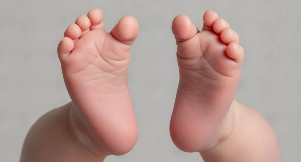
That means if you look at the plantar surface (bottom) of the foot, it appears inverted and adducted, with the heel tilted inward and the ankle pointing downward due to equinus deformity. In severe, untreated cases, children may walk on the lateral border or dorsolateral surface of the foot, but at birth, the sole does not anatomically face upward.
The name Talipes Equinovarus comes from Latin words.
- Talipes is from “Talus (ankle bone) + Pes (foot)”. So talipes means related to the ankle and foot.
- Equino relates to the “horse”, especially the hoof of the horse (pointing down)
- Varus means “bent inward”.

Image of an infant showing clubfoot deformity in the right foot
Together, these 3 terms describe a foot/ankle resembling a horse hoof, as the deformity bends and twists the foot downward and inward.
How common is Talipes Equinovarus?
Clubfoot is a fairly common birth condition that usually affects 1 in 1000 births. In about 50% of cases, the condition affects both legs, which means it can present as either unilateral or bilateral. The condition affects male children more frequently than females, with a ratio of 2:1, although scientists still do not fully understand why this difference exists. Clubfoot involves not only bones but also muscles, tendons, and ligaments, which are shorter and tighter than normal, pulling the foot into its abnormal position.
Early diagnosis and treatment are very important because intervention during the first few weeks of life is most effective when tissues are flexible. With proper management, clubfoot is highly treatable. The Ponseti method, a conservative treatment approach, has revolutionized care and allows most children to achieve normal foot function without major surgery.
Types of Clubfoot
Clubfoot is a complex deformity, and a range of factors are responsible for its occurrence. Based on causes, clubfoot is broadly classified into the following types.
Isolated Clubfoot:
These include idiopathic cases (meaning no obvious cause). This is the most common type of clubfoot overall. The infant is almost normal otherwise, and the clubfoot is entirely a standalone condition. Such cases are usually bilateral and respond very well to the Ponseti method (discussed in the treatment section).
Non-isolated Clubfoot:
Clubfoot can be due to various other reasons as well. They are classified further as follows:
Positional (Postural) Clubfoot:
This type of clubfoot is mainly due to the constrained positioning of the fetus inside the uterus (womb). They are usually correctable with minimal treatment, as they require manipulation and improve with stretching exercises. Unlike structural clubfoot, positional deformities are primarily due to mechanical factors and are typically flexible on examination.
Syndromic Clubfoot:
Certain syndromes can cause clubfoot, and this form usually presents as the most severe and rigid type. Doctors often find that conservative treatment does not work well, so they commonly perform surgery to correct it.
Neuromuscular Clubfoot:
Neuromuscular conditions like spina bifida may lead to clubfoot because they affect muscle control and nerve supply in the lower limbs. The primary reason in such cases is muscle imbalance. Recurrence rate is high in these cases. To manage, it is important to treat both the clubfoot and the associated neurological condition.
Causes of Talipes Equinovarus
Clubfoot has very diverse causes, such as idiopathic (unknown cause), genetic, syndromic, neuromuscular, vascular, maternal/environmental, and developmental defects.
Idiopathic:
Most cases of clubfoot (approximately 70–80%) are idiopathic, meaning doctors have not yet identified a specific cause. In these cases, the deformity develops independently and does not involve any associated syndromes, unlike syndromic clubfoot.
Genetic factors:
They also play an important role in the development of clubfoot. A positive family history increases the risk. Doctors often notice clubfoot occurring within the same family, especially among siblings and close relatives, indicating a possible genetic tendency. This deformity is, in fact, a multifactorial condition, involving the interaction of multiple genes and environmental factors rather than a single gene defect. As mentioned earlier, it is more common in males, which further supports the genetic influence.
Syndromes:
Clubfoot may be part of a Syndrome and thus manifests with additional features of that syndrome. For example,
• Arthrogryposis multiplex congenita (rare, non-progressive condition that affects joints)
• Larsen syndrome (rare, congenital dislocation of large joints)
• Edwards syndrome (Trisomy 18, a very severe developmental condition)
• Diastrophic dysplasia
• Down syndrome
Intrauterine complications:
During pregnancy, some intrauterine complications make it more likely to have clubfoot in the affected fetus. Potential prenatal risk factors of the clubfoot include:
- Intrauterine crowding: It means the fetus has limited space within the uterus due to multiple gestations (twins/triplets) or reduced uterine capacity.
- Oligohydramnios: A pregnancy complication in which there are abnormally low levels of amniotic fluid.
- Breech position: It refers to babies lying bottom first or feet first in the uterus instead of the normal head-first position.
Neuromuscular disorders:
Clinicians observe that spina bifida, myelomeningocele, and, in rarer cases, cerebral palsy contribute to the development of clubfoot. Taking adequate folic acid during pregnancy can prevent spina bifida and myelomeningocele.
Environmental Factors:
Some environmental conditions during pregnancy may raise the chances of a baby developing clubfoot. For example, maternal smoking is a well-established risk factor.
Infections such as Zika virus infection may coexist with foot deformities in affected fetuses. Although they are more strongly associated with CNS abnormalities and arthrogryposis rather than isolated idiopathic clubfoot.
Besides this, some studies have also shown a link with exposure to certain drugs and poor maternal nutrition. However, clear evidence for these associations is lacking.
Vascular disorders:
Some studies show that blood vessel defects, such as abnormal arterial development and reduced blood supply to the foot, can also cause clubfoot. However, vascular theories remain secondary hypotheses, and the exact pathogenesis of clubfoot is still not fully understood.
Symptoms of Clubfoot
Clubfoot is usually noticeable immediately after birth because it is a structural deformity present from development in the womb. The symptoms are primarily related to the appearance and positioning of the foot rather than pain.
Appearance at Birth
As a child is born, the symptoms are appearance-based only. The heel looks smaller than normal and sits higher than usual. The foot is misshapen and is turned inward and downward, with soles pointing to the inside or even upward. The inner border of the foot becomes concave-shaped, while the outer border of the foot gets a convex shape.
Another important feature is that the affected foot and calf are smaller due to hypoplasia (decreased number of cells) of the muscles. These are all about the appearance of the foot and are observable at birth.
Rigidity and Limited Movement
One of the most important clinical features is restricted ankle dorsiflexion due to equinus deformity. The foot cannot be brought into a neutral position easily. Tight Achilles tendons and medial soft tissue contractures contribute to this stiffness.
Functional Impact
Newborns do not experience functional limitations because they are not weight-bearing. However, once the child begins standing and walking, untreated clubfoot leads to abnormal gait. The child may walk on the lateral border or even the dorsolateral aspect of the foot in severe cases. This results in:
- Early fatigue
- Callus formation
- Skin breakdown
- Pain over time
If left untreated, it can cause lifelong disability and difficulty wearing regular footwear
Associated Findings
In syndromic or neuromuscular cases, additional abnormalities may be present, such as joint contractures, spinal defects, or developmental issues. Therefore, a complete newborn examination is essential.
Diagnosis of Clubfoot
The diagnosis of clubfoot is primarily clinical and is often made immediately after birth during routine newborn examination. In some cases, it may be detected before birth on a prenatal ultrasound. Early identification is important because treatment should begin within the first few weeks of life for the best outcomes.
Prenatal Diagnosis
In many cases, clubfoot can be spotted on ultrasound while the baby is still in the womb. This doesn’t change pregnancy care but helps parents and doctors plan treatment after delivery.
Prenatal Ultrasound
Clubfoot can sometimes be detected on a prenatal ultrasound or anomaly scan, usually during the mid-trimester anomaly scan around 18–22 weeks of gestation.
An abdominal ultrasound detects it around the 20th week of pregnancy. A transvaginal ultrasound may even detect the clubfoot around the 13th week, though this is uncommon.
On ultrasound, the finding of persistent inward rotation of the fetal foot relative to the tibia raises suspicion for clubfoot. Early diagnosis through ultrasound helps doctors guide and counsel parents properly. It also prepares them for managing the affected newborn after birth.
Genetic Testing and Amniocentesis
Amniocentesis is not routinely required in isolated clubfoot. Most cases of clubfoot occur without additional abnormalities and do not require invasive genetic testing.
Genetic evaluation, including amniocentesis, may be considered if:
- Ultrasound shows additional structural abnormalities
- There is suspicion of a chromosomal or syndromic condition
- Complex deformities or multiple anomalies are present
In such situations, amniocentesis can help analyze fetal chromosomes and guide parental counseling. For isolated clubfoot, however, genetic testing is usually unnecessary.
Postnatal Diagnosis
Postnatal diagnosis of Talipes Equinovarus is mainly clinical and is usually confirmed during the newborn’s first examination after birth.
Clinical Examination
Clinicians primarily diagnose clubfoot through a clinical examination at birth. They also examine the newborn for signs associated with other syndromes. The clinician confirms whether it is a true clubfoot or a postural (positional) deformity and will assess the severity and rigidity of the defect.
Pirani and Dimeglio Scoring Systems
To assess how severe the deformity is and to monitor the treatment response, the clinicians use the following scoring systems.
Pirani score:
It is a very popular and quick scoring system (clinicians perform it at the bedside when needed). It assesses 6 clinical signs of the deformity. In this system, the maximum score is 6, and the minimum score is 0. The higher the score, the higher the severity of the condition.
Dimeglio classification:
This system is more detailed than the Pirani score. In this system, the maximum score is 20, and the condition is classified into 4 grades, where each grade is equal to 5 points. The higher the score, the higher will be grade and thus the more severe the condition.
Imaging
Imaging is not routinely required in infants because most foot structures are cartilaginous and not fully ossified. Diagnosis remains primarily clinical.
Radiographs may be considered in:
- Atypical cases
- Older untreated children
- Suspected complex or rigid deformities
Ultrasound may occasionally be used to visualize cartilaginous structures, but it is not essential for routine diagnosis.
How is Clubfoot Treated?
The aims of treatment of clubfoot are to achieve a painless, plantigrade (walking with the entire sole of the foot), and flexible foot with good function. Timely treatment (preferably within the first few weeks of life) is very important to achieve maximum functionality later in life.
Ponseti method
This is the treatment of choice for idiopathic clubfoot. It is a non-surgical, conservative, and very effective approach (if started during the early weeks of life). It is a step-by-step method in a proper sequence. The purpose is to manipulate the foot and apply serial casts (usually 5-7 casts) in a standard way, helping to correct deformity.
This correction follows a specific order called CAVE (Cavus, Adduction, Varus, then Equinus). This means the high arch is corrected first, and after that, focus is on correcting the inward turning of the foot. Then the positions of the heel, and finally, the tightness that keeps the foot downward, are addressed.
If the ankle still cannot move up properly, then the doctors perform a minor procedure known as Achilles tenotomy. This percutaneous tenotomy is a routine part of the Ponseti protocol and is not considered major surgery. After this, they apply a final cast for three weeks. Once they remove the cast, they use a special brace to help maintain the correction.
Initially, the brace is applied for the whole day, then reduced to nighttime use until 4–5 years of age. Brace compliance is critical, as recurrence rates (approximately 20–30%) are most commonly due to poor brace adherence. Healthcare providers advise patients to attend follow-up appointments to ensure they correct the deformity properly and prevent any potential relapses.

Step-by-step overview of the Ponseti method for clubfoot treatment.
Surgical Management
If the above conservative approach fails, then surgical options are also available. Doctors perform surgery when the child presents late, when the Ponseti method fails, or when the deformity is very rigid.
In the relatively mild cases, a limited surgical procedure may be enough, such as releasing tight tendons, most commonly the Achilles tendon. On the other hand, if the condition is severe or the child presents too late (neglected case), then the approach is quite extensive for releasing soft tissues that are causing the deformity.
This includes targeting multiple ligaments, tendons, and joint capsules to make maximum correction. After surgical correction, the foot is immobilized using braces and physiotherapy, which helps to maintain the correction.
Although surgical options are important in resistant cases or late presentations, they often cause stiffness later in life, may overcorrect, or cause arthritis later in life. That is why healthcare providers prefer conservative methods such as the Ponseti method. These methods achieve the highest cure rates with minimal drawbacks compared to surgical procedures.
Talipes Equinovarus vs Metatarsus Adductus
Talipes Equinovarus (clubfoot) and metatarsus adductus are two different foot conditions, with clubfoot being more severe and involving the whole foot, while metatarsus adductus is milder and mainly affects the forefoot.
| Feature | Talipes Equinovarus (Clubfoot) | Metatarsus Adductus |
|---|---|---|
| Area affected | The clubfoot affects the whole foot, including the forefoot, midfoot, and hindfoot. | Metatarsus adductus is another condition, but milder, and affects only the forefoot. |
| Heel and ankle position | The heel tilts inward and the ankle points downward, giving the foot its characteristic shape. | The heel and ankle stay in their normal positions. |
| Sole position | The sole of the foot faces medially or upward. | The sole of the foot is normal (pointing downward). |
| Flexibility | Clubfoot makes the foot stiff and hard to move, so it’s not easy to correct by hand. | Metatarsus adductus is much more flexible, and the foot can usually be gently straightened without much effort. |
| Treatment | Requires proper intervention like the Ponseti method, usually within a few weeks of birth for best results; surgery may be required in severe or late-presenting cases. | Requires only reassurance, stretching, and rarely casting. It has good prognosis as it often resolves spontaneously. |
| Prognosis | Although clubfoot is a treatable condition, early diagnosis and management are very important, and when properly treated, it has a good prognosis as well. | Generally excellent because many cases resolve with minimal intervention. |
An illustraion ofMetatarsus Adductus, causing the front foot to turn inward.
Commonly Asked Questions (FAQs)
- Is talipes equinovarus painful?
No, clubfoot is not painful at birth, and the child will usually not feel any pain at the start. But if the condition is left untreated, it may cause pain, difficulty in walking, and disability later in life as the child advances in age.
- Can clubfoot be corrected completely?
Yes, clubfoot is a very treatable condition. The Ponseti method has revolutionized its treatment. If timely treated by this method, in more than 90% of cases, the child fully recovers and can walk, run, play, and enjoy life normally.
- At what age should treatment start?
The ideal time to treat such a case is in the first few weeks of life. The reason for treating it early is that the child’s bones and ligaments are still soft and flexible. Therefore, it is highly recommended to start early treatment for clubfoot.
- Is surgery always required?
No, and in fact, in the majority of cases, the gold standard treatment option is the Ponseti method (conservative approach). Surgery is only required if the conservative therapy fails. The failure is mostly due to severe cases (usually in syndromic cases), late presentation, or atypical cases.
Wrap Up
In conclusion, Talipes Equinovarus is a treatable foot deformity, and early diagnosis with timely intervention offers excellent outcomes. Modern treatments like the Ponseti method have made a huge difference, allowing many kids to walk and play normally without major surgery. Even in more severe cases, timely management greatly improves function and quality of life. With awareness, early diagnosis, and proper treatment, clubfoot is no longer a lifelong limitation but a condition with a very good outlook.
References
[1] Pavone V, Chisari E, Vescio A, Lucenti L, Sessa G, Testa G. The etiology of idiopathic congenital talipes equinovarus: a systematic review. J Orthop Surg Res. 2018;13(1):206.
[2] Clubfoot: an overview and the latest UK guidelines. Br J Hosp Med (Lond). 2022.
[3] Wynne-Davies R. Family studies and the cause of congenital club foot: talipes equinovarus, talipes calcaneo-valgus and metatarsus varus. J Bone Joint Surg Br. 1964;46:445-463.
[4] Mustari MN, Faruk M, Bausat A, Fikry A. Congenital talipes equinovarus: a literature review. Ann Med Surg (Lond). 2022;81:104394.
[5] Rieger MA, Dobbs MB. Clubfoot. Clin Podiatr Med Surg. 2022;39(1):1-14.
[6] Smythe T, Kuper H, Macleod D, Foster A, Lavy C. Birth prevalence of congenital talipes equinovarus in low- and middle-income countries: a systematic review and meta-analysis. Trop Med Int Health. 2017;22(3):269-285.
[7] Clubfoot: Symptoms, Causes & Treatment. Cleveland Clinic. 2026.
[8] Williams Obstetrics. 26th ed. New York: McGraw Hill; 2022
[9] Gelfer Y, Wientroub S, Hughes K, Fontalis A, Eastwood DM. Congenital talipes equinovarus. Bone Joint J. 2019;101-B(6):639-645.
[10] Trout SM, Whitaker AT. Management issues of congenital talipes equinovarus in the neonatal intensive care unit: a systematic review. Foot Ankle Surg. 2021;27(5):480-485.
[11] Wiesel SW, et al. Campbell’s Operative Orthopaedics. 14th ed. Philadelphia: Elsevier; 2021.
[12] Morcuende JA, Dietz FR, Ponseti IV. Treatment of idiopathic clubfoot: the Ponseti method. J Bone Joint Surg Am. 2004;86-A(5):1052-1055.