


What Is Thanatophoric Dysplasia?: Thanatophoric Dysplasia Prenatal
Thanatophoric Dysplasia Prenatal The most prevalent form of lethal skeletal dysplasia is called thanatophoric dysplasia. It is caused by the mutation of a gene, FGFR3 (Fibroblast Growth Factor Receptor 3), resulting in extremely short limbs, a narrow chest, and a large skull. The disease occurs in approximately 1 in 20,000 to 50,000 births worldwide. It is named after the ancient Greek meaning “death-bearing” because this is a serious condition.[1]
This genetic disorder, thanatophoric dysplasia, affects the growth of bones and lungs of the fetus in the womb, creating severe complications that prove fatal in most cases. Underdeveloped lungs due to a narrow cavity between the chest are the main problem that cannot allow proper breathing after birth.[2]
Types of Thanatophoric Dysplasia
Medical experts have identified two different subtypes that possess different characteristics.
Thanatophoric Dysplasia Type 1
The more common form is Type 1, which constitutes the majority of the cases identified.[3]
Thanatophoric Dysplasia Type 2
The Type 2 thanatophoric dysplasia has the same severity but slightly different features.[4]
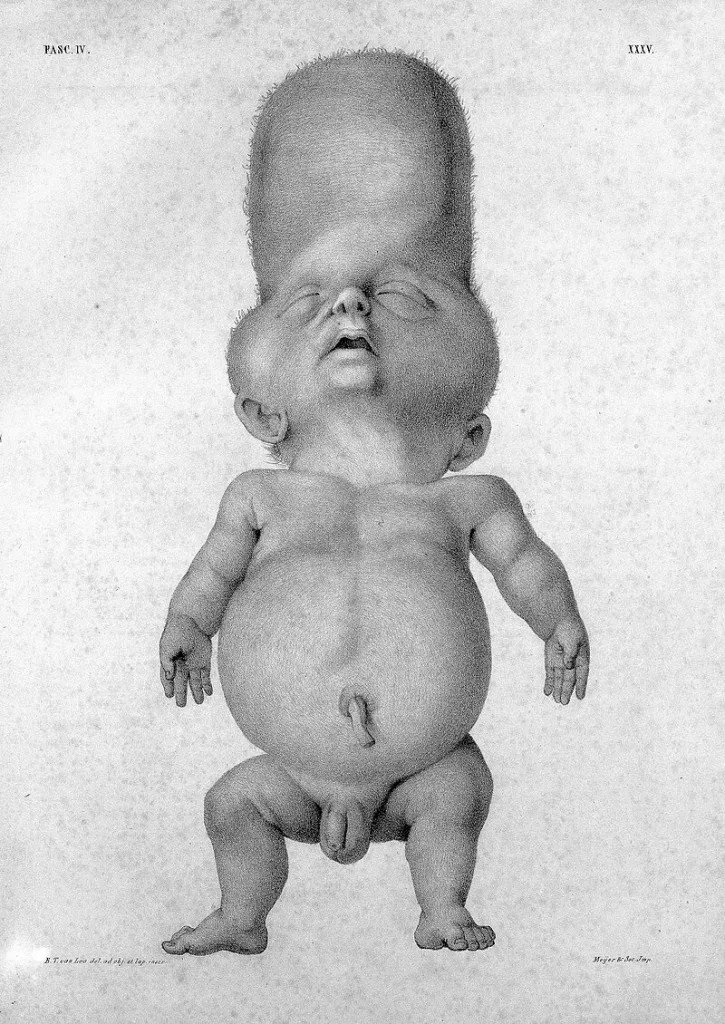
Infant with cloverleaf skull and shortened limbs (thanatophoric dysplasia), https://wellcomecollection.org/works/e93v9br5 CC-BY-4.0
A Comparison of the two types is as follows:
| Feature | Type 1 | Type 2 |
|---|---|---|
| Femur Shape | Curved/bowed (“telephone receiver”) | Straight |
| Skull Deformity | Uncommon, generally normal | Cloverleaf skull (consistent) due to premature fusion of cranial sutures (craniosynostosis) |
| Craniosynostosis | Rare | Always present, moderate to severe |
| Vertebrae | Flat (platyspondyly) | Taller bodies |
| Limb Shortening | More pronounced | Slightly milder |
| Frequency | More common (60-70% of cases) | Less common (30-40% of cases) |
| Prenatal Detection | Often identified by femur bowing | Identified by skull deformity |
Causes and Genetics
Thanatophoric dysplasia is a condition caused by a certain genetic mutation of the FGFR3 gene (fibroblast growth factor receptor-3) on chromosome 4p16.3.skeletal dysplasia severity.American Journal of Human Genetics, 67(6), 1411–1421.” style=”position:relative;color:#309b65;cursor:help;border-bottom:1px dotted #309b65;font-weight:bold”>[5]
In normal conditions, the FGFR3 protein acts as a brake pedal to the growth of bones in the growth plates of the long bones. It controls the endochondral ossification process through which cartilage develops into bone.
In thanatophoric dysplasia, a mutation forms what scientists refer to as a gain-of-function effect. The FGFR3 protein is permanently activated, straining the bone growth brakes excessively and for too long. This causes very short limbs and other skeletal defects.[6]
Thanatophoric Dysplasia vs. Achondroplasia.
The main difference between Thanatophoric Dysplasia (TD) and Achondroplasia lies in the severity of the FGFR3 gene mutation, the clinical outcome, and the extent of skeletal abnormalities. While both are caused by mutations in the FGFR3 gene, they represent different points on the same genetic spectrum; Achondroplasia being non-lethal, and Thanatophoric Dysplasia being lethal in almost all cases. This comparison assists in showing how the variation in the symptoms of skeletal dysplasia could be achieved by different kinds of mutations of the same gene.[7]
| Aspect | Achondroplasia | Thanatophoric Dysplasia |
|---|---|---|
| Incidence | 1 in 15,000-40,000 births | 1 in 20,000-50,000 births |
| Mutation Severity | Moderate gain-of-function | Severe gain-of-function |
| Life Expectancy | Near-normal with proper care | Usually hours to days after birth |
| Limb Shortening | Moderate (rhizomelic pattern) | Severe, extreme micromelia |
| Chest Development | Narrow but functional | Critically narrow, non-functional |
| Lung Development | Normal | Severe hypoplasia (underdevelopment) |
| Intelligence | Normal | Unable to assess due to early lethality |
| Quality of Life | Can live full, active lives | Not compatible with life in most cases |
| Medical Management | Manageable complications | Requires intensive life support |
| Skeletal Features | Shortened limbs, large head | Extreme shortening, severe deformities |
| Respiratory Issues | Occasional sleep apnea | Immediate respiratory failure |
| Survival | Excellent with monitoring | Poor, rarely beyond infancy |
Inheritance Patterns
The condition is not hereditary, and it occurs spontaneously.
Autosomal Dominant de novo origin
Thanatophoric dysplasia has an autosomal dominant genetic inheritance. This implies that the condition can be caused by only a single copy of the mutated gene. In case the gene mutation has been inherited, only one parent has to pass the transformed gene to the child to create thanatophoric dysplasia.[8]
Most cases of thanatophoric dysplasia have de novo mutations.” De novo” means “new” in Latin, and in genetics, it refers to mutations that occur spontaneously for the first time in an affected individual. The mutations do not exist in the genes of parents, and they were not inherited by previous generations.
A few implications of the spontaneous nature of thanatophoric dysplasia are:[9]
- Parents whose child has thanatophoric dysplasia did not inflict the condition upon him or her out of anything that they did or did not do before or during pregnancy.
- The mutation happened by chance during reproductive cell development or during early embryonic development.
- There is no significant increase in the risk of having another child with thanatophoric dysplasia when compared to the general population risk (as a rule, less than 1%).
- There are no more chances of having children with the condition among those who have affected siblings.
The Rare Exception
In very exceptional cases, a survivor with thanatophoric dysplasia who became an adult had the theoretical possibility of passing the gene on to his or her children with a 50 percent chance of each conception. But considering that in this condition, long-term survival is extremely low.[10]
Symptoms and Physical Characteristics
There are several systems that are affected by Thanatophoric dysplasia, but the skeletal and respiratory systems have the greatest effect.[11]
Skeletal Features
The bone defects of thanatophoric dysplasia are severe and involve practically all bones of the body:
Head and Face:
- Macrocephaly (enlarged head)
- Frontal bossing (bulging forehead)
- Flat facial features with a depressed or flattened nasal bridge
- Proptosis (protruding eyes)
- Big anterior fontanelle
- Small bones on the face in comparison to the cranium
- In Type 2, there is a deformity of the skull in the form of a cloverleaf.
Chest and Trunk:
- Extremely constricted chest cavity (thorax)
- Short, horizontal ribs
- Smaller shoulder blades
- Big belly with a stick-out belly button
- Flat vertebral bodies with a short spine
- The small pelvis has typical radiographic appearances
Arms and Legs:
- Very shortening of limbs (micromelia), especially of the upper arms and thighs
- Very short toes and fingers (brachydactyly)
- The limbs develop excess skin folds and look redundant and wrinkled
- Bony perifocal thickened soft tissues
- In Type 1, there is characteristic bowing of long bones, especially the femurs
- Fibula bones are much shorter than tibia bones in the lower legs
- Restricted movement of the bones.
Babies born with thanatophoric dysplasia have a mean length at birth of 40 centimeters (about 15.7 inches) in length, which is a quarter of the normal length of a newborn baby of 48-51 centimeters (19-20 inches). This huge size disparity is the first clinical feature that is easily noticed and is frequently identified by delivery room personnel.
Respiratory Complications
The respiratory system is the most life-threatening symptom of thanatophoric dysplasia. This underdeveloped lung disease (pulmonary hypoplasia) results in serious breathing problems immediately after birth:
- Failure to inflate the lungs sufficiently because of the limited space.
- Immediate postpartum respiratory distress.
- Lack of adequate exchange of oxygen results in cyanosis (blue or purple discoloration of the skin).
- Mechanical ventilation should be done urgently because of respiratory failure.
- The lungs have a reduced number of air sacs (alveoli).
Thanatophoric Dysplasia Diagnosis
Proper diagnosis of thanatophoric dysplasia normally commences during the routine prenatal assessments, but the diagnosis can only be confirmed through specialized tests and knowledge.
Prenatal Detection
A majority of the thanatophoric dysplasia is discovered in the second or third trimester of pregnancy, although it may be difficult to diagnose before 22 weeks. A regular ultrasound could show many alarming symptoms:[12]
Early Indicators (16-20 weeks):
- Considerably reduced femur length measurements, which are significantly lower than the expected ranges.
- Exceptionally small chest cavity on ultrasound.
- Polyhydramnios
- Inequalities in the size of the head in comparison to the body.
Subsequent Ultrasound Findings (24 weeks):
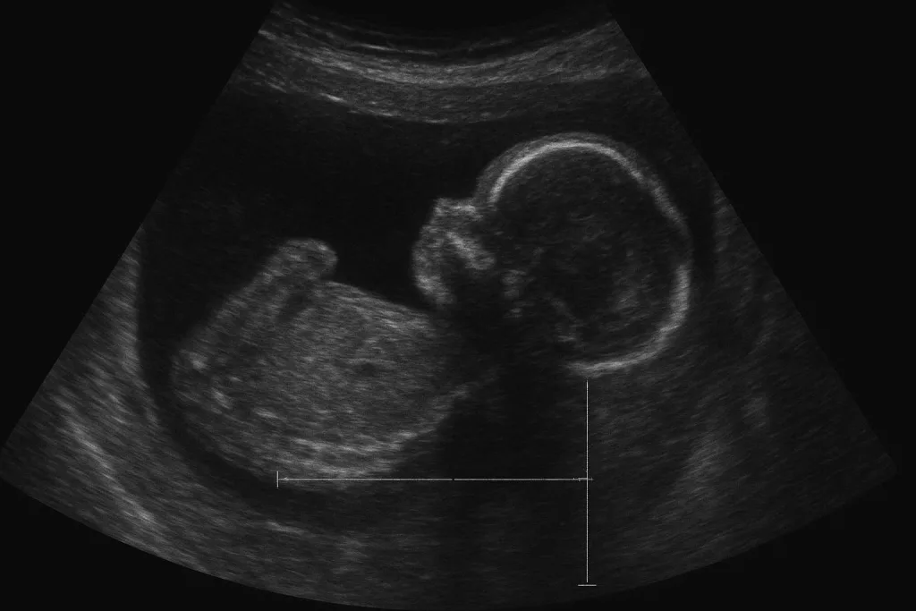
Prenatal ultrasound at 24 weeks demonstrating key findings of thanatophoric dysplasia, including short bowed limbs, cloverleaf skull, small chest cavity, and frontal bossing.
- Severe micromelia (short limbs).
- Type 1 has characteristic bowing of the femurs.
- Appearance of cloverleaf skull in Type 2.
- Very small thoracic cavity and decreased measurements of the chest circumference.
- Massive head with bossing of the forehead.
- Malformed location of limbs.
Healthcare providers who suspect thanatophoric dysplasia based on ultrasound findings usually suggest referral to a specialist in maternal-fetal medicine and the genetic counselor. Further examination may then be scheduled to ascertain the diagnosis.
Genetic Testing Options
FGFR3 mutations can be confirmed in the following two tests:
Chorionic Villus Sampling (CVS):
- Conducted during 10-13 weeks of pregnancy.
- Needs a sample of a little placental tissue.
- Earlier diagnosis than amniocentesis.
- The results are normally accessible within 1-2 weeks.
- Has a low risk of pregnancy complications (0.5-1)
Amniocentesis:
- Carried out at 15- 20 weeks of pregnancy.
- Includes the extraction of a little bit of amniotic fluid, which has fetal cells, which can be used to study genetic mutations.
- It is believed to be very precise in diagnosing genetic disorders.
- The results are normally accessible within 1-2 weeks.
- Has a low risk of pregnancy complications (around 0.1-0.3)
The two procedures are capable of determining the particular FGFR3 defect that causes thanatophoric dysplasia to give a family conclusive results and can enable them to make accurate decisions regarding the management and planning of a pregnancy and delivery.
Postnatal Confirmation
In cases where thanatophoric dysplasia was not diagnosed during the prenatal period, or the diagnosis should be confirmed after birth, several methods of diagnosis can be used:[13]
Physical Examination:
The excessive shortness of the limbs, the narrowness of the chest, the abnormalities of the facial features, and the acute respiratory distress provide a certain clinical image that can be identified in a few minutes by trained specialists after the birth.
X-ray Findings of Thanatophoric Dysplasia Infants
- Appearance of femurs in the Type 1 is “Telephone receiver”.
- Significantly reduced and expanded long bones containing flattened and cupped ends (metaphyses).
- Small, square-shaped pelvis with a typical appearance.
- Lumbar spine short interpediculate distance (between vertebrae)
- Very short metacarpals, metatarsals, and phalanges (hand and foot bones).
- The pelvis has small hypoplastic iliac wings.
- Large gaps between the spinal bones.
- Shorter horizontal ribs.
Other Imaging:
- The extent of pulmonary hypoplasia is evaluated by ultrasound of internal organs, especially the lungs.
- A brain imaging (CT/MRI) can be done to exclude the presence of hydrocephalus or other neurological complications.
- In Type 2, the skull X-rays reveal the cloverleaf deformity.
Molecular Genetic Testing:
A molecular analysis of blood samples obtained after the birth of the newborn could be performed to ascertain the particular mutation of FGFR3, which would validate the clinical and radiographic diagnosis. This genetic confirmation will be significant in offering the proper genetic counseling to the families.[14]
Differential Diagnoses
Such conditions as may be considered include:[15]
- Achondroplasia: Less severe, compatible with life, different mutation pattern.
- Homozygous achondroplasia: Very rare, also lethal, but different inheritance pattern.
- Osteogenesis imperfecta type II: Brittle Bone disease, in which the skeletal appearances vary.
- Campomelic dysplasia: Long bones are bowed, however, in a different manner.
- Jeune syndrome: Asphyxiating thoracic dysplasia with partial similarity in the chest appearance.
Treatment and Management
Although there is no treatment for thanatophoric dysplasia, there is medical management that aims at assisting the family with the diagnosis and, in some unusual instances where life is prolonged beyond the acute postnatal phase, offering aggressive treatment to keep the baby alive and as comfortable as possible.[16]
Immediate Postnatal Care
Infants need urgent intensive ventilation:
- Mechanical ventilation and emergency intubation
- Placement of tracheostomy for long-term airway management
- To keep the lungs inflated, continuous positive airway pressure (CPAP) is used
- Supplemental oxygen
- Bronchodilator medications
- The respiratory assistance needed is usually both long-term and lifelong, and practically all survivors will continue to be ventilator-dependent until death, due to the basic limitation of lung size and capacity
Surgical Interventions for Survivors
The very few children who survive after the newborn stage may need numerous surgeries:[17]
- Suboccipital decompression: This decompresses the skull base.
- Placement of shunts: To deal with the hydrocephalus.
- Orthopedic surgeries: To cure serious deformities of bones.
- Maintenance of tracheostomy: It involves surgery to keep the breathing tube open and control complications.
- Skull expansion procedures: In some cases, to relieve pressure and accommodate brain growth.
Ongoing Medical Support
Long-term survivors need the following care:
- Anti-seizure medications
- Regular neuroimaging
- Communication and hearing aids
- Feeding tubes for nutrition
- Occupational therapy and physical therapy
- Developmental assessments
Pregnancy Management
In the case of thanatophoric dysplasia diagnosed prenatally, obstetrics should avoid complications to the mother and offer compassionate care:[18]
- Polyhydramnios monitoring and management.
- Plan the mode of delivery and the time since positioning of the big head and the stiff neck may complicate vaginal delivery.
- Talking about cesarean section to prevent complications of the birth and a painful birth.
- Having the right staff (neonatology and palliative care) available at the delivery point.
Prognosis and Survival
Babies diagnosed with thanatophoric dysplasia have a poor prognosis, with the majority either born dead or passing away a few hours to days after birth because of respiratory failure. With the knowledge of realistic prognosis, the families prepare themselves emotionally and make wise decisions concerning their care.
Exceptional Long-Term Survivors
The medical literature has reported about nine surviving children after infancy with thanatophoric dysplasia, the age of the individuals at the time of report being between 5 months and 10 years old. The characteristics of these exceptional cases are similar.[19]
Prevention and Risk Factors
When it comes to the prevention of thanatophoric dysplasia, families should know that there is no preventive measure for this condition. The genetic mutation is spontaneous, and hence no intervention can predict or prevent it.[20]
Risk Factors
The condition occurs:
- Equal in all ethnic groups and localities.
- Irrespective of maternal or paternal age.
- In the vast majority of cases, without no association with family history.
- Random, with no family history of other genetic disorders.
Conclusion
One of the most difficult diagnoses that any family can have is that of Thanatophoric dysplasia. The prognosis is poor, and there is no cure; however, with improvements in prenatal diagnosis, families can make informed decisions regarding the management and care of the pregnancy. Healthcare teams, genetic counseling, and palliative care skills, in addition to emotional support services, can enable the families to undergo this challenging process with dignity and compassion. The realization that the condition comes on its own with nothing that parents did is also a critical assurance of an extremely challenging situation.
References
[1] Noe, E. J., Yoo, H. W., & Kim, K. N. (2010). A case of thanatophoric dysplasia type I with an R248C mutation in the FGFR3 gene.Korean Journal of Pediatrics, 53(12), 1022–1025.
[2] MedlinePlus Genetics. (n.d.). Thanatophoric dysplasia.U.S. National Library of Medicine
[3] Kamochi, H., Shimojima, K., Yamamoto, T., & Ozawa, H. (2000). Prenatal diagnosis and genetic analysis of type I and type II thanatophoric dysplasia.Prenatal Diagnosis, 20(8), 656–661.
[4] Shinawi, M., Shaw, C., & Nahum, O. (2009). Genetically confirmed thanatophoric dysplasia with FGFR3 mutation.Clinical Dysmorphology, 18(1), 45–49.
[5] Bellus, G. A., McIntosh, I., Smith, E. A., & Francomano, C. A. (2000). Distinct missense mutations of the FGFR3 lys650 codon modulate receptor kinase activation and skeletal dysplasia severity.American Journal of Human Genetics, 67(6), 1411–1421.
[6] Del Piccolo, N., Placone, J., & Hristova, K. (2015). Effect of thanatophoric dysplasia type I mutations on FGFR3 dimerization.Biophysical Journal, 108(2), 272–278.
[7] Cohen, M. M., & Horton, W. A. (2002). FGFR3 disorders: Achondroplasia and thanatophoric dysplasia.American Journal of Medical Genetics, 112(3), 284–290.
[8] Horton, W. A., & Hall, J. G. (2014). Thanatophoric dysplasia. InGeneReviews®. University of Washington, Seattle.
[9] Genetic and Rare Diseases Information Center (GARD). (n.d.). Thanatophoric dysplasia.National Institutes of Health.
[10] Kocherla, K., & Kocherla, V. (2016). Antenatal diagnosis of thanatophoric dysplasia: A case report and review of literature.International Journal of Research in Medical Sciences, 4(9), 4254–4257.
[11] MedlinePlus Genetics. (n.d.). Thanatophoric dysplasia.U.S. National Library of Medicine.
[12] Nguyen, T. M., et al. (2005). Prenatal diagnosis of thanatophoric dysplasia by mutational analysis of FGFR3 gene.Prenatal Diagnosis, 25(7), 649–653.
[13] Noe, E. J., Yoo, H. W., & Kim, K. N. (2010). A case of thanatophoric dysplasia type I with an R248C mutation in the FGFR3 gene.Korean Journal of Pediatrics, 53(12), 1022–1025.
[14] MedlinePlus Genetics. (n.d.). Thanatophoric dysplasia.U.S. National Library of Medicine.
[15] Horton, W. A., & Hall, J. G. (2014). Thanatophoric dysplasia. InGeneReviews®. University of Washington, Seattle.
[16] Horton, W. A., & Hall, J. G. (2014). Thanatophoric dysplasia. InGeneReviews®. University of Washington, Seattle.
[17] Matsushita, M., et al. (2012). Peptide P3 inhibits FGFR3 signaling and rescues lethal phenotype in mice mimicking human thanatophoric dysplasia.Human Molecular Genetics, 21(26), 5443–5453.
[18] Chaiyasate, S., & Church, V. (2017). Thanatophoric dysplasia: Findings and procedure details.European Congress of Radiology Posters, C-1088.
[19] Horton, W. A., & Hall, J. G. (2014). Thanatophoric dysplasia. InGeneReviews®. University of Washington, Seattle.
[20] Genetic and Rare Diseases Information Center (GARD). (n.d.). Thanatophoric dysplasia.National Institutes of Health.