

Tinea barbae, conventionally known as Barber’s Itch, is a superficial dermatophyte infection that affects the bearded areas of the face and neck. Tinea is a medical term for ringworm, while barbae is the Latin word for beard. Beard ringworm can be passed on by direct contact with infected people or animals, with contaminated objects. Clinically, it presents as inflammatory deep, kerion-like plaques (large, pus-filled sores) or as non-inflammatory superficial plaques resembling tinia corporis. This fungal infection is typically asymptomatic, but moderate itching is common. Delayed diagnosis can result in facial scarring and potential mental health effects.
Although superficial fungal infections are common worldwide, this condition is relatively uncommon. Due to the rarity of the disease, the prediction of its true incidence and risk is difficult. This infection is exclusively seen in adolescent boys and men. However, a case report of tinea barbae in a hirsute woman in Portugal has been reported.
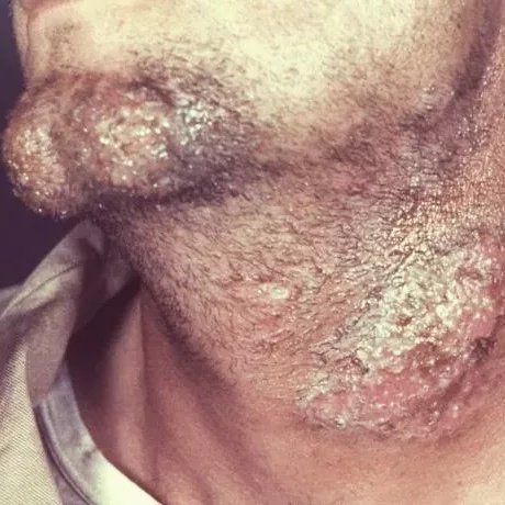
The patient had presented with a dermatophytic fungal infection in the bearded region of his neck, which had been diagnosed as a case of tinea barbae, caused by the fungus Trichophyton mentagrophytes. Image Courtesy: available via PHIL, image ID: 17655.
Causes of Tinea Barbae
Transmission of dermatophytes to humans occurs via direct contact with:
- Anthropophilic (infected humans)
- Geophilic (infected soil)
- Zoophilic (infected animals)
However, research indicates that tinea barbae is exclusively caused by anthropophilic and zoophilic dermatophytes. Fungi called Trichophyton cause tinea barbae. The most commonly reported causative organisms of this condition are:
- Trichophyton rubrum
- T. mentagrophytes
- T. verrucosam
Some other documented causative agents are:
- T. ernacei
- T. interdigitale
- T. tonsurans
- T. schoeleinii
- T. megninii
- T. violaceum
- Microsporum nanum
- M. gypseum
- Epidermophyton floccosum
Domestic animals, pets, and uncommon dermatophytes can also cause this condition. These animals include dairy cows, cats, dogs, pigs, sheep, and horses. Human-to-human transmission is rare but possible. T. tonsurans can cause tinea barbae after shaving with insufficiently disinfected instruments.
The risk factors that increase the likelihood of dermatophyte infection include:
- Age
- Use of steroids
- Immune status
- Trauma
- Diabetes
- Occupational exposures
Pathophysiology
Keratinophilic fungi (dermatophytes) invade keratinized tissues such as the epidermis and hair follicles. Both host and fungal-specific factors contribute to the inflammatory process. The responsible fungal-specific factors include adaptation to the specific host, the release of enzymes, the production of inflammatory factors and toxins, and, lastly, the release of immunomodulatory agents. On the other hand, host-specific factors that play a role in inflammation include the site of entry, the nonspecific defense mechanisms, and the immune response.
Zoophilic dermatophytes cause infection of greater severity than anthropophilic dermatophytes. So, zoophilic organisms are the primary cause of inflammatory kerion-like plaques (they result from a more intense host reaction). Kerion formation usually results from a complex infection from T. rubrum and T. mentagrophytes. The formation of kerion is postulated by two theories.
Theories
- The initial theory proposes that the formation of kerion results from the diffusion of metabolites and toxins from the fungus.
- The second theory proposes that it results from an immunological response to the dermatophyte antigens. This infectious disease begins when spores are inoculated into the keratinized tissue. After inoculation, the carbohydrate microfibril on the fungal spores anchors to the keratinocytes. After being anchored, the spores germinate and produce hyphae. Hyphae spread centrifugally into the deeper layers of the stratum corneum. The invading fungal hyphae produce several enzymes, such as keratinases, elastases, and proteases, and other pathogenic factors into the stratum corneum. The released enzymes aid in the digestion and utilization of keratin and other proteins required for the survival and growth of dermatophytes. The host’s keratinocytes respond by releasing inflammatory mediators such as interleukins and cytokines, which trigger both humoral and cell-mediated immune responses.
Symptoms of Tinea Barbae
Tinea barbae presents reddish, ring-shaped rashes on the epidermis. The rash may be itchy but is not usually painful. These rashes can appear on the cheeks, neck, chin, and moustache areas.
Clinically, tinea barbae presents two different morphologies: inflammatory and non-inflammatory.
Inflammatory Presentations
The classic inflammatory form of tinea barbae (often seen with zoophilic dermatophytosis) develops a characteristic lesion (kerion).
Kerion, an erythematous, tender, boggy, often sterile, weeping plaque or nodule with draining pustules or sinuses. Mostly, patients exhibit solitary plaques or nodules, while multiple nodules are more common. Upper lip involvement is relatively uncommon in this presentation.
Hairs are either damaged or loose. Hair roots and follicles become filled with whitish masses. The indurated surface of the nodule is gradually covered by crust and exudate.
Non-Inflammatory Presentation
Anthropophilic dermatophytes induce non-inflammatory superficial tinea barbae. This variant of barbae is rare. It presents as erythematous patches with raised borders and small follicular pustules (sycosiform variant). Hairs appear brittle, broken, or detached. This type signifies a chronic form of tinea barbae.
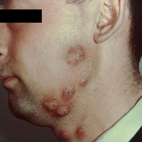
A left lateral view of a patient’s face and neck reveals the presence of a number of chronic erythematous skin lesions, which proved to be due to a dermatophytic fungal infection, commonly referred to as barber’s itch, and specifically tinea barbae. Image courtesy: available via: PHIL, image ID:4807.
Diagnosis of Tinea Barbae
Tinea barbae can be diagnosed clinically. The clinical presentation, along with a thorough history and physical examination, can help with early diagnosis. Simple, inexpensive, in-office testing can establish the diagnosis. These tests include direct microscopic examination of the lesion, skin scraping, fungal cultures, and a skin biopsy.
Direct Microscopic Examination
The healthcare provider obtains skin scrapings and scales from the active border of the lesion, along with epilated hairs, using a scalpel or a moist cotton tip. The provider then transfers the collected sample onto a slide and then adds 10 to 20% potassium hydroxide solution drops. A gentle warm-up of the slide or addition of dimethyl sulfoxide can speed up the emulsification of the skin cells. To highlight the fungal elements, fungal stains are added. Examination of this wet-mount preparation under the microscope reveals rod-shaped, branching fungal filaments with uniform hyphae and minute spores. These indications confirm the diagnosis of tinea barbae. Fungal elements can cover the ectothrix (hair) or invade he endothrix (hair shaft).
Culture
This technique involves inoculating the scraped skin and hair sample into a specialized dermatophyte medium (mycobiotic and mycocel agar). These specialized media contain chloramphenicol and cycloheximide to inhibit the growth of contaminating bacteria and fungi. The fungus takes 7 to 14 days to grow on this medium. If there is no growth for 21 days, then the test indicates negative results.
Skin Biopsy and Histopathological Examination
Fungal cultures and direct microscopic examination are enough to diagnose most of these infections. Skin biopsy and histopathological examination of tissues are performed only when other methods cannot establish a definitive diagnosis. Fungal hyphae, arthroconidia, and inflammatory changes in the epidermis and dermis. In the biopsy section, the inflammatory patterns are consistent with folliculitis (inflammation of hair follicles), perifolliculitis (inflammation of the skin around hair follicles), and microabscesses (localized collections of cells).
Other Diagnostic Methods
Conventional methods are adequate to form a diagnosis of tinea barbae. However, they are not specific enough to identify the exact causative pathogen at the species level. So, new diagnostic modalities such as Polymerase chain reaction (PCR) can be used to identify the fungal species when routine tests are inconclusive.
Treatment and Management of Tinea Barbae
Systemic antifungal therapy is the mainstay of treatment. Topical agents are only supportive and not sufficient alone. The oral antifungal agents with proven effectiveness in the management of this condition are:
- Terbinafine once daily (125 mg to 250 mg)
- Ketoconazole once daily (200 mg to 400 mg)
- Itraconazole once daily (100 mg)
- Fluconazole once daily (200 mg)
Tinea barbae resolves completely within 4 to 6 weeks of therapy with these medications. Common adverse effects of azoles and terbinafine include abdominal pain, nausea, rashes, visual disturbances, and elevated transaminases. Other treatment options include
- Griseofulvin is also a preferred drug but it is rarely used now due to drug resistance, prolonged therapy, rapid clearance of the drug from the skin and its drug resistance.
- There are no well-defined roles of topical agents, such as selenium sulfide, in treating tinea barbae.
- Healthcare providers rarely indicate surgery. They do not recommend drainage of a kerion after incision.
- Photodynamic therapy (5-aminolevulinic acid-based) is a recent alternative that circumvents the adverse effects and drug resistance of these conventional antifungal agents.
Natural Supportive Measures
While antifungal medications are essential, some natural agents may help soothe symptoms when used safely under a doctor’s guidance:
Coconut Oil
Application of direct coconut oil to the affected area can help with ringworm infections due to its antifungal properties.
Diluted Tea Tree Oil
Application of diluted tea tree oil with a few drops of coconut oil to reduce irritation can help resolve the symptoms of tinea barbae due to its antifungal and anti-inflammatory properties. Full-strength oil can be irritating and can cause further damage.
Turmeric
Turmeric possesses antifungal, antibacterial, and anti-inflammatory properties, hence it can inhibit fungal growth.
Tinea Barbae Prevention
Following precautionary measures can lower the risk of getting tinea barbae:
- Wash your hands thoroughly after touching livestock or other potentially infected animals.
- Do not share personal hygiene items or beard grooming tools with others.
- Wash your beard and face daily with a mild cleanser to prevent dirt and oil buildup.
- Ensure your skin is dry, as fungi thrive in moist environments.
Differential Diagnosis
Healthcare providers misdiagnose tinea barbae as follows:
- Allergic contact dermatitis
- Acne vulgaris
- Secondary syphilis
- Contact dermatitis
- Cutaneous candidiasis
- Folliculitis
- Rosacea
- Dermatologic aspects of actinomycosis
Prognosis
Inflammatory lesions resolve spontaneously within a few months. But if left untreated, they can result in scarring. Non-inflammatory tinae barbae lesions are more likely to be chronic and cannot resolve spontaneously. The patients respond well to the oral anti-fungal therapy.
Tinea Barbae versus Tinea Capitis
Tinea barbae is the fungal infection that affects the beard and moustache area, while tinea capitis is a fungal infection of the scalp only.
| Feature | Tinea Barbae | Tinea Capitis |
|---|---|---|
| Location of Infection | Beard and mustache area | Scalp |
| Age group | Adult males | Usually children |
| Mode of transmission | Usually, animal to human. | Usually human to human |
| Clinical forms | Inflammatory and superficial lesions. | Black, dot and grey patches |
Final Remarks
Tinea barbae or beard ringworm is a contagious infection. The condition exclusively affects males and causes ring-shaped skin rashes in the bearded areas, which can lead to scarring and hair loss. Some people develop severe lesions. Oral antifungals can effectively treat this fungal infection.
References
[1] Duarte, B., Galhardas, C., & Cabete, J. (2019). Adult tinea capitis and tinea barbae in a tertiary Portuguese hospital: A 11‐year audit.Mycoses,62(11), 1079-1083.
[2] Rutecki, G. W., Wurtz, R., & Thomson, R. B. (2000). From animal to man: tinea barbae.Current Infectious Disease Reports,2(5), 433-437.
[3] Bonifaz, A., Ramírez‐Tamayo, T., & Saúl, A. (2003). Tinea barbae (tinea sycosis): experience with nine cases. The Journal of Dermatology,30(12), 898-903.
[4] Baumgardner, D. J. (2017). Fungal infections from human and animal contact.Journal of patient-centered research and reviews,4(2), 78.
[5] Hube, B., Hay, R., Brasch, J., Veraldi, S., & Schaller, M. (2015). Dermatomycoses and inflammation: The adaptive balance between growth, damage, and survival.Journal de mycologie medicale,25(1), e44-e58.
[6] Kuruvella, T., Saleh, H. M., & Pandey, S. (2024). Tinea barbae. InStatPearls [Internet]. StatPearls Publishing.
[7] Kuruvella, T., Saleh, H. M., & Pandey, S. (2024). Tinea barbae. InStatPearls [Internet]. StatPearls Publishing.
[8] Furlan, K. C., Kakizaki, P., Chartuni, J. C. N., & Valente, N. Y. S. (2017). Sycosiform tinea barbae caused by trichophyton rubrum and its association with autoinoculation.Anais brasileiros de dermatologia,92(1), 160-161.
[9] Ji, J., Liang, C. T., Zhong, J. J., Kong, X., Xu, H. X., Xu, C. C., & Fu, M. H. (2024). 5-aminolevulinic acid-based photodynamic therapy in combination with antifungal agents for adult kerion and facial ulcer caused by Trichophyton rubrum.Photodiagnosis and Photodynamic Therapy,45, 103954.