

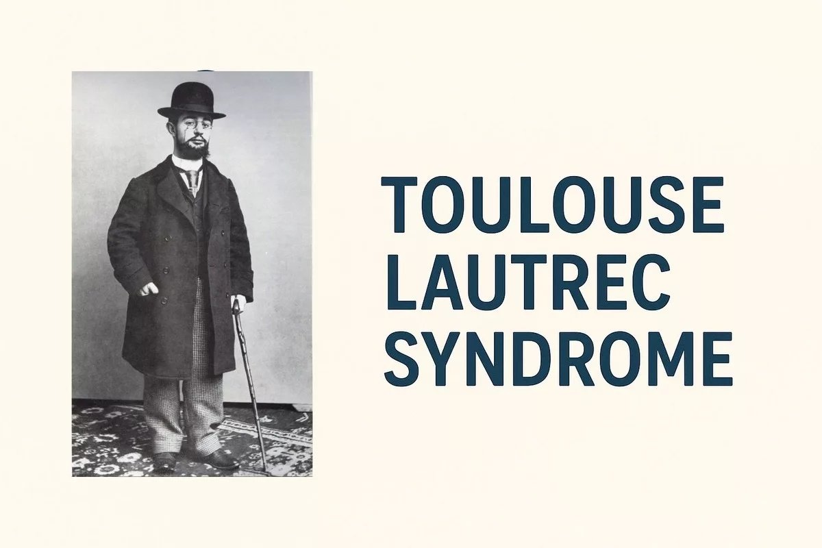
Toulouse Lautrec Syndrome Toulouse-Lautrec syndrome is a rare bone disease that mainly affects your skeletal system. The bones become too dense but remain fragile in this disorder. Besides the skeletal system, the syndrome also impacts your face, hands, and most other physical features.[1]
The prevalence of the disease is 1 in every 1.7 million individuals worldwide. The initially diagnosed individual was a celebrity artist, a French painter, Henri de Toulouse-Lautrec, who lived in the late nineteenth century.[2] The artist gives the disease its name. Families affected by it have significance in healthcare research because only 200 cases in the medical literature have been reported.
Medical professionals use the term pycnodysostosis (abbreviated as PYCD) for Toulouse-Lautrec syndrome. This name was introduced by French physicians Maroteaux and Lamy in 1962. They first described the syndrome as a distinct skeletal dysplasia.
Understanding the Cause: Toulouse Lautrec Syndrome
An enzyme, cathepsin K, is involved in a process of “bone remodeling”. In Toulouse-Lautrec syndrome, some changes in the gene CTSK impair the production of cathepsin K. Because cathepsin K is a lysosomal enzyme, pycnodysostosis is also classified as a lysosomal storage disease.
How Cathepsin K Functions?
Cathepsin K breaks down collagen in your body, which is a protein used in the structural framework of bones. Normally, the destruction and removal of old bone tissue is carried out by specific bone cells known as osteoclasts with the help of cathepsin K to clear the path and allow new and healthy bone to form.[3]
In case of mutations in the CTSK gene, the body is unable to produce functional cathepsin K. In the absence of this important enzyme, osteoclasts find it hard to carry out their critical bone resorption function. The protein of the bone is not decomposed in the right way. This causes excessive deposition of collagen, which makes the bones very dense on medical imaging but not flexible and strong enough to handle normal body forces.[4]
Inheritance Pattern: How Does the Condition Pass Through Families?
Toulouse-Lautrec syndrome has an autosomal recessive pattern. Every individual has two copies of genes, from which they get one from their mother and the other from their father. If a person receives one copy of the mutated CTSK gene, it will not cause pycnodysostosis, because the second gene in him/her is producing normal cathespin K. Such a person is called a carrier.
Risk Calculations for Carrier Parents
When both parents are carriers, their child will inherit both sets of mutated genes. Nevertheless, there are the following possibilities with every pregnancy.[5]
- 25% probability: When the child inherits both normal genes.
- 50% chance: When the child receives one mutated gene and the other normal gene, they become a carrier without symptoms
- 25% chance: The child gets both mutated genes and acquires pycnodysostosis
Risk percentages are the same in every pregnancy. If a female has an affected child, it will not increase the risk in future pregnancies.[6]
Physical Features of Toulouse-Lautrec Syndrome
The most prominent feature of this syndrome is short stature. As bones are dense and fragile so they don’t grow as in normal individuals.
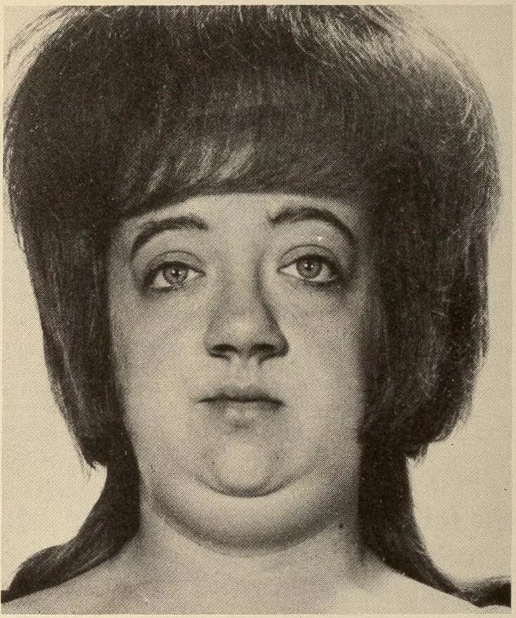
Woman with pycnodysostosis, Sydney S. Gellis & Murray Feingold / Public Domain
Skeletal Features in PYCD
- Men with pycnodysostosis can’t reach more than 150 cm in height (approximately 4 feet 11 inches)
- Women with the disease are shorter
- The proportion of the above and lower trunk is not equal; the legs are shorter as compared to upper body
Other skeletal features are:[7]
- Short distal phalanges (tips of fingers and toes).
- The skin of the fingers is wrinkled, particularly on the back of the fingers.
- Visible grooves (along the length) can be seen on the nails of fingers and toes. Also, these nails are flat.
- Another problem in these patients is collarbone abnormalities. Clavicles are not formed properly, so they appear malformed.
- The collarbone is also more susceptible to fractures.
- Over time, the bones of the fingers and the collarbone become more deteriorated.
Cranial and Facial Features
The distinctive features of skull involvement in pycnodysostosis are:[8]
- Fontanelles or soft spots of your skull remain open for a longer period. Sometimes not close even into adulthood
- In open skull spaces, some extra bones are formed, called Wormian bones.
- The front and back of the head are more prominent (Frontal and occipital bossing)
- The nose appears larger compared to the other face.
- The mid-region of the face is underdeveloped.
- Small, undersized jaw (Micrognathia)
- Narrow or High Arched Palate
Dental Concerns
In individuals with Toulouse-Lautrec syndrome, dental development follows an atypical timeline with the following problems:[9]
- Milk teeth erupt later than normal.
- Then these primary teeth remain in place longer than the age of losing them.
- The next stage of permanent teeth is also affected, as they appear much later than usual.
- Another problem is the irregular spacing and positioning of teeth.
- Some teeth may be missing entirely (hypodontia)
- Jaw abnormalities in combination with a narrow palate usually require a lot of Orthognathicand orthodontic treatment.
Additional Physical Features
- Hepatomegaly is common in some individuals; it is usually without health issues.
- During sleep, abnormal breathing is common
- With the passage of time, vertebral abnormalities result in a curve in the spine, called Scoliosis.
- In most cases, intellectual functions are normal. Only some difficulties in the learning processes are seen.
Bone Fragility and Fractures in Toulouse-Lautrec Syndrome
Bone fragility is the most challenging issue of pycnodysostosis. The brittle bones are more prone to fractures throughout the life of affected persons. Often, very minor injuries cause fractures. The most common sites facing risks are legs, feet, jaw, and collarbones.[10]
Sometimes, small cracks occur from repetitive strains. These are called stress fractures. In an area where multiple fractures occur complicate the diagnosis, because X-rays show overlapping lines of fractures.
Toulouse-Lautrec Syndrome Diagnosis
As the syndrome is a sporadic condition, its diagnosis can be missed. The most important aspect of diagnosis is to consider pycnodysostosis when a child presents with short stature, abnormality in facial structure, and a history of frequent fractures. With the help of clinical evaluation, imaging, and genetic testing, Toulouse-Lautrec syndrome can be accurately diagnosed.
Clinical Evaluation
A detailed medical history and a thorough physical examination are required for diagnosis. Doctors gather family history and information on inherited conditions in relatives for investigations.
Imaging Studies
The characteristic bone density can be easily observed in the radiographic images. The dense state of the bones is manifested as an abnormally white and compact appearance on X-rays. The open skull sutures, Wormian bones, collarbone disorder, shortening of the finger bones, and prior or cured fractures can also be detected in the images.[11]
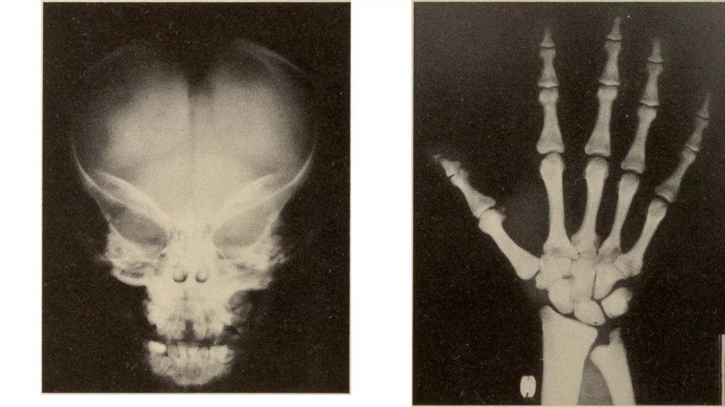
X-rays of the head and hand in pycnodysostosis, showing open fontanelle and shortened distal phalanges, Sydney S. Gellis & Murray Feingold / Public Domain
Genetic Testing
Molecular genetic testing is a conclusive test that gives positive confirmation through the identification of mutations in the CTSK gene. Nonetheless, this test is only undertaken in a few labs due to the rarity of the pycnodysostosis disease, since some laboratories have the expertise and facilities to conduct this type of genetic test.[12]
Toulouse-Lautrec Syndrome vs. Rickets
The following features can differentiate the conditions: [13]
| Feature | Toulouse-Lautrec Syndrome (Pycnodysostosis) | Rickets |
|---|---|---|
| Definition | Uncommon genetic skeletal dysplasia and abnormally dense and brittle bones. | Mineralization-defective metabolic bone disease. |
| Inheritance | Autosomal recessive (CTSK gene mutation) | Varies: X-linked, autosomal recessive, or acquired |
| Primary Cause | Abnormal osteoclast activity; cathepsin K-enzyme abnormality. | Deficiency of vitamin D or phosphate metabolism hereditary disorder. |
| Bone Density | Increased (osteosclerosis)- paradoxically dense and brittle. | Osteopenia (reduced)- soft and feeble. |
| Age of Onset | Childhood; since infancy. | Infancy and early childhood |
| Short Stature | Severe (4-5 feet in adulthood) | Variable; generally less severe |
| Facial Features | Frontal bossing, obtuse mandibular angle, and late eruption of the teeth. | Normal; enamel defects of teeth when severe. |
| Skeletal Deformities | Generalized osteosclerosis, changes of the mandible. | Bowing of long bones, coxa vara/valgum, rachitic rosary |
| Fracture Risk | Increased even in dense bones (paradoxical brittle) | High due to weak mineralization |
| X-ray Appearance | Dense, white bones; “bone-within-bone” appearance | Radiolucent; widened/cupped growth plates; ground-glass appearance |
| Calcium Levels | Normal to elevated | Low (vitamin D deficiency rickets) |
| Phosphate Levels | Normal | Low (vitamin D deficiency rickets) |
| Alkaline Phosphatase (ALP) | Normal to mildly elevated | Markedly elevated |
| Vitamin D Level | Normal | Low in deficiency rickets or normal in hereditary rickets |
| Bone Turnover | Low (hypoformative) | High (hyperformative) |
| Primary Treatment | Supportive care; careful surgical management | Supplements of Vitamin D and calcium; treat underlying causes. |
| Prognosis | Permanent and lifelong; normal lifespan; orthopedic care required. | Excellent with appropriate treatment; preventable if identified early |
Treatment and Management of Pycnodysostosis
At the moment, Toulouse-Lautrec syndrome cannot be cured. Currently, there is no cure for Toulouse-Lautrec syndrome. The interventions are primarily aimed at symptom management, prevention of complications, and improvement of quality of life. This involves collaborative efforts by numerous medical experts who must work as a team.[14]
The Healthcare Team
- A pediatrician
- An orthopedic specialist and orthopedic surgeon
- An endocrinologist to give hormonal treatment
- A dentist and orthodontist
- Oral and Maxillofacial Surgeon
Adults are referred to similar specialists without leaving their primary care physicians, who aid in coordinating the different aspects of their medical care.
Fracture Prevention and Management
The prevention of fractures is one of the main treatment objectives. These include training the affected and their families about the risk of injuries and protective measures. As a precaution, children should not engage in contact sports or high-impact sports that expose them to risks of fracture. The safer alternatives are swimming and cycling, which are very good exercises and cause minimal stress to the bones.[15]
Once these fractures develop, they will require professional orthopedic service. The usual protocols of treating the fractures are applied, yet the recovery may not go as anticipated in people with normal bone density. When there are several fractures in the same bone, the surgeons may advise the insertion of metal rods to offer internal support and prevent future fractures.
Growth Hormone Therapy
Studies have identified that most pycnodysostotic patients have impaired growth hormone secretion, which is one of the factors that makes them short. Linear bone growth has been promoted by growth hormone replacement therapy, which must be closely monitored by an endocrinologist. Although this does not cure the underlying condition, it may aid affected individuals in attaining increased height, which may lead to an increase in both functioning and quality of life.[16]
Dental and Orthodontic Care
Dental defects with pycnodysostosis usually involve massive intervention. Periodic dental checkups, preventive treatment to reduce cavities, and overall orthodontic treatment to correct spacing and alignment problems are all significant in the management.
Living with Toulouse-Lautrec Syndrome
Through proper medical attention and a change in lifestyle, patients with pycnodysostosis have the potential to have a good life. This disorder does not directly shorten life expectancy in otherwise healthy persons.
Lifestyle Adaptations
Some lifestyle changes can make the lives of Toulouse-Lautrec syndrome patients healthier.
- High-risk activities and contact sports should not be done.
- Rather, milder exercises such as swimming are very well-fitting since they are not too risky for the fractures.
- Mindfulness of safety is needed when performing routine activities, even simple falls that may bruise other people, may lead to severe fractures in a person with pycnodysostosis.[17]
Family Planning
The knowledge of the genetic nature of the condition helps the family to make reproductive choices. Genetic guidance gives good advice concerning the risks of inheritance. Careers may also decide to undergo genetic testing to determine whether they are a carrier of the gene, and this factor plays a significant role in determining the chances of having affected children.[18]
Fertility in Toulouse-Lautrec Syndrome
Fertility in individuals with Toulouse-Lautrec syndrome is generally normal. The disorder primarily affects the skeletal system and does not interfere with reproductive organs or hormonal function. Most men and women with pycnodysostosis can conceive naturally and have healthy pregnancies.[19]
However, due to the skeletal fragility and short stature, pregnancy and delivery may require special medical supervision, particularly in affected women. Obstetricians often recommend careful monitoring during pregnancy, as pelvic bone abnormalities may sometimes complicate natural childbirth. Cesarean delivery may be considered in certain cases for safety.
Emotional and Social Support
Being a patient with a chronic condition is an emotional challenge. Finding people with rare bone disorders, having mental health help whenever it is necessary, and being in open communication with the health team all help to improve psychological health.[20]
Conclusion
The Toulouse-Lautrec syndrome, or pycnodysostosis, is a condition in which genetic mutations in a single enzyme can produce extensive body consequences. Although this extremely rare condition poses serious difficulties, especially the bone fragility and short stature, the knowledge of its genetic nature, features, and timely management will allow patients to live a full and healthy life.
Further studies into the role of cathepsin K, bone remodeling, and possible therapeutic solutions provide an opportunity for better treatment in the future. In the meantime, the multidisciplinary approach that involves the combination of orthopedic management, endocrine support, dental care, and lifestyle changes introduces the recent basis of the optimal outcomes.
Genetic counseling, access to special medical facilities, and availability of specialized medical staff continue to be a vital resource to families impacted by this rare condition. Despite the evident problems, pycnodysostosis can be managed, and with proper support and medical attention, the members of the syndrome can enjoy their lives despite the distinctive skeletal traits.
References
[1] Sedano HD, Gorlin RJ, Anderson VE, et al.Pycnodysostosis: Clinical and genetic considerations.Am J Dis Child. 1968;116(1):70–77.
[2] Mujawar Q, Naganoor R, Patil H, Thobbi AN, Ukkali S, Sanadi A.Pycnodysostosis with unusual findings: a case report.Cases J. 2009;2:6544.
[3] Gelb BD, Shi GP, Chapman HA, Desnick RJ.Pycnodysostosis, a lysosomal disease caused by cathepsin K deficiency.Science. 1996;273(5279):1236–1238.
[4] Hou WS, Li W, Keyszer G, et al.Cathepsin K is a novel protease for bone resorption and is deficient in pycnodysostosis.J Clin Invest. 1999;103(5):731–738.
[5] Polymeropoulos MH, Ortiz de Luna RI, Ide SE, et al.The gene for pycnodysostosis maps to human chromosome 1q21.Nat Genet. 1995;10(2):238–239.
[6] Xue Y, et al.Clinical and animal research findings in pycnodysostosis.Orphanet J Rare Dis. 2011;6:20.
[7] Markatos K, Christofi T, Tasopoulou K, et al.Pycnodysostosis: A clinical, genetic, and therapeutic update.Int Orthop. 2018;42(6):1325–1331.
[8] Aynaou H, Lajmi K, Ghailani N, et al.Short stature revealing a pycnodysostosis: A case report.J Orthop Case Rep. 2016;6(2):58–61.
[9] Desmyttere M, Delfosse C, Boon L, Vervaet C, Declerck D.Oral management of two sisters with pycnodysostosis.J Stomatol Oral Maxillofac Surg. 2017;118(3):180–183.
[10] Grewal S, Agrawal A, Arora P, et al.Pycnodysostosis: Case report and review of literature with special emphasis on fracture management.J Orthop Case Rep. 2019;9(3):64–68.
[11] Aynaou H, et al.Short stature revealing a pycnodysostosis.J Orthop Case Rep. 2016;6(2):58–61.
[12] Polymeropoulos MH, et al.The gene for pycnodysostosis maps to human chromosome 1q21.Nat Genet. 1995;10(2):238–239.
[13] Spranger J, Brill PW, Poznanski AK.Bone Dysplasias: An Atlas of Genetic Disorders of Skeletal Development.4th ed. Oxford Univ Press; 2018
[14] Xue Y, et al.Clinical and animal research findings in pycnodysostosis.Orphanet J Rare Dis. 2011;6:20.
[15] Markatos K, et al.Pycnodysostosis: A clinical, genetic, and therapeutic update.Int Orthop. 2018;42(6):1325–1331.
[16] Beighton P.Congenital, Inherited, and Neonatal Skeletal Dysplasias.Springer; 2014.
[17] Aynaou H, et al.Short stature revealing a pycnodysostosis.J Orthop Case Rep. 2016;6(2):58–61.
[18] Xue Y, et al.Clinical and animal research findings in pycnodysostosis.Orphanet J Rare Dis. 2011;6:20.
[19] Alshdefat, A.,et al.(2022). A rare case of pycnodysostosis during pregnancy.Clinical Case Reports, 10(12), Article e6565. https://doi.org/10.1002/ccr3.6565
[20] Beighton P.Congenital, Inherited, and Neonatal Skeletal Dysplasias.Springer; 2014.