

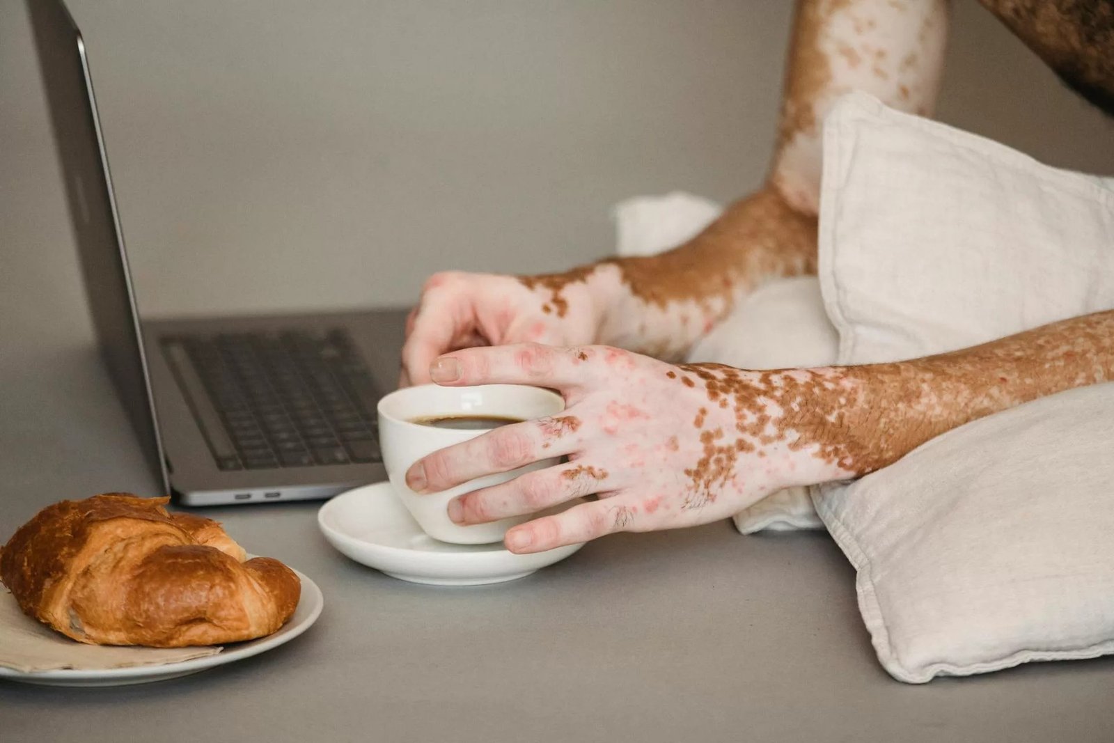
Vitiligo Complete Guide Vitiligo is a common skin disorder characterized by the development of well-demarcated, discolored/white patches on the skin. Medical professionals refer to these patches as “macules.” Vitiligo is one of the most common causes of acquired leukoderma (depigmented patches of skin). The exact cause of vitiligo is unknown, but multiple studies have shown autoimmune-mediated destruction of the pigment-producing cells (melanocytes). Researchers have also found stress, exposure to environmental triggers, and genetic alterations as potential causes for the dermal disorder. Moreover, people with autoimmune disorders (psoriasis, lupus, etc.), anemia, and thyroid disease are at a greater risk of developing macules. Although vitiligo itself is not life-threatening, it is associated with autoimmune comorbidities and can significantly affect psychological well-being. Therefore, most patients seek therapies (like depigmentation/repigmentation therapy, skin grafting, etc.) for aesthetic reasons.
What Is Vitiligo?: Vitiligo Complete Guide
Vitiligo is an acquired skin disorder characterized by the loss of melanocytes, leading to well-defined white (depigmented) patches on the skin. This disease affects males and females equally. It can develop at any age, but most patients notice color changes before the age of 30. The skin disorder affects all races equally; however, the spots are more visible on darker skin.
In some cases, the white spots on the skin are less than 1 cm in width, but you may find patches larger than 1 cm in other cases. The disease affects 0.76-1.11% of US adults, i.e., approximately 1.9-2.8 million cases. Reports suggest that around 40% of adult vitiligo cases in the US go undiagnosed. Globally, vitiligo affects approximately 0.5–2% of the population.[1]
The extent of depigmentation on the skin varies for each person. Some people might notice only a few depigmented areas, while others are worried about the widespread loss of color.
What Are The Different Types Of Vitiligo?
Based on the distribution pattern of the skin lesions, healthcare professionals divide vitiligo into the following types:
- Generalized (Non-Segmental): This is the most common type of the condition (seen in almost 90% of cases), characterized by a widespread symmetrical distribution of macules on both sides of the body.[2]
- Localized: In this type, pigment loss occurs in specific, small areas of the body. It’s commonly seen in children [3] Focal vitiligo and segmental vitiligo are different types of localized vitiligo.
- Focal: In the focal type, the lesions are limited to a small area and generally don’t spread within a couple of years.
- Segmental: White patches affect only one side of the skin (mostly on hands or face).[4]
- Universal: This is a rare type of leukoderma in which pigment loss is seen on more than 80% of the skin, which leads to an entirely white appearance.
- Mucosal: The condition is termed mucosal vitiligo if the white patches appear on your mucosa.
Clinical Variants Of Vitiligo
There are different clinical variants of the disease. The most prevalent variants are discussed below:
Acrofacial:
In this subtype of the skin condition, milky white/depigmented patches are seen on the facial region and the extremities. You can notice lesions on the lips, around the eyelids, and in the nasal region. White spots are also present on hands (fingers), feet (toes), forearms, and trunk.[5]
Marginal Inflammatory:
In the marginal inflammatory type, the white patches are surrounded by an elevated, red border (erythema) of skin. This variant is rare and can cause more problems than other types due to the associated itchiness (pruritus).
Trichorome:
This type is characterized by the formation of a bullseye with a depigmented center surrounded by an area of lighter pigmentation, which is covered by natural skin color. It is called trichrome due to the three different skin colors.
Quadrichrome:
In this type, there are four distinct color shades; thus, the name quadrichrome. The three layers of the lesion are the same as the trichrome type. However, here we see an additional, hyperpigmented margin surrounding the three trichromes. The presence of this fourth shade at the margins indicates active disease and is typically observed in individuals with darker skin tones.
Confetti Vitiligo:
This variant presents as multiple small, confetti-like depigmented macules and is considered a marker of active disease progression.
Symptoms Of Vitiligo
Most youngsters aged below 20 start noticing changes in their skin color. However, there have been reported cases in children as young as 2 years.
White Patches
Most patients start noticing the loss of pigmentation in the skin or mucosa. Pearly-white, well-demarcated patches develop on the skin of the face, hands, arms, and toes, etc. Patients have noted loss of hair pigmentation in the affected area. Hair in the vitiligo patch usually turns silver, gray, or even white.
The most common locations of leukoderma include:
- Face
- Arms
- Hands
- Feet (especially toes)
- Genitals
- Mucosa of the mouth
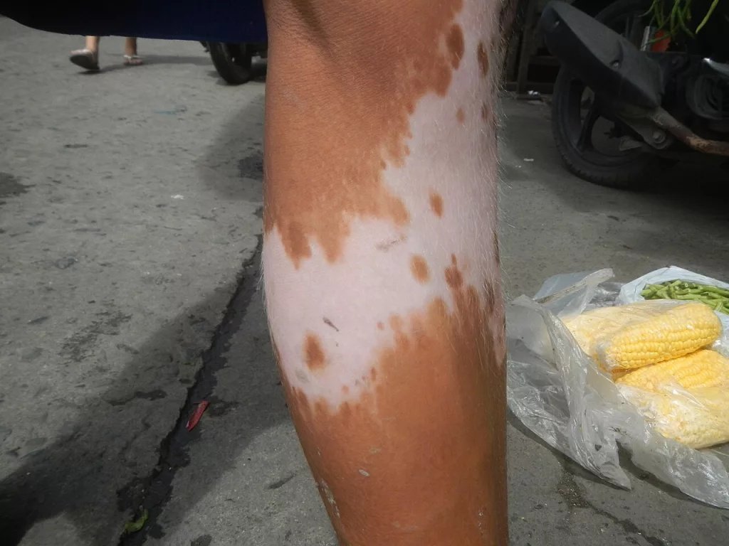
A patient with large vitiligo lesions on the leg.
The progression of vitiligo is unpredictable. Generally, the large patches stay in place for long periods (years). On the other hand, some patients have noticed that the location of the small macules tends to shift over time. This means some areas of skin lose pigment while previous ones regain color.
Koebner Phenomenon (KP)
A salient clinical feature of vitiligo is the Koebner phenomenon (isomorphic response). According to this phenomenon, there is a high risk of vitiligo development in the trauma-prone sites. Patients notice new lesions in the sites of mechanical trauma, like burns, cuts, or abrasions. Isomorphic response in vitiligo is related to chronic friction induced during daily activities.[6]
The presence of Koebner phenomenon often indicates active disease.[7]
Itching
Pruritus is not as commonly seen, but itching with confetti-like depigmentation is considered an initial clinical sign of the disease. Mostly, it indicates active disease.[8]
What Are The Causes Of Vitiligo?
The exact cause of depigmentation is not known. Clinicians believe it is a multifactorial disorder that has a complex pathogenesis. Research over the years reveals both genetic and non-genetic components active in the development of the disease. A common observation in vitiligo skin is the absence of pigment-producing cells, i.e., melanocytes. Different theories try to explain the pathogenesis of the dermal disorder.
Genetics
Variations in over 30 genes are believed to be responsible for the disorder. Researchers note that mutations in the genes NLRP1 and PTPN22 contribute to leukoderma. The NLRP1 gene is responsible for regulating the process of immune-mediated inflammation, and PTPN22 controls the activity of immune cells (especially T cells). Variations in these genes increase the chances of autoimmune attacks on your melanocytes and contribute to increased inflammation.[9]
Neural Cause
According to this hypothesis, stress (physical or emotional) or dysfunctional nerve endings in the skin can cause the death of melanocytes by releasing toxic neurochemical mediators like catecholamines, neuropeptides, and substance P, etc.[10] However, this is considered a supportive hypothesis rather than a primary cause.
Oxidant Accumulation
It is believed that the accumulation of oxidants (more than the body’s ability to neutralize) in the skin potentially leads to vitiligo. Oxidative overload or oxidative stress is a state of excess reactive oxygen species (ROS) in the skin. ROS directly damages the melanocyte DNA, causes functional impairment, and lays the foundation for skin depigmentation.[11] Numerous factors contribute to oxidative stress, including:
- Exposure to UV radiation and ozone
- Exposure to environmental pollutants like polycyclic aromatic hydrocarbons (PAHs)
- Aging
- Mitochondrial dysfunction
- Stress and poor diet (alcoholism)
Intrinsic Melanocyte Defects
Sometimes, the melanocytes are inherently weak and prone to destruction without any external attack or autoimmune mediation. The defective cells are unable to grow and differentiate properly, leading to their early death and subsequent depigmentation of the skin.
Autoimmune (Cytotoxic) Attack
The most probable cause of vitiligo in the majority of cases is an autoimmune attack on melanocytes. A faulty immune system mistakenly identifies healthy melanocytes as pathogenic invaders and begins destroying them. Studies show that immune cells (T cells) play a crucial role in melanocyte destruction. Therefore, most therapies target the activation/action of immune cells.[12]
Risk Factors for Vitiligo
Healthcare professionals have identified several risk factors that increase your propensity to develop vitiligo. Different autoimmune disorders are linked to vitiligo.
Lupus:
There have been comorbid diagnoses of systemic lupus erythematosus in vitiligo cases.[13] Vitiligo can develop secondary to discoid lupus erythematosus.[14]
Psoriasis:
The autoimmune skin disorders are known to co-exist in patients, which is attributed to an altered immune response.[15]
Rheumatoid arthritis (RA):
The painful autoimmune condition of the joints can lay the foundation for this skin ailment. There is a bidirectional association between RA and vitiligo.[16]
Hashimoto’s thyroiditis:
Another malady closely linked to vitiligo is the autoimmune thyroiditis (which causes hypothyroidism). The link between autoimmune thyroiditis and vitiligo is so strong that some studies exclude the thyroid pathology if macules are not seen on the hands.[17]
Anemia:
Pernicious anemia is an autoimmune type of anemia that can co-exist with skin pathology. A 2025 study found that leukoderma is frequently associated with autoimmune conditions like alopecia areataand pernicious anemia. Therefore, vitamin B12 supplementation works in some cases.[18]
Type 1 Diabetes
This type of diabetes is characterized by autoimmune-mediated destruction of the pancreatic cells. Thus, type 1 diabetes is also recognized as a risk factor for vitiligo.
Insufficient production of the hormones cortisol and aldosterone in Addison’s disease can also increase your chances of getting white macules.
Is Vitiligo Contagious?
No, vitiligo is not a contagious disorder. It is mostly caused by an abnormal autoimmune response of the body. It does not spread by touching, sharing utensils, or the exchange of body fluids (saliva).
Diagnosing Vitiligo
Your healthcare provider will perform a physical examination of the macules. He will ask you about your medical history (especially about any autoimmune disorder) and family history (of leukoderma).
For a better diagnosis, he may use a Wood’s lamp to observe your skin. With this specialized lamp, the doctor directs UV light onto your skin, which helps him differentiate vitiligo from other skin pathologies.
Differential Diagnosis
Chemical leukoderma has presentations similar to vitiligo. Therefore, it is important to differentiate between these conditions.
Vitiligo Vs. Leukoderma
Leukoderma is a descriptive term used for white (depigmented) patches on the skin, whereas vitiligo is a specific medical condition and one of the most common causes of leukoderma. While vitiligo occurs due to autoimmune destruction of melanocytes, leukoderma can result from multiple causes, including chemical exposure, post-inflammatory changes, or other skin disorders. Thus, all vitiligo is leukoderma, but not all leukoderma is vitiligo.
Complications Of Vitiligo
Apart from the cosmetic concerns, vitiligo can cause some complications. Therefore, management of the lesions is important. Common complications include:
- Skin sensitivity to sunburns
- Rare ocular involvement, such as uveitis or retinal pigment abnormalities
- Low self-esteem due to skin color
Vitiligo Treatment
Patients seek treatment only for cosmetic reasons, as it doesn’t cause any harm to the body. However, many people visit a dermatologist for emotional peace. The main aim of therapy is to create a uniform skin tone. The choice of treatment depends on the extent, location, and activity (stable vs progressive) of the disease.
Medical Treatment
A simple way of management is to stop the autoimmune attack and promote melanocyte recovery. Corticosteroids help suppress the overactive immune system.
Calcineurin inhibitors not only reduce autoimmune destruction but also promote melanocyte proliferation, which eventually leads to repigmentation. You can get superior results by pairing them with phototherapy.[19]
Topical Janus kinase (JAK) inhibitors, particularly ruxolitinib, are now FDA-approved for the treatment of non-segmental vitiligo and have shown significant efficacy, especially for facial lesions. These agents work by blocking immune pathways responsible for melanocyte destruction.[20]
Phototherapy
Light therapy is a non-invasive treatment modality used to treat various skin disorders. The use of different wavelengths of ultraviolet light on vitiligo skin has some potential benefits. Narrowband UVB (NB-UVB) is considered the first-line treatment for widespread vitiligo due to its safety and effectiveness. It promotes repigmentation, particularly on the face and trunk, although response on hands and feet may be limited.
Phototherapy is often combined with topical agents to enhance treatment outcomes.[21]

Phototherapy (with UVB light) on hand with vitiligo lesions
In earlier times, doctors used the psoralen photochemotherapy to achieve repigmentation, but it was discontinued due to its toxic effects.
Laser Therapy
Medical-grade lasers are used in different skin disorders, including vitiligo. Different types of lasers, like an excimer laser (a type of ultraviolet chemical laser), can be used to treat vitiligo. Excimer laser therapy is particularly useful for localized vitiligo and delivers targeted UVB light to affected areas. It is most effective for lesions on the face and neck but may be less beneficial for acral regions.
Although effective, its use may be limited by cost and availability rather than lack of efficacy.[22]
Depigmentation Therapy
In larger patches of white skin, doctors advise depigmentation of the natural skin to match the color of the affected skin. Monobenzone is the drug used for depigmentation.
Surgery
Surgical treatment is a more permanent solution for skin depigmentation. It is reserved for stable vitiligo (no new lesions or progression for at least 6–12 months). In most cases, doctors take a skin graft from a normal/unaffected part of your body and replace the affected skin part.
A minimally invasive surgery is the suction blister grafting, used to cover small macules. In this technique, doctors create thin blisters on the donor site (mostly thighs) while removing the top layer of the affected skin using dermabrasion. The blister from the healthy skin (which has active melanocytes) helps repigment the skin. It has proven to be an effective treatment with high repigmentation rates and a good safety profile.[23]
Wrapping Up
Vitiligo is a skin disorder characterized by the formation of white patches (macules) on the skin. We frequently see well-demarcated patches of pearly-white color on the skin of the face, hands, and feet. However, you may see macules on other body parts too. Generalized vitiligo is the most common type, which is characterized by a symmetrical development of depigmentation patches on both sides of the body.
The exact cause of vitiligo is not known, but the condition is believed to arise from autoimmune-mediated attack on the pigment-producing melanocytes. Genetics/genetic mutations play a role in the onset of the disease. Among other theories of disease onset, the neural theory suggests that stress releases neurochemical mediators that disrupt melanocyte function. Oxidant-antioxidant theory suggests that oxidative stress (accumulation of reactive oxygen species) causes functional impairment of the pigment producers. However, in some cases, inherently defective melanocytes have suboptimal performance and a higher tendency to disintegrate.
Individuals with autoimmune disorders like lupus, psoriasis, alopecia areata, rheumatoid arthritis, Hashimoto’s disease (autoimmune thyroiditis), and type 1 diabetes are at a higher risk of vitiligo. Treatment includes repigmentation with medicines (Janus kinase inhibitors and calcineurin inhibitors), phototherapy (UVB light therapy), and laser therapy. For large depigmented areas, doctors go for depigmentation of neighboring healthy skin (via monobenzone) to match the shade. For a more permanent solution, doctors abrade the top layer of affected skin with dermabrasion and replace it with a fine blister taken from a healthy donor site. It offers good repigmentation due to the supply of healthy melanocytes in the region.
References
[1] Gandhi, K., Ezzedine, K., Anastassopoulos, K. P., Patel, R., Sikirica, V., Daniel, S. R., … & Pandya, A. G. (2022). Prevalence of vitiligo among adults in the United States.JAMA dermatology,158(1), 43-50.
[2] Seneschal, J. (2023). Clinical features of vitiligo and social impact on quality of life.Dermatology Practical & Conceptual,13(4 Suppl 2), e2023312S.
[3] Mulekar, S. V., Al Eisa, A., Delvi, M. B., Al Issa, A., & Al Saeed, A. H. (2010). Childhood vitiligo: a long‐term study of localized vitiligo treated by noncultured cellular grafting.Pediatric Dermatology,27(2), 132-136.
[4] Lin, X., Meng, X., & Lin, J. (2025). Segmental vitiligo: autoimmune pathogenesis, neuronal mechanisms, and somatic mosaicism.International Journal of Dermatology,64(3), 490-498.
[5] Šitum, M., Bulat, V., Kovačević, M., & Goren, A. (2020). A Patient with Acrofacial Depigmentation and Chronic Fatigue. InClinical Cases in Pigmentary Disorders(pp. 29-32). Cham: Springer International Publishing.
[6] Zhang, Y., Ding, X., Wang, F., Li, M., & Du, J. (2023). Clinical significance of Koebner’s phenomenon in vitiligo: a hospital-based epidemiological investigation from China.Chinese Medical Journal,136(4), 502-504.
[7] Tang, X., Hao, C., Fan, M., Dong, P., Qi, Y., Gao, J., … & Wang, W. (2025). Correlation between the koebner phenomenon and clinical features in vitiligo.Clinical, Cosmetic and Investigational Dermatology, 97-103.
[8] van Geel, N., Grine, L., De Wispelaere, P., Mertens, D., Prinsen, C. A. C., & Speeckaert, R. (2019). Clinical visible signs of disease activity in vitiligo: a systematic review and meta‐analysis.Journal of the European Academy of Dermatology and Venereology,33(9), 1667-1675.
[9] Męcińska-Jundziłł, K., Tadrowski, T., Jundziłł, A., Witmanowski, H., & Czajkowki, R. (2023). Evaluation of polymorphisms and expression of PTPN22, NLRP1 and TYR genes in vitiligo patients.Advances in Dermatology and Allergology/Postępy Dermatologii i Alergologii,40(2), 225-233.
[10] Al Abadie, M. S., & Gawkrodger, D. J. (2021). Integrating neuronal involvement into the immune and genetic paradigm of vitiligo.Clinical and experimental dermatology,46(4), 646-650.
[11] Chang, W. L., & Ko, C. H. (2023). The role of oxidative stress in vitiligo: an update on its pathogenesis and therapeutic implications.Cells,12(6), 936.
[12] Chang, W. L., Lee, W. R., Kuo, Y. C., & Huang, Y. H. (2021). Vitiligo: an autoimmune skin disease and its immunomodulatory therapeutic intervention.Frontiers in cell and developmental biology,9, 797026.
[13] Li, H. O. Y., Bailey, A. M. J., Pastukhova, E., Tan, M. G., & Kirchhof, M. G. (2023). Comorbid diagnosis of systemic lupus erythematosus in vitiligo: a systematic review and meta-analysis.British Journal of Dermatology,188(3), 440-441.
[14] Jin, S., Wang, S., Jin, S., Tang, C., & Wang, P. (2025). A case of vitiligo secondary to discoid lupus erythematosus treated with tofacitinib.Medicine,104(26), e43118.
[15] Marasca, C., Fornaro, L., Martora, F., Picone, V., Fabbrocini, G., & Megna, M. (2021). Onset of vitiligo in a psoriasis patient on ixekizumab.Dermatologic Therapy,34(5), e15102.
[16] Cohen, N. T., Schonmann, Y., & Kridin, K. (2024). A bidirectional autoimmune cluster between vitiligo and rheumatoid arthritis: a large-scale population-based study.Archives of Dermatological Research,316(7), 366.
[17] Chivu, A. M., Bălășescu, E., Pandia, L. D., Nedelcu, R. I., Brinzea, A., Turcu, G., … & Ion, D. A. (2022). Vitiligo—Thyroid Disease Association: When, in Whom, and Why Should It Be Suspected? A Systematic Review.Journal of Personalized Medicine,12(12), 2048.
[18] Khan, S. (2025). Exploring the link between pernicious anemia and vitiligo: Digging Deeper.Scholastic Orthopaedics,1, 01-02.
[19] Chang, H. C., Hsu, Y. P., & Huang, Y. C. (2020). The effectiveness of topical calcineurin inhibitors compared with topical corticosteroids in the treatment of vitiligo: A systematic review and meta-analysis.Journal of the American Academy of Dermatology,82(1), 243-245.
[20] Utama, A., Wijesinghe, R., & Thng, S. (2024). Janus kinase inhibitors and the changing landscape of vitiligo management: a scoping review.International Journal of Dermatology,63(8), 1020-1035.
[21] Bouceiro Mendes, R., Alpalhão, M., & Filipe, P. (2022). UVB phototherapy in the treatment of vitiligo: State of the art and clinical perspectives.Photodermatology, photoimmunology & photomedicine,38(3), 215-223.
[22] Li, C., Hu, Y., Mu, Z., Shi, L., Sun, X., Wang, X., … & Li, X. (2024). Comparison of various excimer laser (EL) combination therapies for vitiligo: a systematic review and network meta-analysis.Journal of Dermatological treatment,35(1), 2302064.
[23] Shi, X., Wang, F., Sun, Y., Du, J., & Ding, X. (2024). Long-Term Effects and Prognosis Following Suction Blister Epidermal Grafting in Vitiligo Patients.Journal of Cutaneous Medicine and Surgery,28(3), 264-268.