
 is a rare disorder of the breathing apparatus, where the cartilage of the trachea...)

Tracheomalacia Symptoms Treatments Tracheomalacia (TM) is a rare disorder of the breathing apparatus, where the cartilage of the trachea (windpipe) is so soft, weak, or flaccid that it collapses, resulting in breathing obstruction. The condition most commonly exists as a congenital disorder in infants, which often improves or resolves as the baby grows. However, you can also acquire tracheomalacia due to injury or prolonged intubation. Mild cases present with noisy breathing (stridor, wheeze), breathing difficulties, and recurrent respiratory infections. However, in severe cases, there may be life-threatening breathing obstruction and cyanosis.
In some patients, tracheomalacia is accompanied by bronchial weakness, and the condition is called tracheobronchomalacia. According to one study, the incidence of tracheomalacia and tracheobronchomalacia in young patients was found to be approximately 1 in 2,100 children.[1] Doctors treat the disorder mainly with nonsurgical therapies (like physical therapy and CPAP) and rarely with surgery.
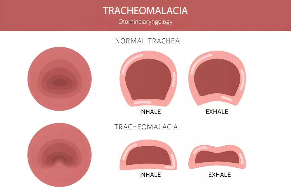
The illustration shows the size of the tracheal lumen in a normal person vs a tracheomalacia patient.
Tracheomalacia Types: Tracheomalacia Symptoms Treatments
Generally, tracheomalacia is divided into two types:
Congenital Tracheomalacia:
This type of cartilage weakness/flaccidity is present at birth. The congenital form arises due to the incomplete growth of the windpipe during fetal development (in the uterus). It is often associated with other congenital airway anomalies, such as laryngomalacia, which can cause significant respiratory distress in infants.[2]
Acquired Tracheomalacia:
This type develops later in life in response to damage from some external factors. Common causes of acquired tracheomalacia include injury, surgery, or prolonged respiratory intubation (for mechanical ventilation).
Another type of classification used in pediatric patients is:
Type Ⅰ
Tracheal weakness develops due to congenital abnormalities in the breathing structures. It may be associated with developmental anomalies like esophageal atresia (lack of connection between the esophagus and the stomach) or tracheoesophageal fistula (abnormal communication between the esophagus and the trachea).
Type Ⅱ
Some extrinsic factors cause disease rather than intrinsic factors (like weak tracheal cartilage). In this type, the cause is a congenital anomaly where the aorta forms a ring around your trachea (and esophagus), which compresses the pipes. This abnormal ring of the vessel is known as a vascular ring.[3]
Type Ⅲ
The third type is the acquired type of tracheomalacia, which develops later in life due to some external factor. The most common causes of type Ⅲ tracheomalacia include prolonged intubation,chronic tracheal inflammation, and sometimes, disorders like relapsing polychondritis (an autoimmune disorder that causes damage to the body’s cartilages).[4]
Tracheomalacia Symptoms
The collapsing trachea poses numerous problems for newborns and adults. Generally, congenital tracheomalacia is diagnosed at ages between 4 and 8 weeks.
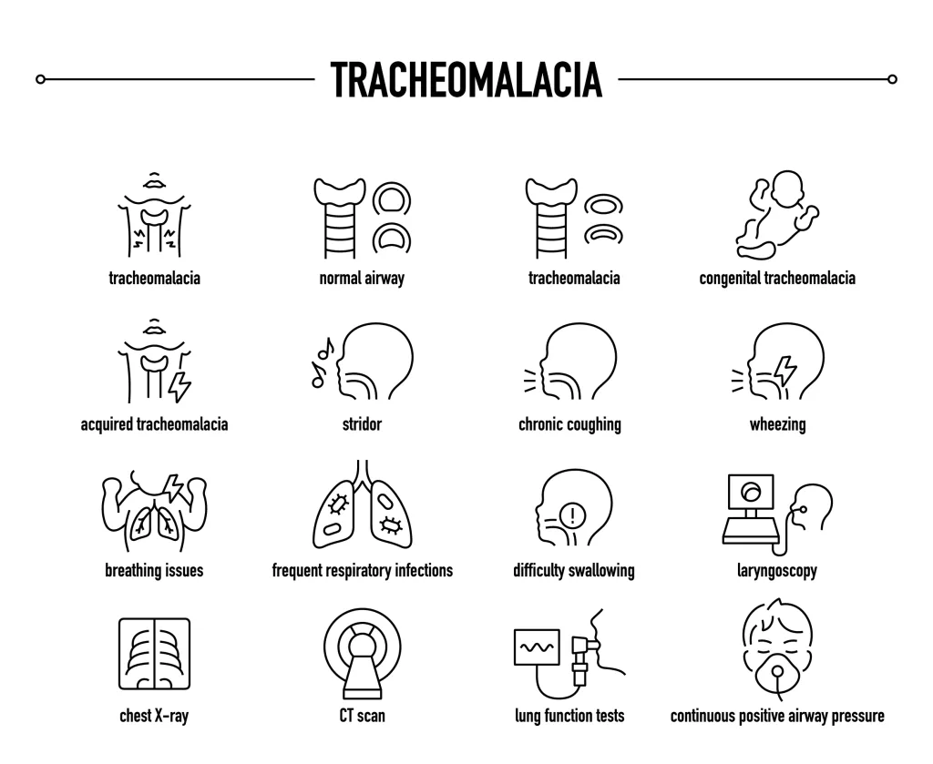
Tracheomalacia symptoms and management.
The most prevalent symptoms of tracheomalacia include:
Breathing sounds:
Infants with tracheomalacia have abnormal breathing sounds. Wheeze (whistling/rattling sound) is a common presentation of tracheomalacia. Doctors observe a typically a single-pitched expiratory wheeze (fixed wheeze) during exhalation in most patients.[5]
A stridor is a similar high-pitched sound produced during inhalation. Laryngomalacia and tracheomalacia are the most common causes of inspiratory stridor in infants.[6]
Bronchoscopic analysis of 88 infants revealed that the most common presentation of infant tracheomalacia was wheeze (58%), followed by stridor (42%) and cough (38.6%).[7] Breathing sounds are also present in adults.
Cough:
Coughing is a salient feature of infantile tracheomalacia. It is only second to abnormal breathing sounds. In children, the collapse of the tracheal lumen in tracheobronchomalacia presents with wheezing, dyspnea, and a barking cough.[8] Young children suffering from airway malacia (weakening) constantly try to clear the airways. This leads to the development of a chronic cough.[9] Clinicians have observed barking seal cough in adults, too.[10]
Shortness of breath:
As this disease leads to partial closure of the airways, shortness of breath becomes an issue. Dyspnea is known to worsen when the child coughs, cries, or even during feeding. Therefore, dyspnea is an important feature of the disorder.[11]
Hoarseness:
If you have acquired tracheomalacia following surgery or injury, you might end up having hoarseness. There have been cases where persistent dyspnea and hoarseness turned out to be due to tracheomalacia.[12] The narrowing of the tracheal lumen potentially leads to improper airflow through the voice box, which consequently leads to voice changes.
Feeding/Swallowing Difficulties:
Infants with the disorder suffer from feeding difficulties. In adults, swallowing difficulties develop secondary to tracheal anomalies. In patients, the size of the oesophagus increases with swallowing, which compresses the trachea. Thus, exacerbation of symptoms is seen immediately after (or even during) eating. This adds to the patient’s eating avoidance. Long-standing esophageal atresia and tracheoesophageal fistula are linked to honking-type cough and dysphagia (swallowing difficulty).[13]
Developmental delays:
Due to the respiratory problems, children with tracehobroncomalacia often have problems performing basic physical activities that their counterparts can do easily. This, paired with the poor respiration and growth, leads to potential developmental delays. Kids with esophageal atresia and tracheomalacia often exhibit growth retadation and developmental delays.[14] Tracheal flaccidity is also linked to issues like heart defects and Ehlers-Danlos syndrome.
Cyanosis:
Severe cases of tracheomalacia can lead to life-threatening consequences such as cyanosis (a condition in which the skin around the lips and nails turns blue in response to a lack of oxygen in the body).
Many times, doctors have to perform emergency maneuvers and interventions in acute phases of apnea and consequent cyanosis. According to one clinical study, a 2-month-old male baby developed recurrent episodes of apnea (worsening episodes) and central cyanosis as a consequence of airway malacia. Doctors performed aortopexy to manage symptoms.[15]
The disease can rarely cause cyanosis in adults; however, there have been several recorded clinical cases.[16]
Recurrent infections:
Patients of all ages are exposed to recurrent respiratory infections. Infants and adults seem to develop repeated pneumonia infections. Thus, in many cases, this is the foundation for a surgical intervention, as pulmonary infections lead to multiple hospitalizations and compromised quality of life. Plus, there are chances of life-threatening events with severe lung infections.[17] Additionally, you may also find associated bronchitis in such patients.
Tracheomalacia Causes
Congenital tracheomalacia
Tracheomalacia in newborns develops when there is incomplete or improper development of the tracheal cartilage. This incorrect development leads to a windpipe that is floppy and flaccid. Thus, the airways collapse under air pressure, leading to symptoms.
Acquired tracheomalacia
Several pathologies, conditions, and procedures can lead to abnormal closure of your trachea’s lumen, some of which are mentioned below:
Surgery:
Certain surgical procedures and iatrogenic damage during surgical procedures can lay the foundation for airway malacia. There is a chance that you may develop TM after a total thyroidectomy procedure for a multinodular goiter, lung transplant, or tracheoesophageal fistula repair surgery.[18]
It can also be a complication of long-term intubation with tracheostomy.[19]
Health conditions:
You can also fall prey to airway malacia secondary to different underlying health conditions. Gastric problems like chronic gastroesophageal reflux disease (GERD) andobesity are identified as risk factors.[20]
Chronic inflammatory conditions like bronchitis (inflammation of the bronchi), chronic chest infections, and relapsing polychondritis can lay the foundation for TM.[21] External tracheal compression due to tumors, cysts, or aortic aneurysms can also contribute to TM.
Developmental abnormalities like tracheoesophageal fistula and esophageal atresia are strongly linked to windpipe malacia. Moreover, a vascular ring can also compress the tracheal lumen.
Tracheomalacia Diagnosis
Your healthcare provider will start by taking a history of your symptoms, followed by a physical examination. He will listen to the breathing sounds (stridor, wheeze) and further investigate to identify the underlying cause of the labored breathing.
Diagnostic tests:
To reach a conclusive diagnosis, your doctor will look into the inside of your windpipe and airways. A laryngoscopy procedure involves visualizing your voice box (larynx) with the help of a laryngoscope, i.e., a lighted tube that has a camera attached to it. For direct laryngoscopy, the laryngoscope is inserted via the throat.
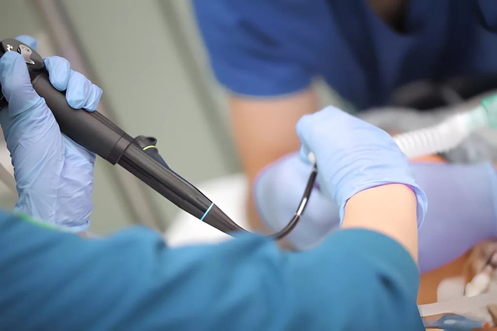
The image shows a professional performing a bronchoscopy procedure with a bronchoscope.
However, the most accurate and reliable test in TM diagnosis is bronchoscopy. It is similar to laryngoscopy; the only difference is that the tube goes deeper into the airways (bronchi). Flexible fiberoptic bronchoscopy is the gold standard for diagnosis.
Imaging tests:
Doctors may also use different types of imaging tests for diagnosis. Ultrafast computed tomography (CT) imaging alone provides a quick assessment of your airways. At times, this is paired with bronchoscopy. MRI scans can also be used. Modern clinicians may use cine-MRI, which makes a video of the organs by stitching together images. In abarium swallow (esophagram), the doctor visualizes the esophagus (with X-rays) after giving the contrast liquid containing barium. This imaging test is also used in the diagnosis of tracheomalacia.
Pulmonary function tests help identify the extent of respiratory compromise. It is done in conjunction with other tests.
Differential Diagnosis
Tracheomalacia Vs. Laryngomalacia
In tracheomalacia, there is weakness of the tracheal cartilage that leads to noisy breathing, which is most noticeable during expiration. On the other hand, laryngomalacia is characterized by weakness in the laryngeal (voice box) cartilage that causes noisy breathing during inspiration.
Tracheomalacia Treatment
Congenital tracheomalacia and mild acquired disease resolve spontaneously (without any intervention). Thus, in such cases, doctors only monitor the patient.
When significant symptoms are present, the treatment modalities for tracheomalacia depend on the severity of the disorder. Patients with mild breathing difficulties can be managed with simpler non-surgical therapies. However, for severe cases that can potentially turn life-threatening, surgical intervention is the only option.
Prescribing medicines is also part of the conservative management of the disease. Doctors frequently prescribe mucolytics and corticosteroids to soften/thin out mucus and reduce inflammation, respectively. Bronchodilators are also given to relax the tracheal muscles so that breathing is improved although these may worsen tracheal collapse in some cases. As tracheomalacia patients are plagued with respiratory infections, antibiotics are a part of most of the drug regimens.
Nonsurgical Management
The main aim of non-interventional strategies is to enhance the patency of the airways. Most commonly adopted therapies include:
Physical therapy: Conservative management with chest physical therapy can improve symptoms of tracheomalacia. However, there is low clinical evidence for its efficacy.
Humidifiers: Individuals with collapsing trachea have impaired clearance of the airways and thus, mucus accumulation further aggravates the issue. The use of humidified air (via a humidifier) can soften the mucus and make clearance easier for you.
CPAP: Continuous positive airway pressure (CPAP) is employed in the management of numerous respiratory conditions (COPD, sleep apnea, etc.). Clinical studies reveal that patients report notable improvements in symptoms with CPAP, thanks to the pressurized air that forcefully keeps the trachea open.[22]
Surgical Management of Tracheomalacia
Surgical intervention is reserved for severe cases that do not respond to conservative approaches. The most common procedures include:
Aortopexy
It’s done to keep the trachea from collapsing due to extrinsic factors. The surgeon raises your aorta up and away from your airways and attaches it to the sternum (breastbone). When performed in children, aortopexy has been shown to have good outcomes with a good safety profile.[23]
Aortopexy is ideal for anterior vascular compression (by the aorta). However, it doesn’t work well for posterior issues. Thus, doctors prefer tracheopexy in such cases.
Tracheopexy
In tracheopexy, the surgeon connects your windpipe (a part of it) to a neighboring structure, like the sternum or the soft tissues of your spine. This provides support to the airway structures and helps keep the trachea open. Posterior tracheopexy has proven to be a safe and effective treatment for tracheomalacia. Thus, in modern times, it is a frequently adopted surgical procedure for TM.[24]
Stenting
In severe cases, doctors place a hollow tube in the trachea, known as a tracheal stent, to prevent it from collapsing. They are usually reserved for severe, selected cases due to risks like granulation tissue or stent migration. However, stents have been proven to be feasible and effective in the management of pediatric tracheomalacia. Now you can get advanced removable metal stents for children that work superbly without side effects.[25]
Tracheomalacia Complications
You can observe the following complications:
- Severe airway obstruction
- Aspiration pneumonia
- Failure to thrive in infants (now known as growth faltering)
Wrapping up
Tracheomalacia is a condition in which the trachea (windpipe) collapses and constricts the lumen. This is attributed to different causes, like congenital weakness/softness of the tracheal cartilage, improper communication between the esophagus and trachea, anatomical abnormalities (vascular rings), and damage from chronic infection and surgery. The collapsed trachea presents with stridor, wheezing, dyspnea, chronic coughing, hoarseness, and swallowing difficulties. Pediatric patients are prone to recurrent respiratory infections and developmental delays. Severe hindrance of gas exchange can lead to cyanosis, which requires immediate medical attention.
Doctors diagnose it with airway studies like tracheoscopy and bronchoscopy. Congenital tracheomalacia and mild conditions resolve without intervention. When symptomatic, experts manage it with CPAP, physical therapy, and air humidifiers. Surgical interventions like tracheopexy, aortopexy, and stenting are reserved for severe cases.
References
[1] Boogaard, R., Huijsmans, S. H., Pijnenburg, M. W., Tiddens, H. A., de Jongste, J. C., & Merkus, P. J. (2005). Tracheomalacia and bronchomalacia in children: incidence and patient characteristics.Chest,128(5), 3391-3397.
[2] Varela, P., & Schweiger, C. (2021, June). Congenital airway anomalies. InSeminars in Pediatric Surgery(Vol. 30, No. 3, p. 151055). WB Saunders.
[3] Porcaro, F., Ciliberti, P., Petreschi, F., Secinaro, A., Allegorico, A., Coretti, A., & Cutrera, R. (2023). Long term respiratory morbidity in patients with vascular rings: a review.Italian Journal of Pediatrics,49(1), 24.
[4] Winter, G., Löffelmann, T., Chaya, S., Kaiser, H., Prenzler, N. K., Warnecke, A., … & Schwerk, N. (2024). Relapsing polychondritis with tracheobronchial involvement: a detailed description of two pediatric cases and review of the literature.Klinische Pädiatrie,236(02), 97-105.
[5] Wallis, C., Alexopoulou, E., Antón-Pacheco, J. L., Bhatt, J. M., Bush, A., Chang, A. B., … & Priftis, K. (2019). ERS statement on tracheomalacia and bronchomalacia in children.European Respiratory Journal,54(3).
[6] Shanthakunalan, K., Wijayasingam, G., Sudarshan, P., Prayle, A., & Daniel, M. (2023). Investigation and management of stridor in neonates.Paediatrics and Child Health,33(4), 91-96.
[7] Vijayasekaran, D., Balasubramanian, S., Sivabalan, S., & Vindhiya, K. (2018). Clinical characteristics and associated congenital lesions with tracheomalacia in infants.Indian Pediatrics,55(10), 883-884.
[8] Weinberger, M. (2023). Chronic cough and causes in children.Journal of Clinical Medicine,12(12), 3947.
[9] Fahy, A. S., & Chiu, P. P. (2021, June). Airway clearance in tracheomalacia. InSeminars in pediatric surgery(Vol. 30, No. 3, p. 151061). WB Saunders.
[10] Ferrante, F., Onorati, I., Radu, D. M., Herve-Carrega, A., Didier, M., Huet, O., & Martinod, E. (2024). Unexplained disabling and long-lasting cough: a case report.Journal of Cardiothoracic Surgery,19(1), 648.
[11] Alsalihi, Y., Yousef, N. M., Grewal, S., & Teitelbaum, B. (2023). Tracheomalacia in adults: an uncommon cause of dyspnea.Cureus,15(11).
[12] Yusur, A., Yousef, N. M., Sundeep, G., & Teitelbaum, B. (2023). Tracheomalacia in Adults: An Uncommon Cause of Dyspnea.Cureus,15(11).
[13] Kovesi, T., & Rubin, S. (2004). Long-term complications of congenital esophageal atresia and/or tracheoesophageal fistula.Chest,126(3), 915-925.
[14] Tytgat, S. H., van Herwaarden-Lindeboom, M. Y., van Serooskerken, E. S. V. T., & van der Zee, D. C. (2018). Thoracoscopic posterior tracheopexy during primary esophageal atresia repair: a new approach to prevent tracheomalacia complications. Journal of Pediatric Surgery,53(7), 1420-1423.
[15] Pertzborn, M., Renno, M. S., Lyons, K., Fett, J., & Agarwal, A. (2021). Apnea and Cyanosis Due to Severe Tracheobronchomalacia in the Setting of Congenital Short Trachea with Resolution by External Stenting and Aortopexy. InTP77. TP077 PEDIATRIC CONGENITAL LUNG LESIONS(pp. A3461-A3461). American Thoracic Society.
[16] Kabbabe, G. M., Daradkeh, B., Almustafa, S., Jama, A., Gamma, O., Shbat, M., & Ahmad, R. (2025). Uncommon clinical manifestation of tracheomalacia in a 29-year-old adult: a rare case report.Annals of Medicine and Surgery,87(6), 3902-3905.
[17] Møller, H. H., Schmidt, M. N., Ifaoui, I. B., Thyregod, H. G., Christiansen, E. S., Muthialu, N., … & Nielsen, K. G. (2025). Tracheomalacia and surgical options: A case series perspective.Acta Paediatrica,114(1), 59-64.
[18] Soewoto, W., & Ardianti, M. (2024). Tracheomalacia following a total thyroidectomy in a patient with a large non-toxic goiter: A case report.International Journal of Surgery Case Reports,116, 109211.
[19] Wallace, S., & McGrath, B. A. (2021). Laryngeal complications after tracheal intubation and tracheostomy.BJA education,21(7), 250-257.
[20] Kandaswamy, C., Bird, G., Gill, N., Math, E., & Vempilly, J. J. (2013). Severe tracheomalacia in the ICU: identification of diagnostic criteria and risk factor analysis from a case control study.Respiratory care,58(2), 340-347.
[21] Peters, S. M. (2020). An unexpected cause of wheezing and shortness of breath.JAAPA,33(2), 54-56.
[22] Syamil, R. F., & Azman, M. (2025). Acquired Tracheomalacia Following Tracheostomy: A Case Report and Literature Review.Cureus,17(5).
[23] Zanini, A., Mazzoleni, S., Arcieri, L., Giordano, R., Marianeschi, S., & Macchini, F. (2025). Aortopexy for tracheomalacia in children: a systematic review and meta-analysis.Journal of Clinical Medicine,14(4), 1367.
[24] Sumida, W., Yasui, A., Shirota, C., Makita, S., Okamoto, M., Ogata, S., … & Uchida, H. (2024). Update on aortopexy and posterior tracheopexy for tracheomalacia in patients with esophageal atresia.Surgery Today,54(3), 211-219.
[25] Lopez-Minguez, S., Rodriguez-Zapater, S., Bonastre, C., Rodriguez, J., De Gregorio, M. A., Guirola, J. A., & Serrano-Casorran, C. (2022). A new removable helical metallic stent for the treatment of tracheomalacia in children: study in pathological animal model.Journal of Clinical Medicine,11(22), 6757.