
 is a common condition in which the nail itself separates from its nail...)

Onycholysis Symptoms Treatment Onycholysis (pronounced on-ih-KOL-uh-sis) is a common condition in which the nail itself separates from its nail bed. The term is a Greek word that is used for loosening or separation of nails (Onycho means nail, and lysis means loosening). Both fingernails and toenails can be affected, typically beginning at the tip and progressing toward the base. Typically, onycholysis is painless, although when left untreated, it may result in secondary infections. Cosmetic concern is also a big problem.
Anatomy of the Nail: Onycholysis Symptoms Treatment
By knowing the nail structure, you will be able to know more about the disease.
- Nail Plate: This is a part that we term the nail. The nail plate is made up of the keratin protein.
- Nail Bed: This is the skin found under the nail plate. The normal color of the nails is pink due to the blood vessels present in this region.
- Nail Matrix: This is where a new nail cell is made and nail growth starts under the nail cuticle, which is the base of the nail.
- Hyponychium: This is the covering between the skin and nail plate, and it acts as a protective coating against infection.
- Nail Folds: This refers to the skin on the sides and base of the nail that helps secure it.
[Image: Detailed diagram of the anatomy of the human nail to better understand the nail plate, nail bed, and nail matrix in conditions like onycholysis.]
Detailed diagram of the anatomy of the human nail. By Hariadhi – Own work, CC BY-SA 4.0, https://commons.wikimedia.org/w/index.php?curid=149984130
In onycholysis, the attachment between the nail plate and the nail bed is disrupted, allowing debris and microbes to collect beneath the nail.
What is Onycholysis?
Onycholysis can be described as the painless detachment of the nail from the nail bed. The condition typically begins at the tip of the nail and gradually spreads to the base, although in some cases it starts around the cuticle. The detached part of the nail is usually white, yellow, green, or grayish in contrast to the normal pink one of the attached nails.
The disorder is not a disease but is a sign of some underlying problem. It impacts across all ages and races, but it is predominant in females. To treat and prevent recurrence, it is important to understand what is the root cause.
Onycholysis Stages
Onycholysis generally follows several stages:
Early Stage (Mild Onycholysis)
In the early stage, only a small area (less than 25%) of separation is seen at the nail tip without any pain or discomfort. This region is normally white in color or slightly opaque. The nail plate is smooth.
Moderate Stage
Separates further up to the base of the nail, and there is an observable gap between the nail plate and the nail bed. The color turns yellow, green, or white. This phase might begin to disrupt normal life. The separation is now 25-50%
Severe Onycholysis
You are now at the stage of losing the nail, and there is a significant separation (more than 50% of the nail). There is a discoloration with a higher risk of secondary bacterial or fungal infections. At this stage nail may become thick and brittle. Nail deformity or complete nail loss is possible
The course of progression is different according to the cause. Onycholysis induced by trauma can be stable, as soon as the injury is healed, whereas cases caused by infections can progress quickly without treatment.
Onycholysis Causes
We can divide the causes following:
Trauma and Injury
Onycholysis is most commonly caused by physical trauma. An injury to the nail may be caused by frequent minor traumas, e.g., typing, playing musical instruments, and tapping nails. Other traumas causing this condition are:
- Accidental or impacted direct injury.
- Tight shoes are exerting some pressure on the toenails.
- Violent manicures or pedicures.
- Placing manicure tools deeper under the nail.
- Excessive use of nail files.
- The use of artificial nails that press the natural nails.
Even minor and constant trauma may lead to a gradual weakening of the nail-bed attachment.
Fungal and Bacterial Infections
- Fungal Infections (Onychomycosis): Dermatophytes and yeasts (especially Candida), as well as non-dermatophyte molds, are the possible invaders of the nail unit. These fungi cause the tissue beneath the nail plate to swell and therefore lift. Fungal infections usually make the nails yellow or white and also lead to nail thickening.
- Bacterial Infections: Pseudomonas bacteria can result in green nail syndrome or chloronychia, whereby the affected part turns green. It normally occurs when the moisture is confined beneath the raised nail.
Skin Conditions
Onycholysis is caused by some dermatological conditions, such as Psoriasis (approximately half of all cases develop onycholysis), Eczema,Lichen planus, and Contact dermatitis.
Chemical Exposure
Exposure to different chemical substances may cause onycholysis:
- Nail polishes and removers
- Nail hardeners
- Cleaning agents and detergents
- Industrial chemicals
- Solvents and degreasers
Medications
Certain medications are associated with onycholysis, including:
- Tetracyclines
- Chemotherapy drugs
- Fluoroquinolone antibiotics
- Oral contraceptives
- Psoralens
- Retinoids
Systemic Diseases
Onycholysis is also seen in some medical conditions:
- Hyper and hypothyroidism
- Iron deficiency anemia
- Diabetes mellitus
- Connective tissue diseases
- Chronic renal failure
- Yellow nail syndrome
Other Causes
- Long exposure to moisture (dishwasher, cleaner, healthcare worker occupational hazard).
- Pregnancy
- UV nail lamp exposure
- Idiopathic (unknown cause)
Onycholysis Symptoms
The first sign of onycholysis is the apparent distance between the nail and the bed. Nevertheless, the condition can be manifested by different related features.
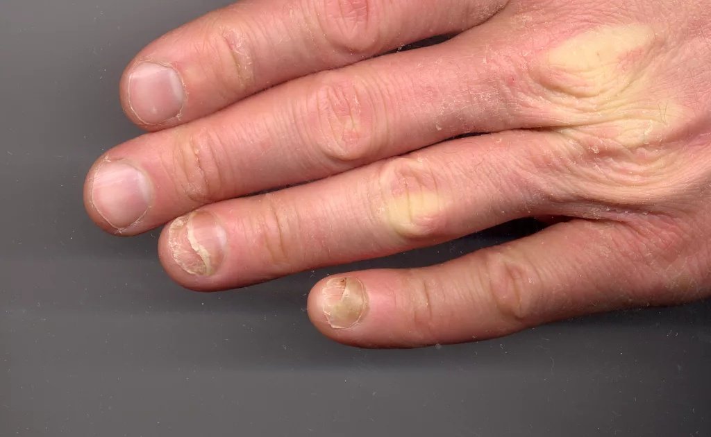
Onycholysis on CopperKettle’s left hand. The ring and little fingers are affected. By CopperKettle – Own work, CC BY-SA 3.0, https://commons.wikimedia.org/w/index.php?curid=21190610
Primary Symptoms
Nail Separation: The peculiarity is the distance between the nail plate and the nail bed, which usually begins at the free edge and advances in the direction of the proximal area.
Color Changes: The detached part of the nail changes its normal pink color to white or opaque. It can also be yellow or yellowish brown i some cases. In case of infections, it becomes green, and if there is bleeding beneath the nail or a fungal infection occurs, then the color will be dark gray.
Irregular Border: The border between the attached and detached portions may be:
- Linear, smooth.
- Curved or wavy
- Jagged with spikes or projections (fungal infection)
Associated Symptoms
1. Discoloration and Thickening of the whole nail may occur in addition to the white color of the affected area (subungual hyperkeratosis).
2. Secondary Infections: The isolated zone will give a home to the growth of bacteria or fungi, which may result in:
- Foul odor
- Increased discoloration
- Grounding up of the adjacent tissue.
- Pus formation
3. Nail Pitting
4. Lengthwise or crosswise lines
5. Pain or Discomfort: While onycholysis itself is typically painless, discomfort can occur from:
Onycholysis Diagnosis
History and clinical examination help to make most of the diagnosis. During your check-up, the doctor will visually inspect your nails and the surrounding skin, noting how they have separated and any other changes, like thickening or discoloration. They will also enquire about your symptoms, in case there are any recent traumas, about your daily routine, and general medical history, to aid in diagnosing the cause.
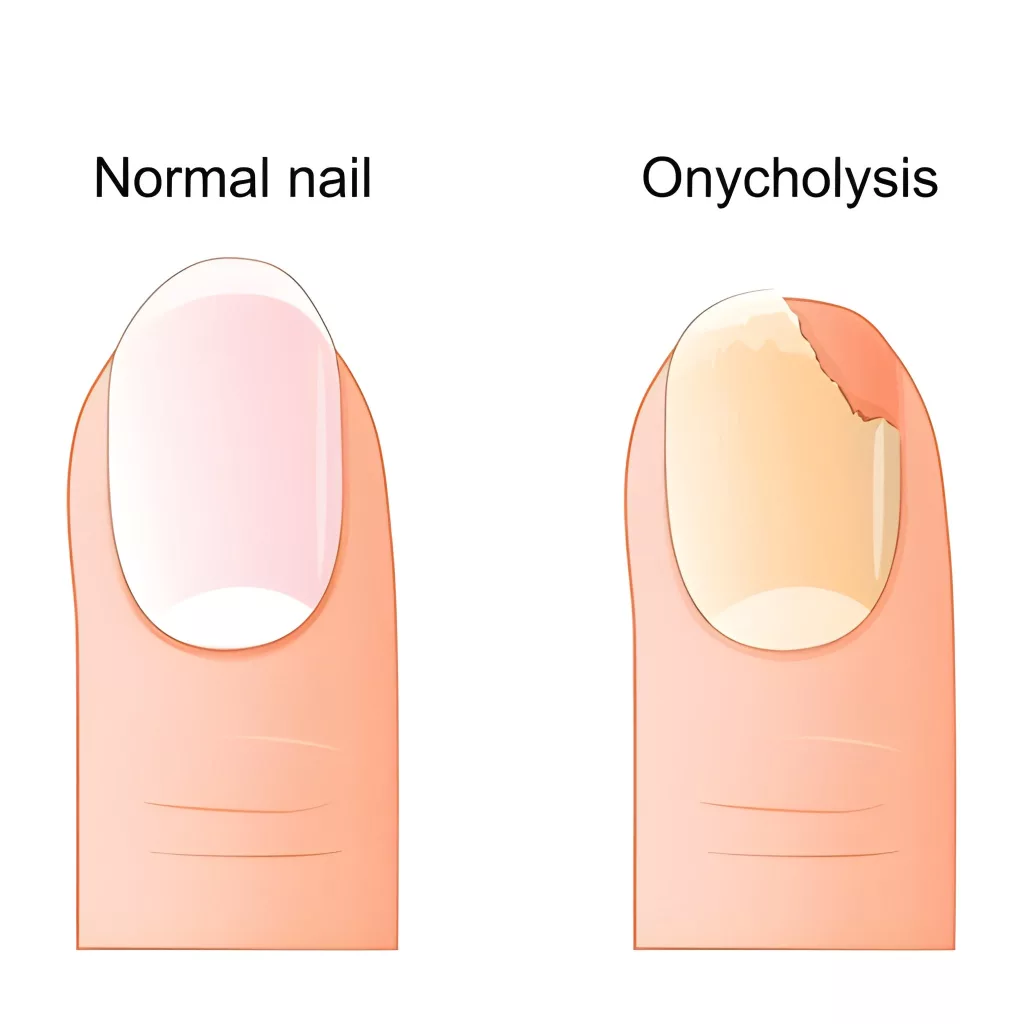
Onycholysis. Comparison and difference between a healthy fingernail and the detachment of the nail plate from the nail bed.
Diagnostic Tests
Mycology (Fungal Culture):
In this culture, you’ll submit Nail clippings/ nail scrapings to a lab to determine the fungus. The test may require weeks to get the results.
KOH (Potassium Hydroxide) Test:
It is a fast office test that involves the nails, which are dissolved in KOH solution, and the fungal elements are visible under a microscope.
PAS (Periodic Acid-Schiff) Stain:
It is a type of histological stain that is very accurate in detecting fungal elements in nail tissue.
Bacterial Culture:
Samples may be cultured in case of bacterial infection in order to determine the organism.
Blood Tests:
- Thyroid function (TSH, T3, T4)
- Iron and complete blood count
- Vitamin deficiencies
- Markers of systemic disease
Nail Biopsy:
It can be done in case:
- The diagnosis is unclear
- A tumor is suspected
Dermoscopy:
Doctors do this with an instrument that enables a better magnified view of the nail that can help tell the difference between the various causes of onycholysis.
Differential Diagnosis
It’s important to distinguish onycholysis from similar conditions:
| Condition | Someof the Major Distinguishing Characteristics |
|---|---|
| Onychomadesis | Separation starts at the proximal nail (near the cuticle) rather than the distal edge; complete nail shedding |
| Subungual Hematoma | Dark red or black discoloration from blood under the nail; history of acute trauma |
| Nail Psoriasis | Pitting, patches of salmon, oil drop sign; other psoriatic manifestations. |
| Nail Trauma | Clear history of trauma; local to the site of trauma |
Onychomycosis vs. Onycholysis: How to Tell the Difference?
These two terms can confuse you; however, they mean two different things:
Key Distinguishing Features
| Feature | Onycholysis (non-fungal) | Onychomycosis |
|---|---|---|
| Edge of separation | Usually smooth, linear border | Serrated edge having spikes facing the cuticle |
| Nail thickness | Typically normal or slightly thin | Severely thickened, with subungual hyperkeratosis |
| Progression | May stabilize when the cause is taken off | Progressive worsening without treatment |
| Distribution | It can affect a single nail | Often affects multiple nails, especially toenails |
| Associated findings | Depends on cause (e.g., pitting in psoriasis) | Crumbling, yellowish debris, rough surface |
| Diagnostic confirmation | Clinical examination | Requires fungal culture or KOH test |
Onycholysis Treatment
Regrettably, the detached fragment of the nail cannot be reattached. Treatment is done to see that new growth is attached to the nail bed.
General management and Home remedies.
Whatever the reason, the measures below are universally practical:
- Keep Nails Short: Cut the separated part of your nail with care, using a clean clipper. Always trim straight to prevent nail growth.
- Keep Clean and Dry: keep the affected area dry and clean. Humidity encourages bacterial and fungal infections in the gap between the nail.
- Avoid Trauma: Prevent further damage to nails. Be careful in everyday life. It is a long process, which on average takes 4-6 months for fingernails and 12-18 months for toenails to grow completely.
- Reduce Chemical Exposure: Reduce/ avoid coming into contact with harsh chemicals, nail products, and irritants. The nail can be healed by changing to hypoallergenic alternatives and using moisturizers. In case of inflammation, the doctor will suggest a topical steroid.
- Wear Protective Gloves: Always use vinyl-lined gloves during wet work. Do not wear gloves for long durations since they retain moisture.
- Avoid Nail Products: The patient should stop using nail polish, artificial nails, and nail enhancements till the condition clears.
- Hygiene Practice: Wash hands on a regular basis, but do not expose water to too much.
Cause-Specific Treatments
The type of treatment that you will have is dependent on the cause of your nail separation.
- Antifungal medication is necessary in case of a fungal infection. In the mild cases, your doctor will treat the nail with a daily prescription lacquer or solution applied to the nail. In case of more common infections, oral drugs are more efficient; use them for a week or a month. Combining oral medication, topical treatment, and periodic professional trimming of the infected nail is the most successful technique.
- In the case of Bacterial Infections, the treatment aims at eradicating the bacteria. This can be in the form of applying a topical antibiotic cream or more severe cases which require oral antibiotics. It is essential to ensure that to keep the nail dry to heal. Treatment of one of the types that makes it appear green (Pseudomonas) is special antibiotic drops.
- The management of Psoriatic Onycholysis focuses on the management of the inflammation in the skin. Treatment options are to apply very strong corticosteroid creams to the nail fold, steroid injections directly into the nail area, or systemic drugs or light therapy in the worst-case scenario.
- For Medication-Induced Onycholysis, you should consult the doctor who prescribed it. They can probably change your dose or place you on another. When you have to continue the medication, use protective measures to control the symptoms.
- In the case of Systemic Disease-Related Onycholysis, the nail separation is an indicator of a more serious health problem, like a thyroid disease or nutritional deficiency. The main aim is to treat and diagnose the underlying medical condition. When you become more generally healthy, your nails will tend to become so.
The Prevention of Secondary Infection.
Since the space between the separated nail may trap water, it is a place where bacteria and fungi multiply. To prevent this, you can use daily antimicrobial soaks, such as a diluted vinegar solution, to keep the area clean.
Conclusion
Onycholysis is a common phenomenon having numerous possible causes ranging from simple injuries to complicated systemic infections. Although you cannot replace the fallen nail, with good diagnosis and treatment of the cause, a new nail can grow in good health and attach to the nail bed.
The most important thing in effective management is to find out the underlying cause, which could be recurring trauma or fungal infection, psoriasis, exposure to chemicals or medicines, or an underlying medical condition.
Nails take time to heal; in fact, it takes 4-6 months before the fingernails and 12-18 months before the toenails are fully healed. Good nail health, taking the care prescribed by your medical practitioner, and taking precautionary measures are key requirements at this time to ensure the best results.
References
[1] de Berker DAR. Disorders of Nails. In: Griffiths C, Barker J, Bleiker T, Chalmers R, Creamer D, editors.Rook’s Textbook of Dermatology.9th ed. Wiley-Blackwell; 2016. p. 65.1–65.75.
[2] Rapini RP. Nail anatomy and disorders. In:Dermatology: 2-Volume Set.Mosby; 2007. p. 1071–1075.
[3] Viecelli JD. Onycholysis.JAMA Dermatol.2014;150(12):1353.
[4] Navarro‐Triviño FJ, Sánchez‐Carazo JL, Monteagudo B. Patterns and classification of onycholysis.J Cosmet Dermatol.2021;20(3):737-745.
[5] Gupta AK, Paquet M. Onychomycosis and Onycholysis: Epidemiology, Diagnosis, and Management.Clin Dermatol.2013;31(5):544-554.
[6] Jadhav VM. Nail-pitting and onycholysis.Indian J Dermatol Venereol Leprol.2009;75(6):581-584.
[7] Tosti A, Baran R. Nail Disorders: Common Signs and Symptoms.Clin Dermatol.2013;31(5):531-533.
[8] Westerberg DP, Voyack MJ. Onychomycosis: Current Trends in Diagnosis and Treatment.Am Fam Physician.2013;88(11):762-770.
[9] English JC, Zirwas MJ. Nail biopsy and diagnosis of nail disorders.Am J Clin Dermatol.2006;7(5):297-305.
[10] Piraccini BM, Alessandrini A. Onychomycosis: A Review.J Fungi (Basel).2015;1(1):30-43.
[11] Baran R, Dawber RP. Physical and chemical management of nail disorders.Clin Dermatol.2013;31(5):553-559
[12] Baran R, Haneke E.An Atlas of Diseases of the Nail.3rd ed. London: Taylor & Francis; 2012. p. 45-49.
[13] Berker DAR. Successful management of chronic onycholysis with avulsion and topical therapy.Clin Exp Dermatol.2009;34(5):611-613.