

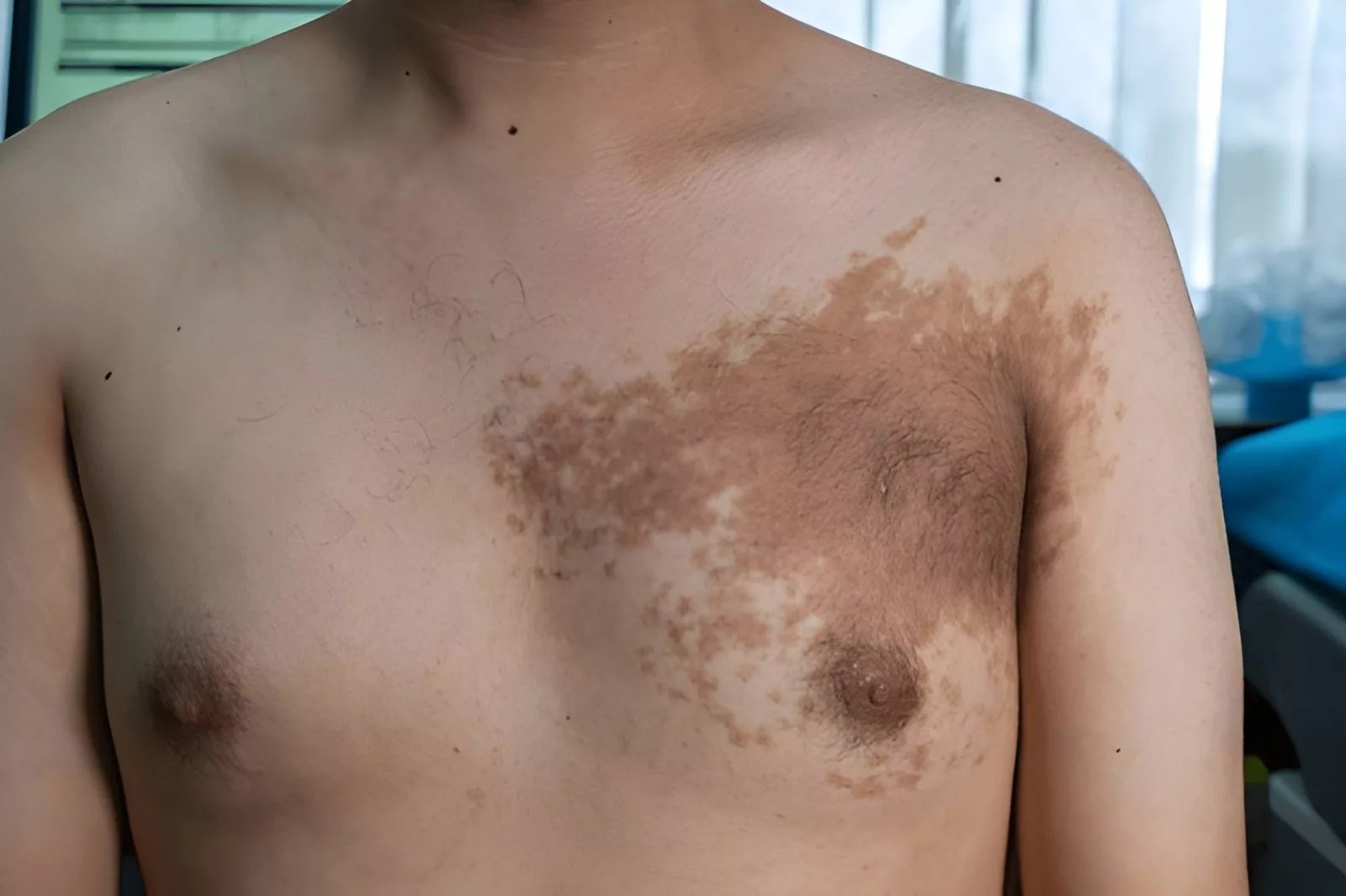
Becker’s nevus If you notice a large, irregular brown patch on your shoulder or chest, it may be a Becker’s nevus. Hair may begin growing over it during adolescence. It is usually asymptomatic, persists long-term, and does not disappear spontaneously. Becker’s nevus is a benign cutaneous hamartoma that occurs in approximately one out of every 200 men.
Though quite widespread, the nevus of Becker is not well known outside dermatology circles. A large number of individuals pass through years without adequate diagnosis. Other people are too concerned about cancer. And in some, more so with females, a late diagnosis may cause complications that could otherwise be avoided by early intervention.
What Is Becker’s Nevus?: Becker’s nevus
Becker’s nevus (or Becker melanosis, Becker pigmentary hamartoma, or pigmented hairy epidermal nevus) is a benign skin growth. It is classified as a benign skin condition that mainly affects men, which usually presents itself as an atypical hyperpigmented patch on the chest or upper arm, which slowly grows larger, and in most cases, it is covered by hair.
The condition belongs to a group of lesions known as epidermal hamartomas, which are abnormal but non-malignant overgrowths of normal skin tissue. It occurs as a hyperpigmented, occasionally hypertrichotic plaque or patch on the chest and shoulder, and it falls under the category of benign cutaneous hamartoma.
The lesion was originally described by American dermatologist Samuel William Becker in 1948, and hence the name. There is still a relative lack of medical awareness of the condition, especially in relation to other skin disorders, due to its low occurrence rates and the fact that it is not a serious health problem by itself.
How Common Is It?
It is more prevalent in men and has a male-to-female ratio of approximately 5:1, and Becker’s melanosis has a prevalence of about 0.5 percent in men. A study of close to 20,000 males in France established that the prevalence was approximately 0.52% and thus more frequent than many rare skin disorders, but much less frequent than normal moles.
What Does Becker’s Nevus Look Like?
Not all brown patches are the Becker nevus. Knowing about it will be useful in differentiating among other skin disorders, some of which are also urgent. Being aware of the particular characteristics will allow separating it from other dermal diseases, including those that require immediate care.
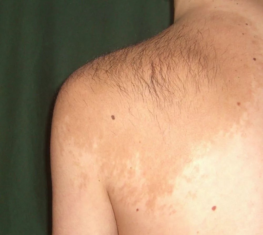
Irregular, light-brown patch typical of Becker’s nevus on the upper back. Image credit:Siller– Own workCC BY-SA 3.0
Key Physical Features
A typical Becker nevus can be presented with:
- A tan to dark brown, flat or raised patch with irregular and splashed edges.
- A lesion size ranging from a few centimeters to more than 20 cm in diameter.
- Most of the time, located in the upper chest, shoulder, back, or upper arm.
- The patch is covered by coarse, dark hair (hypertrichosis), particularly after puberty.
- Intermittent acne-like spots in and around the affected region.
In short, BN appears as a unilateral, isolated, sharply defined tan to brown hyperpigmented patch or slightly raised plaque, frequently hypertrichotic, with irregular edges, and may be located in abnormal locations, such as the face or lower extremity.
When Does It Appear?
Time is one of the most indicative hints. The lesion typically occurs in the puberty stage, though in rare cases, it has been reported at birth or early childhood, and also familial. The lesion was found at a younger age (under 10 years) in about half of the cases that were studied. In the other half, it developed between ages 10 and 20.
It is commonly believed that sun exposure causes or aggravates it, but studies indicate that this can only be a cause in approximately a quarter of the cases.
What Causes Becker’s Nevus?
The actual reason is yet to be determined; however, there are two primary reasons, which are androgen sensitivity and genetic mutations.
The Androgen Connection
The observation that Becker is predominant in males, and commonly around puberty, and that it usually leads to hair growth has led researchers for decades to androgens (male sex hormones). The androgen involvement has been observed through male preponderance, peripubertal development, hypertrichosis, the presence of acneform lesions on the patches, and the presence of more androgen receptors in the involved skin.
In essence, it is observed that the skin cells in the area affected are more sensitive to androgens as compared to normal skin. At puberty, the excess production of testosterone triggers the hyperirritable state of this part of the body, which results in increased pigment, increased hair follicles, and increased skin tissue.
Genetic Mutations
More recently, genetic research has identified a molecular cause. Recurrent mutations in the actin-beta (ACTB) gene have been demonstrated within arrector pili muscles. The ACTB gene encodes beta-actin, an important structural protein involved in cell growth, migration, and cellular organization. These mutations may activate pathways such as Hedgehog signaling, influencing hair follicles and smooth muscle proliferation within the lesion.
These mutations are postzygotic, or in other words, they develop after fertilization or at an early stage of cell division. They are not inherited from parents in most cases. This explains why the condition usually appears on only one side of the body.
Diagnosing Becker’s Nevus: Clinical and Microscopic Methods
It is mostly a clinical diagnosis, which is to say that a dermatologist may often diagnose it by examining the lesion along with the history of the patient. Nevertheless, tools such as dermoscopy and biopsy can provide some certainty.
Dermoscopy
A Becker nevus has a characteristic pattern under a dermoscope. Pigment network, perifollicular and focal hypopigmentation, hypertrophic hair follicles, skin furrow, and vessels are the main dermoscopic features. The pattern assists in differentiating it from other brown lesions, such as the cafe-au-lait macules, congenital melanocytic nevi, and nevus spilus.
Histopathology
In cases where a skin biopsy is required, the microscopic results are very specific. Histopathological changes of Becker nevi are epidermal acanthosis, normal elongation and fusion of rete ridges, hyperpigmentation of the basal layer, hyperkeratosis, and dermal smooth muscle hyperplasia.
One encouraging observation: malignant change is not seen in the nevus of Becker. This is a stark contrast to such conditions as dysplastic nevi or melanoma, where cancer risk is a genuine issue.
Differential Diagnosis
| Condition | Onset | Hair Growth | Location | Malignant Potential |
|---|---|---|---|---|
| Becker’s Nevus | Childhood/Puberty | Common | Trunk, shoulder | None |
| Congenital Melanocytic Nevus | Birth | Sometimes | Anywhere | Low to moderate |
| Café-au-Lait macule | Birth/Early childhood | Rare | Anywhere | None (unless linked to NF1) |
| Nevus Spilus | Birth/Childhood | Rare | Trunk, limbs | Very low |
| Smooth Muscle Hamartoma | Birth/Infancy | Common | Trunk | None |
Becker’s Nevus in a Female: Why Is It Different?
The majority of the literature on Becker nevus is written about males, yet women can and do have this condition. It is clinically significant to recognize it in a female for another reason: its association with breast development.
The belief that it is more frequent in males has recently been shown to be partially misleading, and the misconception is thought to be due to the lesions being less visible in females because of their androgen dependence. The lesion is less pigmented and less hairy in females and is therefore easier to ignore.
When the lesion develops on the chest or upper trunk in females, clinicians should evaluate for underdevelopment of the breast on the same side. In such situations, the condition may have functional and developmental implications rather than being purely cosmetic.
Becker’s Nevus Syndrome
Becker’s nevus syndrome is a separate but related diagnosis. It was formally defined in 1997 by Happle and Koopman as the association of a Becker’s nevus with ipsilateral breast hypoplasia or, more rarely, skeletal, cutaneous, or muscle deformities.
What Characterizes Becker’s Nevus Syndrome?
Other signs and symptoms that may accompany the nevus are ipsilateral breast hypoplasia, skeletal anomalies, including hypoplasia of the shoulder girdle, scoliosis, fused ribs, and ipsilateral shortness of the arm, among other features.
Other associations reported are:
- Supernumerary nipples
- Absence or underdevelopment of the pectoralis major muscle
- Spina bifida
- Segmental odontomaxillary dysplasia (jaw andtooth abnormalities)
- Limb length discrepancies
A systematic review of 84 cases of Becker’s nevus syndrome reported ipsilateral breast hypoplasia as the most commonly described malformation, observed in 56% of cases.
The Importance of Early Detection
In female patients with breast hypoplasia, early detection of Becker nevus syndrome is essential, which should be followed by proper medical care. Spironolactone may even prevent breast hypoplasia when used early enough.
That is why any girl or young woman who presents with a chest-area Becker’s nevus must be screened to identify Becker’s nevus syndrome. One dermatology visit at the right moment can alter the treatment process completely.
Becker’s Nevus Treatment
The nevus of Becker has no cure in the classical sense. It is benign and, therefore, it is usually not medically necessary to treat it. But for many patients, particularly teenagers and young adults, the cosmetic impact on self-confidence is significant and real.
Laser Therapy
The most commonly researched treatment for Becker nevus is laser therapy, with mixed outcomes.
The results of laser removal are debatable, with Er: YAG 2940 nm being regarded as more effective than Q-switched Nd: YAG 1064 nm in pigmentation, but Nd: YAG can be more effective on darker skin. Alternatives are low-fluence Q-switched Nd: YAG lasers and long-pulsed Nd: YAG lasers, which can treat pigment and hair growth during the same treatment course.
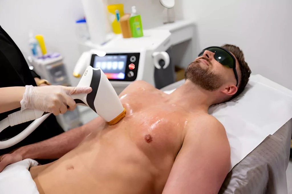
Laser therapy is being used to reduce pigmentation in Becker’s nevus
A key drawback: recurrence is common. Q-switched lasers have shown acceptable efficacy in treating hyperpigmentation in Becker’s nevus but are followed by a considerable chance of recurrence.
Topical Medications
Topical flutamide has been used, leading to a satisfactory reduction of hyperpigmentation. This anti-androgen cream directly targets the hormonal mechanism driving pigmentation in the lesion.
Hair Removal Options
For patients bothered primarily by the hair growth:
- Shaving and trimming provide short-term relief
- Laser hair removal and electrolysis offer longer-lasting results
- Diode lasers are also effective, specifically for the hypertrichosis component
Dermabrasion
Sandpaper dermabrasion is a relatively inexpensive procedure to remove skin lesions and may be employed as an effective and practical approach to the treatment of Becker’s nevus, especially when laser treatment has failed.
Specific Treatment Of Becker’s Nevus Syndrome
When the condition extends beyond the skin to the Becker’s nevus syndrome spectrum, treatment becomes multidisciplinary.
Spironolactone is used in female patients with breast hypoplasia with remarkable breast development, while breast lipofilling is a good alternative, leading to improvement in breast volume and hyperpigmentation. There are more severe cases that necessitate breast reconstruction surgery.
In case of skeletal problems such as scoliosis, orthopedic consultation is essential. The management must be individualized based on the symptoms and severity of the patient.
Follow-up: Practical Considerations
Individuals with Becker nevus complain of self-consciousness, especially in adolescence when the lesion is most noticeable. Others do not swim, wear sleeveless garments, or do anything that exposes the skin.
A few practical points worth keeping in mind:
- Sun protection matters. Ultraviolet exposure may darken the lesion further. You can prevent worsening pigmentation by using a broad-spectrum sunscreen on the affected area daily.
- It will not transform into cancer. This is the one most reassuring fact to most patients.
- Reassurance is often the first treatment. In simple cases without related abnormalities, it can be of great assistance to describe what the lesion is and what it is not.
- Follow-up is still wise. Although there is no specific treatment, a dermatologist must investigate the patient with Becker nevus syndrome, especially in women.
Conclusion:
Becker’s nevus is not dangerous in itself. It will not develop into melanoma. It is not contagious, and it does not produce any physical damage. However, you should take it seriously medically, particularly when it is present in a female patient on or around the chest, since it sometimes is the superficial manifestation of an underlying syndrome with structural implications.
See a dermatologist in case you or someone you know has a large, irregular, hairy brown patch that developed during adolescence and has not disappeared. Getting a proper diagnosis is the first step, whether the goal is reassurance, cosmetic improvement, or ruling out Becker’s nevus syndrome. It is completely possible to manage this condition with the help of the right specialist and the right information.
References
[1] Patil A, Kumar A, Dalave K, Deora MS, Pol D. Clinicohistopathological and dermoscopic features of Becker nevus: An analytical observational study from southwestern India.Pigment Int.2023;10:24–29.
[2] Glinick SE, Alper JC, Bogaars H, Brown JA. Becker’s melanosis: associated abnormalities.J Am Acad Dermatol.1983;9(4):509–514. doi:10.1016/S0190-9622(83)70161-1
[3] Tymen R, Forestier JF, Boutet B, Colomb D. Late Becker’s nevus. One hundred cases.Ann Dermatol Venereol.1981;108(1):41–46.
[4] Rasi A, BerenjiArdestani H, Tabaie SM. Hypertrichosis is not so prevalent in Becker’s nevus: analysis of 47 cases.ISRN Dermatol.2014;2014:953747. doi:10.1155/2014/953747
[5] Person JR, Longcope C. Becker’s nevus: an androgen-mediated hyperplasia with increased androgen receptors.J Am Acad Dermatol.1984;10(2 Pt 1):235–238. doi:10.1016/s0190-9622(84)70029-6
[6] Cai ED, Sun BK, Chiang A, Rogers A, Bernet L, Cheng B, et al. Postzygotic mutations in beta-actin are associated with Becker’s nevus and Becker’s nevus syndrome.J Invest Dermatol.2017;137(8):1795–1798. doi:10.1016/j.jid.2017.03.017
[7] Patil A, Kumar A, Dalave K, Deora MS, Pol D. Clinicohistopathological and dermoscopic features of Becker nevus: An analytical observational study from southwestern India.Pigment Int.2023;10:24–29.
[8] Glinick SE, Alper JC, Bogaars H, Brown JA. Becker’s melanosis: associated abnormalities.J Am Acad Dermatol.1983;9(4):509–514.
[9] Grande Sarpa H, et al. Androgen receptor expression patterns in Becker’s nevi: an immunohistochemical study.J Am Acad Dermatol.2008;59(5):834–838.
[10] Happle R, Koopman RJ. Becker nevus syndrome.Am J Med Genet.1997;68(3):357–361. doi:10.1002/(SICI)1096-8628(19970131)68:3<357::AID-AJMG20>3.0.CO;2-R
[11] Scheuer EM, et al. Becker nevus syndrome and ipsilateral breast hypoplasia: a systematic literature review.Eur J Plast Surg.2017;40:537–545. doi:10.1007/s00238-017-1327-6
[12] Jung JH, Kim YC, Park HJ, Cinn YW. Becker’s nevus with ipsilateral breast hypoplasia: improvement with spironolactone.J Dermatol.2003;30(2):154–156. doi:10.1111/j.1346-8138.2003.tb00365.x
[13] Al-Bakaa MK, Al-Dhalimi MA, Dube P, Khalaf FK. Evaluating the roles of different types of laser therapy in Becker’s nevus treatment.J Clin Med.2022;11(14):4230. doi:10.3390/jcm11144230
[14] Taheri A, Mansoori P, Sandoval LF, Feldman SR. Treatment of Becker nevus with topical flutamide.J Am Acad Dermatol.2013;69(3):e147–e148. doi:10.1016/j.jaad.2013.03.026
[15] Ebrahimi M, Salehi Fadardi J. Successful treatment of Becker’s nevus with dermabrasion by sandpaper: a case report.Dermatol Reports.2021;13(2):9006. doi:10.4081/dr.2021.9006(or the equivalent PMC version: PMC8405417)
[16] Ho Quoc C, Fakiha M, Meruta A, et al. Breast lipofilling: a new treatment of Becker nevus syndrome.Ann Chir Plast Esthet.2015;60(4):336–339. doi:10.1016/j.anplas.2014.10.006