
 is a life-threatening manifestation of systemic sarcoidosis. It can be defined as: “a...)
Cardiac Sarcoidosis Sarcoidosis Cardiac Sarcoidosis (CS) is a life-threatening manifestation of systemic sarcoidosis. It can be defined as: “a rare inflammatory condition characterized by the formation of granulomas, or clusters of immune cells, in the heart tissue; leading to disruptions in the normal functioning of the heart, potentially causing arrhythmias, heart block, heart failure, or even sudden cardiac death.”[1] There is currently no definitive cure for sarcoidosis; therefore, management focuses on controlling inflammation, treating symptoms, and preventing complications such as heart failure with the help of different medications and cardiac devices.
What is a Cardiac Sarcoid? : Cardiac Sarcoidosis Sarcoidosis
A sarcoid, the lesion characteristic of sarcoidosis, is essentially a “non-caseating granuloma“. Granuloma is defined in pathology as a focal, compact collection of chronic inflammatory cells, primarily composed of activated macrophages (epithelioid cells), surrounded by a collar of lymphocytes and plasma cells.[2] Noncaesating refers to the absence of dead tissue in the middle of the granuloma. When these granulomatous lesions involve the myocardium, conduction system, or other cardiac structures, the condition is termed cardiac sarcoidosis.
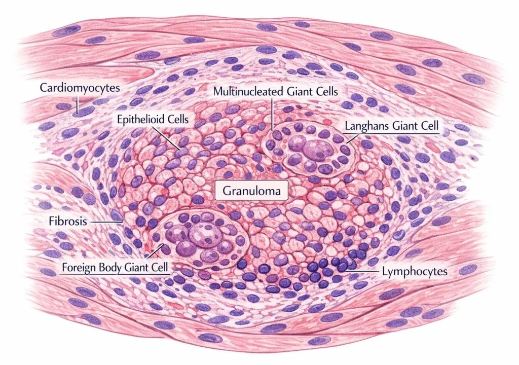
A histopathology diagram showing a non-caseating granuloma in myocardial tissue. The granuloma contains epithelioid macrophages and multinucleated giant cells surrounded by lymphocytes. Heart cells are intact, and early fibrosis surrounds the granuloma. No central necrosis is present, typical of cardiac sarcoidosis.
Who is at risk?
Although it can happen at any age, middle-aged individuals (30-50 years) are more commonly affected. If you suffer from systemic sarcoidosis, you have a risk of developing sarcoidosis in your heart as well. Clinically apparent cardiac involvement occurs in about 5% of patients with systemic sarcoidosis, although autopsy studies suggest cardiac involvement in up to 25–30% of cases.[3] Females have a slightly higher risk than men. CS has a racial predisposition towards African American (Black), Northern European, and Japanese people. In addition, family history may increase the risk of developing CS.[4]
What Causes Cardiac Sarcoidosis?
The exact cause of Cardiac Sarcoidosis is unknown. However, experts believe that inflammation is the key factor that drives sarcoid formation. Various reasons cause the inflammation:
- Genetic Factors: Sarcoidosis is considered an immune-mediated inflammatory disease rather than a classic autoimmune disease, and genetics play a crucial role in its manifestation. Gene studies have not yielded conclusive results; however, the human leukocyte antigen (HLA) group of genes, which are expressed in granuloma formation, is suspected.[5]
- Environmental Factors: Excessive exposure to mold, fungus, silicadust, insecticides, or any similar antigen that can trigger our immune system.[6]
- Occupational exposure to such toxins plays an important role as well, such as with farmers, construction workers, and firefighters.
- Infectious Agents: Bacteria such as Cutibacterium acnes (formerly Propionibacterium acnes) and Mycobacterium species are associated with sarcoidosis, although a direct causal relationship has not been definitively proven.
- Immune Dysfunction: Overactivation and overexpression of the immune system in response to these exogenous factors can lead to immune cells accumulating and forming granulomas, the characteristic feature of sarcoidosis.
Pathophysiology – How does it happen?
When the exogenous toxin stimulates the immune system, it results in overexpression and drives the excessive release of immune cells. The first in line white blood cells are the macrophages that take up the antigen and present it to the T-helper cell. Even though these lymphocytes (CD4+ T-helper cells) don’t destroy the pathogen directly, they instead release a cascade of inflammatory mediators, including the cytokines, interleukins (IL), and tumor necrosis factors (TNF). Consequently, these inflammatory mediators stimulated other immune cells, such as B-lymphocytes or cytotoxic T-cells, to participate in the immune reaction.[7]
The accumulated lymphocytes, macrophages, and other immune cells form a “granuloma,” which is characteristic of a sarcoid lesion. In normal conditions, the inflammatory response is resolved with the elimination of the foreign particle. However, in sarcoidosis, the response is abnormal, and the immune cells are hyperactive and dysregulated, consequently leading to more and more recruitment of activated cells into the lesions. This subsequently results in a cyclic cascade, leading to the inflammation persisting for a long time.
These granulomas then infiltrate into the myocardium, driving more inflammation and causing damage to the heart muscles and architecture. Consequently, prolonged inflammation, known as “Granulomatous myocarditis,” leads to fibrosis and scar tissue formation. The presence of scar tissue in the heart impairs its function and presents as a risk of heart failure and sudden cardiac death.
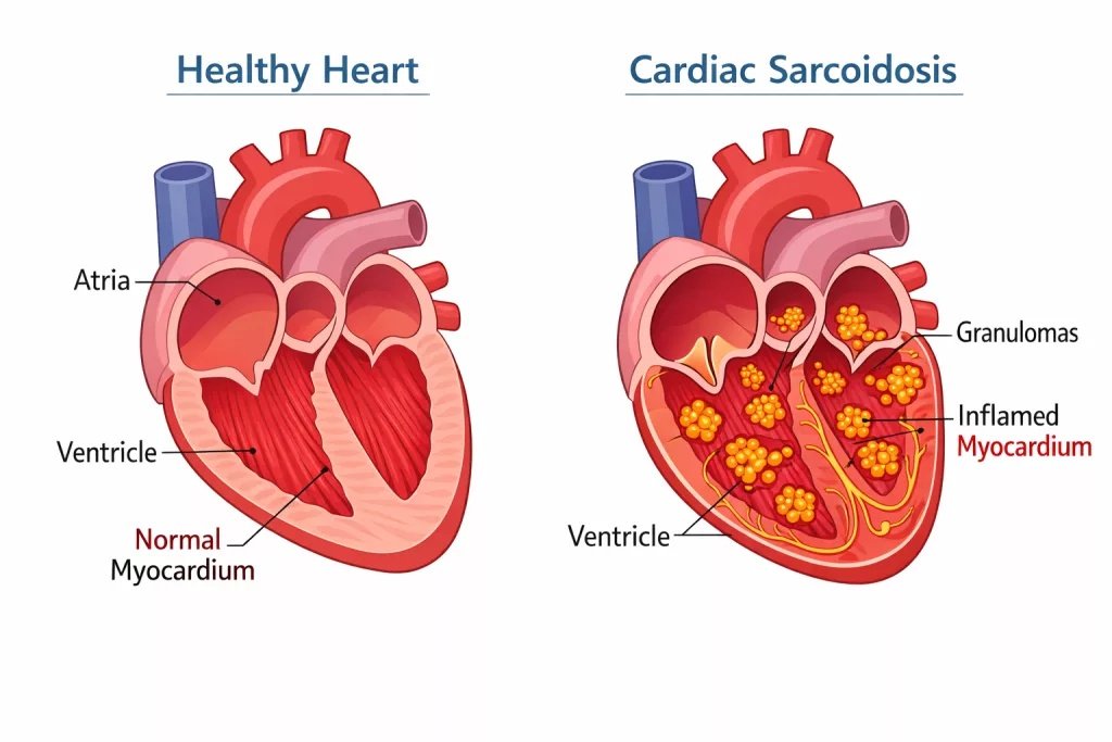
An illustration comparing normal myocardial structure versus cardiac involvement in sarcoidosis. The affected heart shows multiple granulomatous clusters within the myocardium, representing inflammatory infiltration characteristic of cardiac sarcoidosis.
Symptoms of Cardiac Sarcoidosis
It is important to know that you may not experience any symptoms until the disease has significantly progressed. Since the inflammation affects the conduction of the electrical heart, you may present to the clinic with:
- Arrhythmias: You may feel your heart beating at an abnormal pace, maybe too fast, too slow, or skipping beats. It can feel like a pounding, fluttering, shaking, or skipping sensation in the chest.
- Chest Pain: You may feel a dull discomfort in your chest, and the pain may radiate to your left arm or left shoulder.
- Fatigue and Dizziness: Having extreme fatigue and feeling very tired can be a symptom. A dysregulated heartbeat can lead to inadequate blood supply to the body, resulting in low energy levels.
- Shortness of breath: Experiencing unexplained breathlessness, even at a state of rest, can be a sign of heart damage.
- Fainting / Syncope: Low heart rates consequently lead to less oxygen supply to the brain, and you may faint.
- Swelling: Fluid retention and edema in the body can lead to swelling in the legs or abdomen.
Evaluation and Clinical Assessment
History Taking
The clinical presentation of a patient with sarcoidosis is vague and diverse, ranging from occasional palpitations to heart failure symptoms. Hence, a detailed history is important to differentiate CS from other cardiac diseases. One should ask about the symptoms mentioned above, including chest pain, palpitations, syncope, and general fatigue. Unexplained symptoms in young individuals are an indicator of investigating for CS.
Asking about the family history of hereditary heart disease can play a key role in making a diagnosis. The physician should also ask if the patient is already suffering from systemic sarcoidosis or if they have a blood relative who was diagnosed with sarcoidosis. A history of sarcoidosis in the patient himself or a family member raises suspicion for further testing.
Physical Examination
The physician will perform a physical exam to check for an irregular pulse, tachycardia (fast heartbeat), bradycardia (slow heartbeat), swelling in the feet or abdomen, or bulging of the jugular veins in the neck. Vital signs such as blood pressure and breathing rate will be recorded to assess your situation.
Initial Diagnostic Testing:
- ECG:[8] ECG is the initial screening test of CS. However, the results are not specific to CS. They vary depending on the area of granulomatous inflammation and the extent of progression of the disease. The most significant findings include conduction abnormalities, ventricular arrhythmias, and repolarization changes. These translate into atrioventricular (AV) blocks, QRS complex widening, bundle branch blocks, ventricular tachycardia, ST, and T-wave abnormalities.
- Echocardiogram:[9] It is also a first-line screening tool that uses sound waves to create live images of the heart. But it has low sensitivity in diagnosing CS. The echo may appear normal in mild stages of the disease, but it can still indicate abnormalities in the myocardium (e.g., thinning of the basal septum). Thinning of the basal septum indicated infiltration of granulomas into the myocardium. It is useful as a screening tool for patients suffering from systemic sarcoidosis.
Imaging Techniques
- MRI:[10] Cardiac MRI is an advanced and highly sensitive imaging technique used to detect CS. It shows heart wall thinning, the function of the ventricles, and the size of the heart chamber. The technique “Late Gadolinium Enhancement (LGE)” is used to identify scar tissue, which is characteristic of cardiac sarcoidosis. A gadolinium dye is injected via IV, and the MRI scan is done “late” after 10-20 minutes. After that time has passed, the dye has washed out from the normal tissues of the heart and only remains in the scarred tissue. Therefore, the scar tissue is imaged and identified, indicating the presence of a late stage of CS.
- FDG-PET Scan:[11] This is a nuclear imaging technique that involves injecting a radioactive tracer, fluorodeoxyglucose (FDG), into the bloodstream. It acts as a substitute molecule for glucose. Active inflammatory cells have a high metabolic rate and take up FDG. Therefore, the inflammatory cells and sarcoid granulomas present in the myocardium take up high levels of FDG and appear as “hot spots” on the scan.
Endomyocardial Biopsy
The endomyocardial biopsy is an invasive procedure, and it involves the physician taking a sample of your heart and investigating it. An MRI or a PET scan can be used to guide the catheter to the abnormal areas of the heart, increasing diagnostic accuracy. After that, the sample is examined under the microscope to identify the presence of noncaseating granuloma formation.[12]
Cardiac Sarcoidosis Diagnostic Criteria
The diagnosis of CS is done based on the criteria provided by the Heart Rhythm Society (HRS)[13]. This criterion requires a histologic diagnosis proving the presence of non-caseating granulomas in the myocardium, along with one or more of the clinical signs present:
- Immunosuppressant/corticosteroid-responsive cardiomyopathy or heart block
- Unexplained reduced Left Ventricular Ejection Fraction (LVEF) <40%
- Unexplained sustained ventricular tachycardia
- Patchy FDG uptake on a dedicated cardiac PET in a pattern consistent with CS
- LGE (Late Gadolinium Enhancement) on cardiac MRI in a pattern consistent with CS
- Positive ⁶⁷Ga (Gallium-67) uptake in a pattern consistent with cardiac sarcoidosis
- And other causes have been reasonably excluded
Progression of Cardiac Sarcoidosis
The stages of cardiac sarcoidosis based on the progression of the disease can be understood as follows:
| Pathobiology | FDG-PET Scan | MRI | Symptoms | |
|---|---|---|---|---|
| 1.Inflammatory Stage | Inflammatory cells infiltrate and accumulate. | Focal FDC uptake | T2 hypersensitivity | – Chest pain– New-onset AV block |
| 2.Granulomatous Stage: | Granuloma formation | Persistent but less intense | – Established (LGE)– Residual T2 edema | – Increased arrhythmia– Ventricular tachycardia |
| 3.Fibrous Stage: | Granuloma replaced by fibrous tissue and scarring | No significant uptake | Extensive LGE | – Chronic heart failure– Permanent AV block |
What is the Prognosis?
The prognosis of the disease depends primarily on its early detection. Early detection, when the inflammation has just started to form, can prevent fibrosis and scar tissue formation of the heart. With modern treatment, the overall prognosis has improved, although patients remain at risk for ventricular arrhythmias and progressive heart failure.
Cardiac Sarcoidosis Treatment
The treatment of Cardiac Sarcoidosis aims to reduce inflammation, suppress the hyperactive immune system, and treat the condition of the heart. If detected early, immune suppression and reducing inflammation can help to ‘reverse’ the disease. However, if the disease is advanced, scarring and fibrous tissue are not reversible, and management is focused on slowing disease progression and improving heart function.
Immunosuppressive Therapy:
- Corticosteroids are the first-line drugs that physicians use to combat and suppress inflammation. The drug “prednisone” is the standard medicine of choice for treating active inflammation.
- Steriod-Sparing drugs, such as methotrexate and azathioprine, are also immunosuppressants and are used to treat chronic inflammation. They are given in conjunction with corticosteroids to prevent high dosages of them, in return, reducing the adverse side effects of steroids.
- Other immunosuppressive medications that may be used in refractory disease include mycophenolate mofetil or tumor necrosis factor-alpha inhibitors such as infliximab.
Arrhythmia Management:
- A pacemaker is helpful in people with abnormal rhythms. It is implanted when the disease causes AV blocks or ventricular tachycardia.
- An implantable cardioverter-defibrillator (ICD)[14] is the go-to device for abnormalities caused by CS, since they can adapt to both slow and fast rhythms. The patient is at risk of developing sudden cardiac death, and the design of the ICD helps supplement that.
Heart Failure Management:
- Guideline-directed medical therapy (GDMT) uses drugs such as beta-blockers, angiotensin receptor blockers, and ACE inhibitors to improve the function of the heart.
- A left ventricular assist device (LVAD) is used to support the function of the left ventricle, the main chamber that delivers blood to the whole body.
- Heart Transplant: In severe forms of heart failure and excessive fibrosis of the myocardium, a heart transplant can be considered for the patient.
Combination Therapy:
Treating Cardiac Sarcoidosis is a combination of immunosuppressive drugs, reducing inflammation, along with placement of cardiac devices, and heart failure management. Regular monitoring via the FGD-PET scan, every 6 months, is important to keep a check on the progression of the disease in the long run.
Conclusion
Cardiac Sarcoidosis is a chronic inflammatory disease, characterised by the formation of non-caseating granulomas in the myocardium, consequently leading to impaired heart function. CS imitates other heart diseases and can be difficult to diagnose, but advanced imaging modalities and biopsy can provide a definitive diagnosis. The prognosis of the disease is dependent on early detection, supplemented with effective management. Treatment aims to reduce inflammation and also improve heart function via various medicines and cardiac devices.
References
[1] Cardiac Sarcoidosis [Internet]. Yale Medicine. 2026 [cited 2026 Feb 22].
[2] Williams O, Fatima S. Granuloma [Internet]. PubMed. Treasure Island (FL): StatPearls Publishing; 2022.
[3] Ribeiro Neto ML, Jellis CL, Culver DA, et al.Cardiac sarcoidosis.Clin Chest Med.2024;45(1):105-118.
[4] Hussain Haider Shah, Syeda Alishah Zehra, Shahrukh A, Waseem R, Hussain T, Muhammad Sheheryar Hussain, et al. Cardiac sarcoidosis: a comprehensive review of risk factors, pathogenesis, diagnosis, clinical manifestations, and treatment strategies. 2023 May 19;10.
[5] Spagnolo P, Maier LA. Genetics in sarcoidosis. Current Opinion in Pulmonary Medicine. 2021 Jun 28;27(5):423–9.
[6] Judson MA. Environmental Risk Factors for Sarcoidosis. Frontiers in Immunology. 2020 Jun 26;11.
[7] Vagts C, Ascoli C, Jacobson JR. Immunopathogenesis of Sarcoidosis. Seminars in respiratory and critical care medicine [Internet]. 2025 Jun;10.1055/a2716–5737.
[8] Willy K, Dechering DG, Reinke F, Bögeholz N, Frommeyer G, Eckardt L. The ECG in sarcoidosis – a marker of cardiac involvement? Current evidence and clinical implications. Journal of Cardiology. 2020 Sep
[9] Au K, Wong D, Newbigin K, Allen R, Scalia GM. Multimodality Imaging to Diagnose Isolated Cardiac Sarcoidosis and Determine Regional Inflammatory Activity Levels. CASE. 2017 Oct;1(5):171–5.
[10] Blankstein R, Waller AH. Evaluation of Known or Suspected Cardiac Sarcoidosis. Circulation: Cardiovascular Imaging. 2016 Mar;9(3).
[11] Hicham Skali, Schulman AR, Sharmila Dorbala. 18F-FDG PET/CT for the Assessment of Myocardial Sarcoidosis. Current cardiology reports [Internet]. 2013 Apr;15(4):352
[12] Ikeda U. Editorial: Diagnosis of cardiac sarcoidosis – What is the role of endomyocardial biopsy? Journal of Cardiology Cases. 2015 Sep;12(3):72–3.
[13] Cheng RK, Kittleson MM, Beavers CJ, Birnie DH, Blankstein R, Bravo PE, et al. Diagnosis and Management of Cardiac Sarcoidosis: A Scientific Statement From the American Heart Association. Circulation. 2024 Apr 18;149(21).
[14] ELREFAI M, MENEXI C, ROBERTS P. Device Therapy in Cardiac Sarcoidosis: Current Review, Challenges, and Future Prospects. Journal of Innovations in Cardiac Rhythm Management. 2024 Nov 1;15(11):6088–94.