
 containing pus or semi-solid material...)
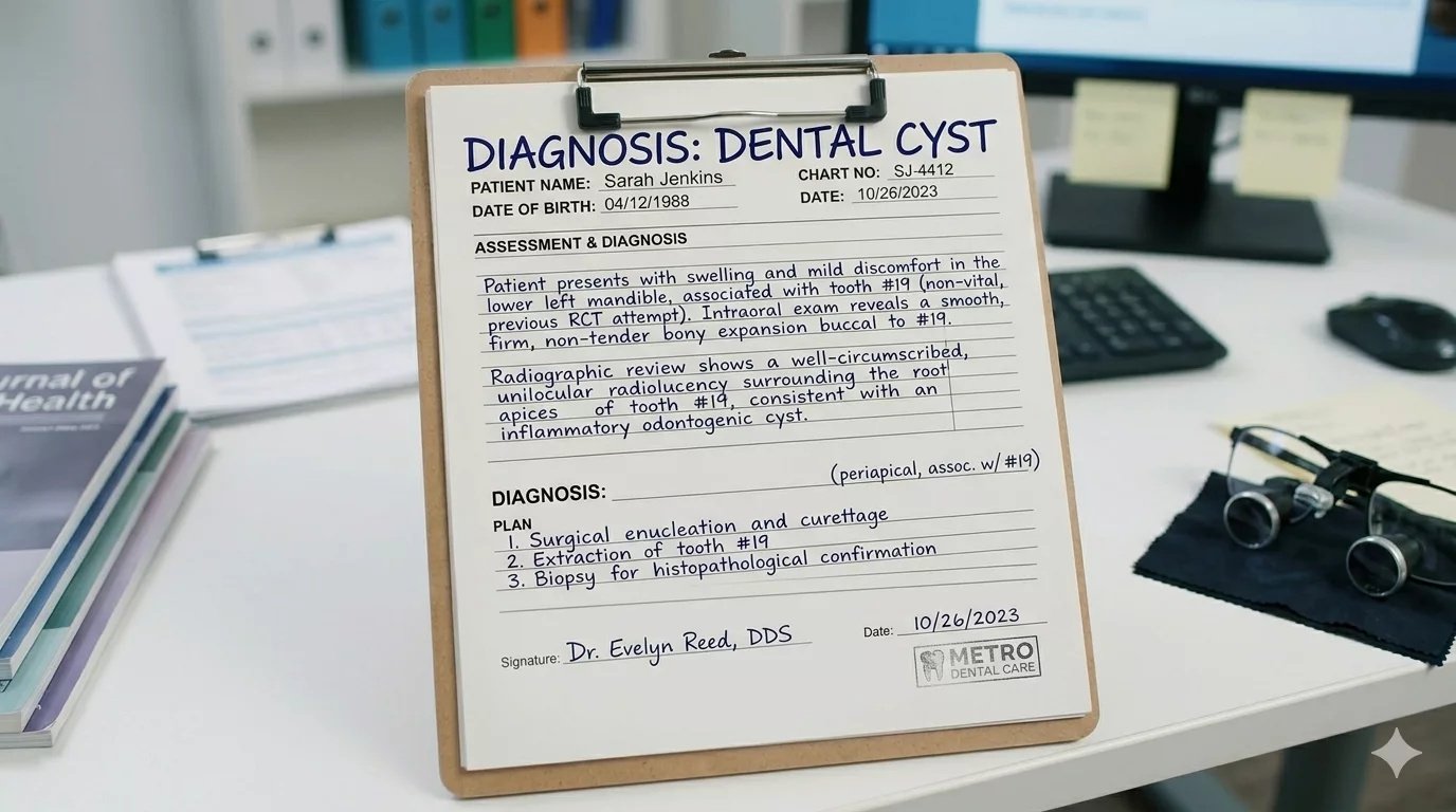
Dental Cysts Symptoms “Dental cyst” is an umbrella term that refers to any enclosed cavity (with a sac) containing pus or semi-solid material within the oral cavity. Different types of dental cysts exist. Most of these growths are benign, but their aggressive growth can cause a lot of trouble.
Cysts arising from tooth-forming tissues are called odontogenic cysts. Non-odontogenic cysts are rare as compared to odontogenic cysts. As per the latest reports, the global prevalence of odontogenic cysts is 13.8%.[1] Doctors treat the dental cysts according to their types. Some cysts just require regular monitoring, while others need surgical intervention.
What Is a Dental Cyst?: Dental Cysts Symptoms
A cyst is an abnormal cavity lined by epithelium (a type of body tissue that covers internal and external surfaces of your body). This epithelium-lined cavity can exist anywhere in the body, but when these anomalies form within the mouth, they are called dental cysts. Furthermore, if the epithelial lining of the cyst is derived from the tooth (odontogenic) epithelium, it is called an odontogenic cyst.
Types Of Dental Cysts
Based on the tissues (epithelium) forming these cysts, experts divide dental cysts into:
Non-Odontogenic Cysts
These types of cysts are rare and generally asymptomatic. As the name indicates, non-odontogenic cysts do not arise from dental tissues. Rather, they are developmental lesions that arise due to the trapping of embryonic tissue remnants in the oral region.[2] Common types include:
- Nasopalatine duct cyst
- Nasolabial cyst
- Palatal cysts of the neonate
- Oral lymphoepithelial cyst
- Epidermoid cyst
Odontogenic Cysts
These cysts are composed of the very structures that formed your tooth. Structures involved in their creation include:
- Remnants of dental lamina: A dental lamina is a thickened band of oral lining (epithelium) present in embryonic life that lays the foundation for tooth development.
- Enamel organ: It is an epithelial structure responsible for forming the outer hard layer of the tooth, i.e., enamel.
- Reduced enamel epithelium (REE): It refers to a thin layer of epithelium that remains over/around the tooth’s crown after completion of the enamel. REE plays an important role in tooth eruption.
Types Of Odontogenic Cysts
Based on their mode of development, experts divide odontogenic cysts into two categories: developmental and inflammatory.
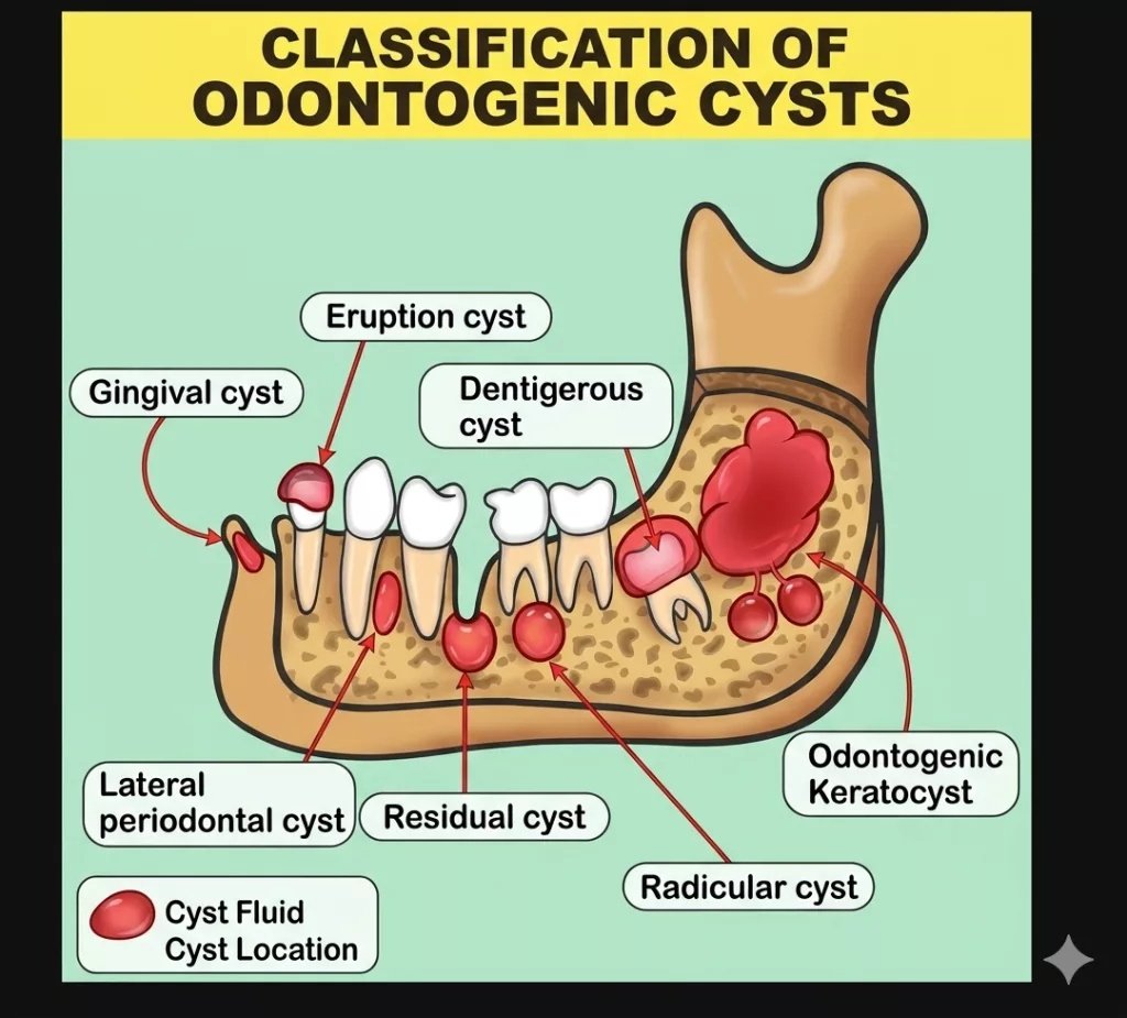
Different types of odontogenic cysts in their common anatomical locations
Developmental Cysts:
These are non-inflammatory cysts formed during tooth development. Developmental cysts grow slowly and are usually asymptomatic until they become large enough to interfere with neighboring structures. Common types include:
- Dentigerous cyst
- Eruption cyst
- Odontogenic keratocyst (OKC)
- Lateral periodontal cyst
- Gingival cyst of the newborn
- Gingival cyst of the adult
- Calcifying odontogenic cyst
Inflammatory Cysts:
This type of lesion contains fluid (pus) and inflammatory cells. Inflammatory odontogenic cysts arise due to infection (and consequent pulpal necrosis), trauma, or caries. Common types of inflammatory dental cysts include:
- Periapical (radicular) cyst
- Residual cyst
- Paradental cyst
Dental Cyst Causes, Symptoms, And Treatment
For a better understanding, we discuss the causes, symptoms, and treatment options of all cysts individually.
Nasopalatine Duct Cyst
Nasopalatine duct cyst is the most common type of non-odontogenic cyst that forms on the roof of your mouth. It accounts for 1% of maxillary cysts.[3]
Symptoms And Treatment
Most patients notice a swollen bump behind the upper front teeth. Due to its proximity to the nose, it can cause nasal obstruction. Mostly, these cysts are asymptomatic. However, it can be symptomatic, in which case, patients notice burning pain and pus discharge (if the cyst gets infected).
Researchers believe that it arises due to the retention of some part of the nasopalatine duct. The nasopalatine duct is a transient structure in the mouth and nose region that forms and disappears during embryonic life. In the majority of cases, an oral and maxillofacial surgeon removes the cyst surgically.
Nasolabial Cyst
A nasolabial cyst appears as a painless swelling in your smile line (nasolabial fold). The rare fluid-filled lesion elevates your nose/lip region. The exact cause of its development is unknown, but many believe that it develops from remnants of the nasolacrimal duct. The nasolacrimal duct is a hollow, tube-like structure that carries tears from the eye (lacrimal sac) into the nose.
Symptoms And Treatment
In general, the swelling is painless but can lead to nasal obstruction if infected. Doctors completely remove the entire cyst (lining and its contents) in a surgical process called enucleation. A complete endoscopic removal is an effective treatment modality with a rare chance of recurrence.[4]
Palatal cysts are more common in infants and resolve themselves. Oral lymphoepithelial cysts occur on the border of the tongue or the floor of the mouth and are managed by conservative strategies. Epidermoid cysts are slow-growing masses on the floor of the mouth and are treated with surgical enucleation (removal of the whole cyst).
Dentigerous Cyst
A dentigerous cyst is a fluid-filled cavity that forms over the crown of an unerupted permanent tooth. We most commonly see this type of cyst affecting impacted wisdom teeth; however, they can exist over any tooth. According to a detailed study conducted in 2021, the prevalence of dentigerous cyst was found to be 1.8%, with the most preferred site being the posterior mandible (molar side of the lower jaw). The most commonly involved tooth was the mandibular third molar.[5] However, it can also form on the maxillary canine.
The cyst usually forms on the tooth while it’s still trapped in the jawbone. It is attached near the neck of your tooth, i.e., the area where the crown meets the root. Most of the time, this cyst is diagnosed in routine radiological examinations.
Cause
In normal developmental stages, the tooth forms inside a follicle, which ruptures when the tooth erupts. However, sometimes, the follicle fills up with fluid and fails to rupture, leading to cyst formation. The failure of tooth eruption keeps the cyst and the tooth trapped inside the jawbone.
Symptoms
Typically, the cysts are asymptomatic, but their sheer ability to grow to large sizes potentially leads to serious adverse effects. Patients often notice gum swelling. The slowly growing cysts grow to large sizes that erode nearby structures (bone and teeth). Therefore, huge cysts are linked to the displacement of teeth, consequent induction of gaps/spaces between the affected teeth, and destruction of the bone.[6]
Moreover, the bone erosive property of the cyst increases your chances of getting a jaw fracture. As per clinical evidence, there have been cases of mandibular fractures associated with dentigerous cysts.[7]
Treatment
Oral surgeons surgically remove entire cysts that are small in size via a process called enucleation. However, large cysts destroy a significant amount of neighboring bone, requiring extensive treatment strategies.
Marsupialization is a minor surgical procedure to drain a large cyst to reduce its size. In this, the surgeon makes a window in the cyst wall via an incision and evacuates the contents of the cyst, which is followed by suturing of the cyst lining to the oral mucosa (gums). Following the procedure, the cyst reduces in size, and bone regenerates (especially in pediatric patients). Complete resolution can take many months. Studies show marsupialization yields good results and uneventful healing.[8]
For cysts that have caused extensive bone damage, doctors perform a bone graft to cover the defect. Your dentist will also prescribe antiseptic mouthwashes to prevent surgical site infection.
Eruption Cyst
This cyst occurs on an erupting tooth and is frequently seen in pediatric patients. However, both adults and babies can get them. In most cases, a fluid-filled lump (clear or blue-colored) appears in the oral cavity just before the eruption of a new tooth.
Cause
An eruption cyst emerges when fluid gets trapped between the dental follicle and an erupting tooth. The dental follicle is a connective sac that surrounds a developing tooth. On eruption, this follicle transforms into a tooth-supporting structure called the periodontium. An eruption cyst forms when it fails to transform and traps fluid.
Symptoms And Treatment
The majority of eruption cysts are asymptomatic and resolve without any intervention. Rarely, they can cause problems like swelling, pain, bleeding, and infection. Generally, treatment is needed if the cyst persists for more than 2 weeks. Doctors go for a “wait and watch” approach. If not resorbing on its own, doctors perform a small incision to deroof (and evacuate) the cyst, which allows normal tooth eruption.
Odontogenic Keratocyst (OKC)
This is a benign lesion that grows aggressively. This is the third most common dental cyst after radicular and dentigerous cysts. Derived from the dental lamina, this cyst type is generally asymptomatic but has the potential to cause havoc in the jawbone. The dental lamina (also known as the rests of Serres) is a band of epithelial tissue that serves as the initial structure for tooth formation. OKCs are prevalent in individuals aged 20-40 years. The most frequent site of OKCs is the posterior mandible.
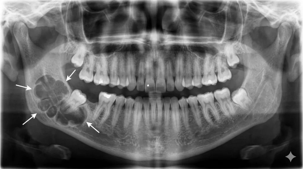
Radiographic image of an odontogenic keratocyst (multilocular lesion) on the right mandibular molar of the patient.
Cause
During the early stages of development, the cells of the dental lamina disintegrate into small epithelial clusters, which resorb with time. Incomplete resorption of these clusters leads to cysts. OKC can exist as a single chamber (unilocular) or multi-chambered (multilocular) cyst. Odontogenic keratocysts are often found in relation to syndromes like Basal cell nevus syndrome and Gorlin-Goltz syndrome. Approximately 25-40% of OKCs are associated with an impacted tooth.
Symptoms And Treatment
This particular cyst is notorious for expanding aggressively and rapidly. Mostly asymptomatic, large cysts can present with edema (swelling), lockjaw (trismus), pain, sensory alterations, and infection (with pus drainage).
Doctors select the ideal treatment plan depending on the location and size of the cyst. Small cysts are dealt with by enucleation.
As they cause significant destruction to the jaw bones by growing in the medullary spaces, oral surgeons perform procedures like posterior ostectomy with marsupialization to manage large OKCs. In this surgery, the surgeon removes 1-2 mm of bone surrounding the cyst after enucleation to achieve healthy bone margins. It is considered an effective treatment option.[9]
Despite the effective treatments, OKCs have a high recurrence rate of up to 62.5%.[10] Recurrence is attributed to the formation of daughter cysts (satellite cysts/microcysts) that form in the surrounding connective tissue.
Lateral Periodontal Cyst
A rare dental cyst that develops on the side of a healthy (vital) tooth is the lateral periodontal cyst (LPC). The non-inflammatory cyst is usually seen between the mandibular canines and premolars. Typically seen in individuals aged 40-70 years.
Cause
Clinicians don’t know the exact cause of its development. However, the slow-growing cyst likely develops from REE, remnants of dental lamina, or remnants of epithelial rests of Malassez. Rests of Malassez are small clusters of residual epithelial cells found in the ligaments that hold your tooth, i.e., the periodontal ligament.
Symptoms And Treatment
It is mostly asymptomatic, but bone (cortical) expansion can present with localized gingival swelling. Conservative surgical enucleation is the preferred treatment for most cases. A typical LPC has a low recurrence rate (upto 3%). However, a modified form, i.e., botryoid odontogenic cyst (BOC), is a multilocular periodontal cyst that has a high recurrence rate (about 21.7%).[11]
Gingival Cyst Of The Newborn
Also known as dental lamina cyst or Epstein’s pearls, gingival cysts of the newborn are common, tiny nodules (yellow-white colored) found on a newborn’s gums or palate. They develop from the remnants of the dental lamina and are pretty common in infants. These small keratinized cysts do not cause any discomfort or feeding issues and resorb themselves within a few weeks (to three months) without treatment.[12]
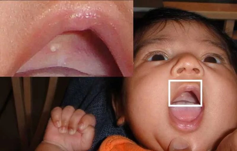
Epstein pearls are visible on the palatal mucosa in a 5-week-old infant. Sghael, Public Domain, viaWikimedia Commons
Gingival Cyst Of The Adult
It is a very rare odontogenic cyst that grows slowly in the soft tissues (gums) of middle-aged/aged adults (50-60 year olds). Clinicians mostly see this cyst in the mandibular canine-premolar region. It appears as a bluish dome in the gums. It originates from the epithelial rest of Serres. Generally, it doesn’t cause any symptoms or bone expansion. Treatment involves surgical excision/enucleation of the cyst.[13]
Calcifying odontogenic cyst is a rare odontogenic lesion that is now designated as calcifying cystic odontogenic tumor. It is also known as a Gorlin cyst.
Periapical Cyst
Also known as an apical cyst or radicular cyst, a periapical cyst is a slow-growing inflammatory odontogenic cyst that forms at the apex (tip) of a tooth root. It is the most common odontogenic cyst (accounts for about 60% of odontogenic cysts), typically developing from an untreated tooth that has undergone infection, decay, or trauma.
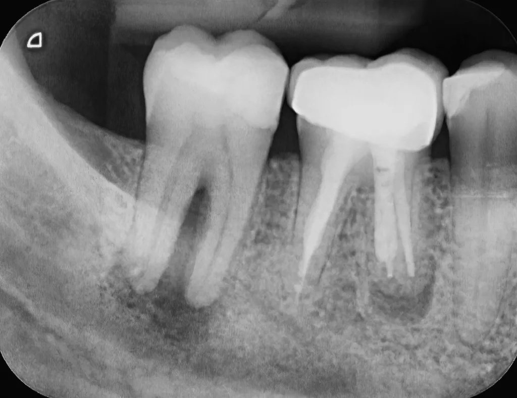
The dark shadows at the bottom of the root represent periapical cysts on two neighboring teeth following infection. The cyst on the first molar persists even after a root canal treatment.
Causes
Trauma or infection leads to pulp necrosis. Death (necrosis) of the dental pulp (part of the tooth that contains the nerves and blood vessels) results in a reduction of the blood supply, which ends up in radicular cyst formation.
Symptoms And Treatment
In the early stages, the cyst is asymptomatic. However, as it grows in size, the following symptoms arise:
- Tenderness and pain on chewing
- Persistent toothache
- Swelling in gums
- Facial swelling (in advanced/severe cases)
- Tooth shifting and mobility
- A draining fistula that appears like a pimple or bump on the gum
In the vast majority of cases, dentists treat the cyst (and the infected/decayed tooth) with endodontic (root canal) treatment. In this procedure, the dentist removes the infected pulp and cleans the canal. Most cysts resorb after appropriate endodontic treatment.[14]
For cysts not resolving with root canal therapy, doctors opt for minimally invasive endodontic therapy. Cystectomy andapicectomy are the processes of surgically removing the radicular cyst with 1/3rd of the affected root (to which the cyst is attached). Root canal therapy with cystectomy has good long-term results.[15]
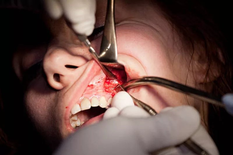
A dentist performs a cystectomy and apicectomy on a patient.
In cases where the infection and the cyst can not be resorbed with conservative therapies, doctors go for tooth extraction.
Residual Cyst
In most cases, a periapical cyst is removed with the extraction of the tooth. However, sometimes the cyst can stay inside the jaw after tooth removal, in which case, it is called a residual cyst. Usually, these lesions are asymptomatic, but the inflammatory cyst can continue to grow slowly and cause bone destruction. Thus, patients can notice jaw swelling, bone resorption, and some facial deformity associated with the cystic lesion.
Residual cysts are frequently seen in the anterior maxillary region. They are discovered during routine dental radiographs and managed with surgical removal of the cyst. According to studies, they are most prevalent in elderly, edentulous patients (people with no teeth). Surgical enucleation is an effective treatment option with a very low recurrence rate.[16]
Paradental Cyst
A paradental cyst is a rare inflammatory odontogenic cyst that develops on a partially erupted healthy (vital) tooth. The most common site for a paradental cyst is the mandibular third molar. However, they can also be seen associated with first and second mandibular molars. This particular type of cyst develops at the junction of the crown and root part (cervical margin). It grows on the cheek side (buccal aspect) and is linked with pericoronitis (swelling of gum tissue overlying wisdom teeth).
The buccal bifurcation cyst is a rare variant of the paradental cyst that mainly affects pediatric patients (4-14 year olds). It is typically seen on the buccal (cheek) aspect of the lower first molar and can hinder eruption.
Cause
The underlying cause is unknown, but it’s believed to arise from the reduced enamel epithelium.
Symptoms And Treatment
Paradental cysts are asymptomatic in general. However, large cysts can present with gum swelling, pain, and tenderness on the buccal side. Patients with infected cysts can see pus discharge too. Most patients are annoyed by recurrent episodes of pericoronitis. Foul smell may also be linked to infection of the cyst.
Doctors adopt the extraction of the tooth and the attached cyst for the 3rd molars. However, surgical enucleation is preferred for cysts on the 1st and 2nd molars.[17]
Diagnosis of Dental Cysts
Most of the time, dental cysts are diagnosed during routine dental examination because the vast majority of these lesions are asymptomatic. In case of symptomatic presentations, doctors take a history of the symptoms and correlate it with the clinical picture of the patients. Location of pain, tenderness makes it easier, as most cysts are frequently seen in their common locations.
Imaging Tests
When a dental cyst is suspected, doctors go straight away for imaging studies. Conventional X-rays help diagnose the condition. However, advanced CBCT scan is emerging as the more accurate, detailed, and reliable form of radiographic imaging.
On a radiograph, dental cysts appear as dark (radiolucent) lesions that are well defined with white borders. Oral cysts can be unilocular (single nodule) or multilocular (multiple nodules).
Final Word
A dental cyst is a benign lesion present in the oral region. A cyst has its own epithelial lining and carries liquid or semi-solid material inside. Dental cysts are not found very commonly. They may arise from developmental faults in non-tooth (non-odontogenic) structures and from tooth-making structures (odontogenic). Non-odontogenic cysts include nasopalatine and naslobial cysts. Odontogenic cysts develop from tooth structures like the dental lamina, reduced enamel epithelium, and enamel organ.
Odontogenic cysts unrelated to inflammation include dentigerous, eruption, lateral periodontal, odontogenic keratocyst, and gingival cysts (in neonates/adults). On the other hand, inflammatory cysts include periapical, residual, and paradental cysts. Generally, cysts are mostly asymptomatic. When present, patients experience symptoms like pain, swelling, tenderness, bone destruction, and tooth displacement, etc. Doctors manage cysts conservatively via processes like marsupialization. However, surgical removal (enucleation) is the most effective solution.
References
[1] Salihu, B., Ahmedi, J., Ademi Abdyli, R., Reçica, B., Shkreta, M., & Jerliu, N. (2026). Global prevalence of odontogenic cysts: A systematic review.The Saudi Dental Journal,38(3), 23.
[2] McKinney, R., Menicucci, C., & Brizuela, M. (2025). Non-Odontogenic Cysts. InStatPearls [Internet]. StatPearls Publishing.
[3] Hasan, S., Popli, D. B., Ahmad, S. A., Sircar, K., Mansoori, S., & Dua, K. (2022). Nasopalatine duct cyst with impacted inverted mesiodens: a rare case report and literature review.Case Reports in Dentistry,2022(1), 5981020.
[4] Almutairi, A., Alaglan, A., Alenezi, M., Alanazy, S., & Al-Wutayd, O. (2020). Nasolabial cyst: case report and review of management options.BMC surgery,20(1), 10.
[5] Noujeim, Z., & Nasr, L. (2021). The prevalence, distribution, and radiological evaluation of dentigerous cysts in a Lebanese sample.Imaging science in dentistry,51(3), 291.
[6] Austin, R. P., & Nelson, B. L. (2021). Sine qua non: dentigerous cyst.Head and neck pathology,15(4), 1261-1264.
[7] Kouhsoltani, M., Mesgarzadeh, A. H., & Khiavi, M. M. (2015). Mandibular fracture associated with a dentigerous cyst: report of a case and literature review.Journal of dental research, dental clinics, dental prospects,9(3), 193.
[8] Abu-Mostafa, N., & Abbasi, A. (2017). Marsupialization of a large dentigerous cyst in the mandible with orthodontic extrusion of three impacted teeth. A case report.Journal of clinical and experimental dentistry,9(9), e1162.
[9] Khalil, A., Albash, Z., Sleman, N., & Sayegh, W. (2023). Marsupialization and peripheral ostectomy for the management of large odontogenic keratocyst: a case report.Journal of Surgical Case Reports,2023(3), rjad119.
[10] Fidele, N. B., Yueyu, Z., Zhao, Y., Tianfu, W., Liu, J., Sun, Y., & Liu, B. (2019). Recurrence of odontogenic keratocysts and possible prognostic factors: Review of 455 patients.Medicina oral, patologia oral y cirugia bucal,24(4), e491.
[11] e Silva, L. F. D. C., Lima, C. F., Cabral, L. A. G., Brandão, A. A. H., & Almeida, J. D. (2011). Lateral periodontal cyst: a case report and literature review.Journal of Oral & Maxillofacial Research,1(4), e5.
[12] Moda, A. (2011). Gingival cyst of newborn.International journal of clinical pediatric dentistry,4(1), 83.
[13] Mota, M. E., Oliveira, D. M. D. A., Medeiros, Y. D. L., Moreira, M. S., Lopes, R. N., Alves, F. A., … & Prado, J. D. (2023). Gingival cyst of the adult.Autopsy and Case Reports,13, e2023454.
[14] Abbott, P. V. (2022). Pulp, root canal, and periradicular conditions.Endodontic advances and evidence‐based clinical guidelines, 85-116.
[15] Zamaliauskiene, R., & Veberiene, R. (2023). Successful treatment of cystic lesion combining cystectomy, nonsurgical endodontics and vital pulp therapy of mature permanent mandibular molars: a case report with 19 months follow-up.Journal of Oral & Maxillofacial Research,14(1), e4.
[16] Titinchi, F., & Morkel, J. (2020). Residual cyst of the jaws: A clinico-pathologic study of this seemingly inconspicuous lesion.Plos one,15(12), e0244250.
[17] Li, P., Zhao, Y., You, Y., Lin, L., Yu, D., & Zhao, W. (2023). Current perspectives on paradental cyst: a literature review.Dentistry Journal,11(12), 281.