
 is a dark spot in the eye. Much like the...)
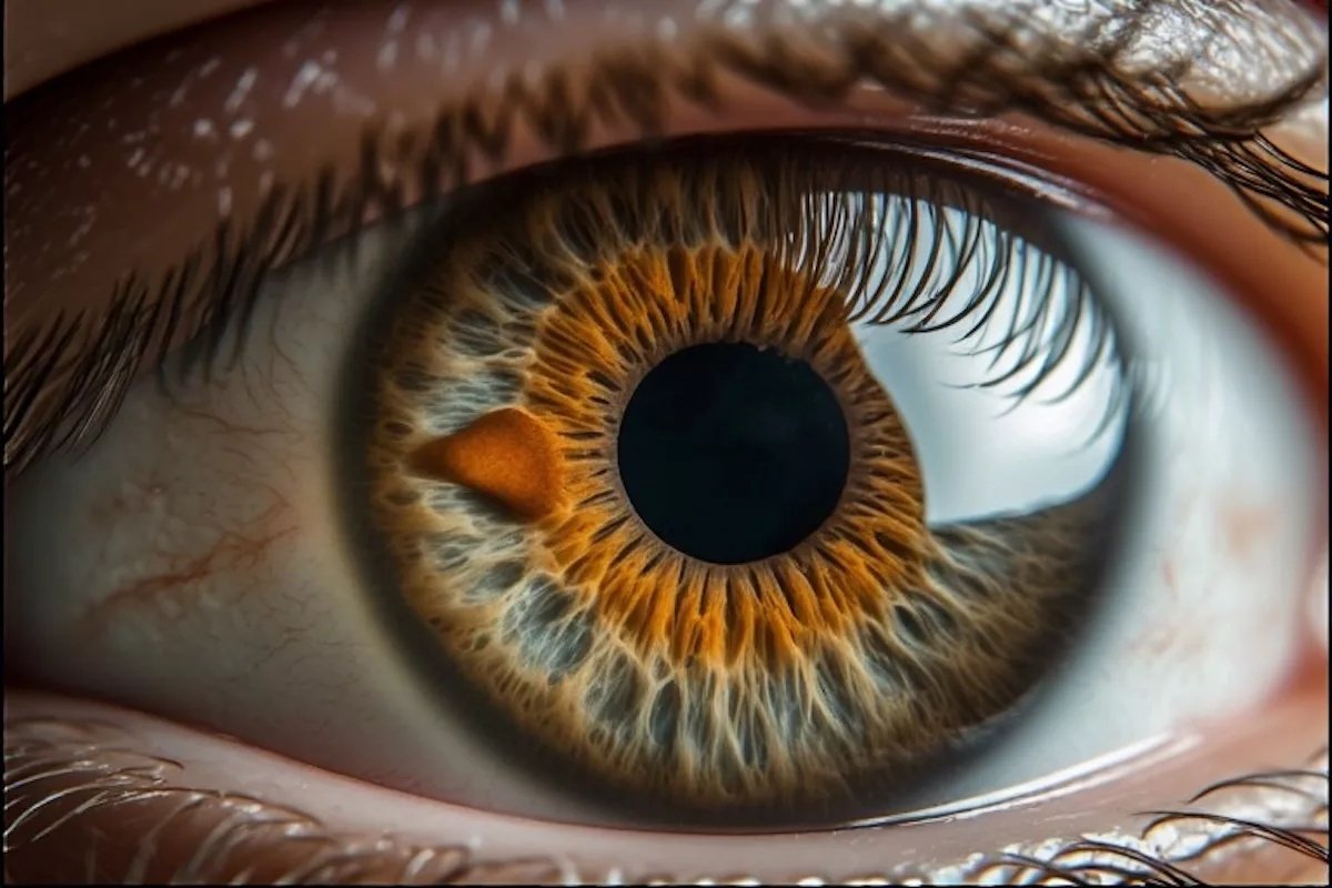
Eye Freckle that In simple words, an eye freckle (or eye nevus, as it is called in the clinical world) is a dark spot in the eye. Much like the freckles on your skin, most eye freckles are benign clusters of melanocytes (pigment cells). However, because these same cells can cause cancer (melanoma), we cannot afford to ignore them. In 2026, we have better imaging than ever to track these spots, but the first line of defense is still going to be your own observation.
What is an Eye Freckle: Eye Freckle that
Just like a freckle on your arm, an eye freckle is a cluster of pigment-producing cells called melanocytes. While the term eye freckle is often also used for an Iris Ephelis, that is technically just a flat patch of melanin overproduction, usually from sun exposure. In contrast, a nevus is a true cellular cluster with the potential to thicken or grow.
They can appear in different parts of the eye, and their location determines how they are monitored and their potential for complications. The three main locations are on the iris, conjunctiva, or in the choroid:
Iris Nevus
These are clinically visible pigmented lesions of the iris and are less common than choroidal nevi but easier to detect because of their location. They appear on the iris (colored part of the eye). While they are visibly prominent, they are usually benign. Unless one spot starts to grow or changes the shape of your pupil, there’s no requirement for immediate intervention.
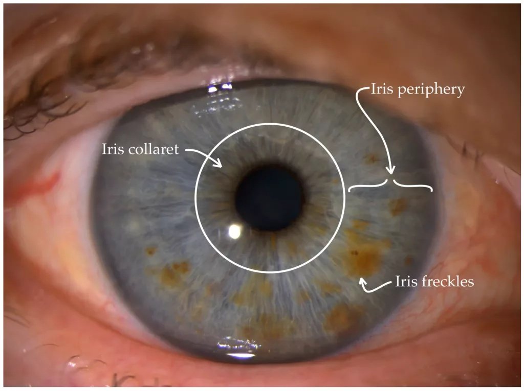
Freckles/nevi on the iris, along with anatomical landmarks of the iris used to monitor the location and growth of eye freckles. (Image Courtesy: Šemeklis, L., Kapitanovaitė, L., Butrimas, G., Briedė, K., Dubinskaitė, A., Žemaitienė, R., & Valiukevičienė, S. (2024). Iris Pigmented Lesions and Risk of Cutaneous Melanoma: Case–Control Study in Lithuania. Journal of Personalized Medicine, 14(5), 530. Available fromMDPIand licensed under CC by 4.0)
Conjunctival Nevus
These appear on the conjunctiva (white of the eye). They often start as a tannish (or yellowish) patch but can darken during puberty or pregnancy due to hormonal changes.[1] On slit-lamp examination, many conjunctival nevi contain small intralesional cysts, which are reassuring signs of benignity. While these are also usually harmless, they obviously are more visible, so they are frequently removed for aesthetic reasons if they become large or elevated.
Choroidal Nevus
These are the hidden freckles. The location is in the choroid (the layer of blood vessels under the retina), so they are invisible to the naked eye. You won’t see these in the mirror; most patients only discover them during a routine dilated eye exam. Choroidal nevi are actually the most common type of ocular nevus, occurring in approximately 5–10% of the Caucasian population. While most remain stable for life, they carry a small but real risk of transformation into choroidal melanoma.[2]
Eye Freckle vs. Melanoma
The risk for a freckle turning into cancer is real, specifically, Ocular Melanoma. While rare, it is a serious condition. Naturally, ruling this out is the biggest concern for any patient.
Most eye freckles will never become cancerous. However, a small percentage can transform into a malignancy. The estimated annual risk of transformation of a choroidal nevus into melanoma is approximately 1 in 8,000 to 1 in 9,000.[3] Which nevus has the potential to turn cancerous is impossible to detect by the naked eye, which is why a baseline exam with an ophthalmologist is non-negotiable.
In 2026, ophthalmologists use the ‘TFSOM-DIM’ mnemonic to evaluate the risk of a choroidal nevus turning into a choroidal melanoma. It means ‘To Find Small Ocular Melanoma Doing IMaging,’ and each letter stands for the following risk factors:[4]
- T (Thickness more than 2mm, measured by ultrasound)
- F (Subretinal Fluid leaking around the spot)
- S (Symptoms: blurred vision, flashes, or floaters)
- O (Orange tint (lipofuscin) on its surface)
- M (Margin within 3mm of the optic disc)
- D (Diameter > 5mm based on fundus photography)
- I (Multimodal imaging)
- M (Melanoma: acoustic hollowness on ultrasound)
The presence of multiple risk factors significantly increases the likelihood of malignant transformation and typically warrants referral to an ocular oncology specialist.
Biopsy is not routinely performed for typical choroidal nevi and is generally reserved for cases where the diagnosis is uncertain.
Why Do Eye Freckles Form?
Sun exposure is a major factor. Just as UV rays damage the skin, they can also trigger melanocytes in the eye. This is why ophthalmologists advocate for UV-rated sunglasses, even on cloudy days when you might think you won’t have exposure to the sun. However, the direct causal relationship between UV exposure and choroidal nevi or melanoma is not as clearly established as it is in cutaneous melanoma.
Similar to skin cancer, genetic predisposition plays a role here as well; if you have fair skin and a history of skin freckles, you are at a higher risk for an eye nevus.
Eye Freckles in Children
While most nevi appear in adulthood, they can be congenital (present at birth) or appear during childhood.
The Rule for Parents: If your child has a new spot in their eye, do not wait for their next school vision screening. Children’s eyes grow rapidly, and any pigment change needs a baseline photograph immediately. Although ocular melanoma is extremely rare in children, documentation of size and appearance allows appropriate long-term monitoring.
Management of Eye Freckles
The eye exam for an eye freckle will be a bit different from a standard vision test. Your pupils will be widened so the doctor can see the back of the eye and check for hidden choroidal nevi.
If your ophthalmologist determines the freckle is benign, the treatment is usually just serial monitoring.
- We take high-resolution photos of the nevus to serve as a baseline.
- Annual or, in some cases, 6-monthly checkups are scheduled, during which we compare new images to the baseline. Even a 1mm change can be significant.[5]
- In some cases, we use Optical Coherence Tomography (OCT). It uses light waves to create high-resolution cross-sectional images of the retina and can detect subretinal fluid or structural changes.
B-scan ultrasonography is used to measure lesion thickness and assess internal acoustic characteristics. Even small documented changes in size or associated fluid can be clinically significant.
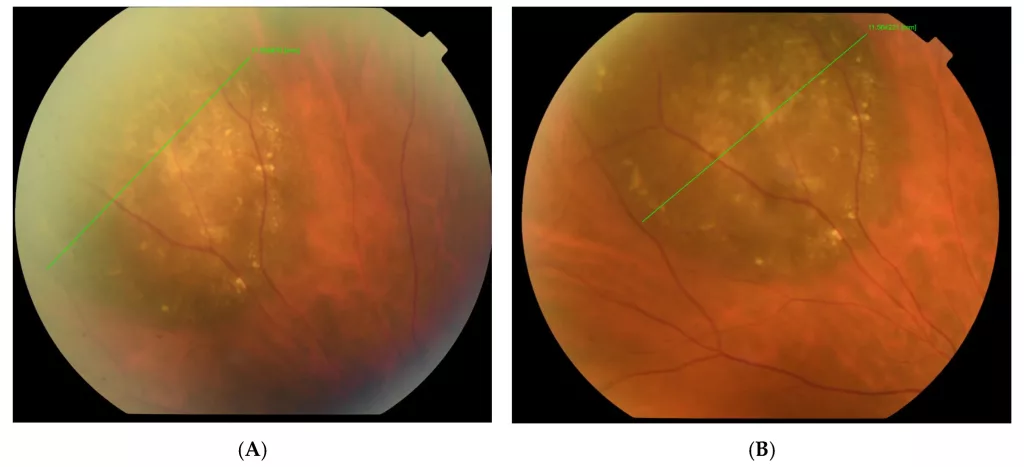
Photography of the fundus over a 12-month period showing a stable choroidal nevus with negligible growth 11.55—11.56 mm (0.01 mm) (Image Courtesy: Stoyanova, N. S., Atanassov, M., Mitkova-Hristova, V. T., Basheva-Kraeva, Y., & Kraeva, M. (2025). Giant Choroidal Nevus—A Case Report. Reports, 8(2), 41. Available fromMDPIand licensed under CC by 4.0)
How Can an Eye Freckle Be Removed?
If an eye freckle is visible, patients naturally ask for removal for aesthetic reasons. Any intervention with the eye is risky in terms of vision, though, so whether it can be done or not depends on the location of the freckle.
For conjunctival nevi (on the white of the eye), surgical excision is possible. We call this process a ‘surgical shave’, and it allows us to obtain a sample to be sent for biopsy.[6] But it’s not completely risk-free. The procedure can leave a small scar or a localized area of chronic redness (vessels) in the area.
Another option is Plaque Brachytherapy, a form of localized radiation wherein a small radioactive disc is temporarily attached to the eye to starve the tumor. However, it is not used for benign nevi. It is a treatment reserved for confirmed cases of ocular melanoma.
In non-malignant cases, we prioritize saving the patient’s life and vision over aesthetics.
Laser Treatment
If a nevus is suspected of being an early-stage melanoma, ophthalmologists may use a laser (typically an argon laser) to ‘seal’ the blood vessels feeding the growth. This is done to starve a potential tumor.[7] However, laser photocoagulation is not a standard treatment for choroidal melanoma.
It’s worth mentioning that some clinics offer laser treatment to remove these freckles for purely aesthetic reasons. But using a laser to break up melanin in the iris is risky, and this is not a procedure specialists recommend unless necessary. Basically, when a laser breaks up the pigment in the iris, the microscopic particles are released into the eye’s internal fluid. This can
- Clog the eye’s drainage system, causing raised intraocular pressure (IOP) and permanent optic nerve damage,
- Trigger chronic inflammation (uveitis),
- Or accidentally damage the iris muscles.
Preventive Care with UV Protection
We have clear evidence linking UV radiation to the development and darkening of eye nevi. Just as you wear sunscreen to protect your skin, your eyes need physical barriers.
- UV400 Sunglasses block 99-100% of UVA and UVB rays.[8]
- Wraparound styles prevent peripheral leakage of UV rays that often strike the side of the eye where conjunctival nevi form.
While UV protection is advisable for overall ocular health, it should not be viewed as a guaranteed method of preventing choroidal nevi or melanoma.
Final Word
Should you be worried about eye freckles? Well, finding one is surely a reason to book an eye exam, but not a reason for panic. Most nevi stay dormant for a lifetime. However, it’s recommended to get it examined by your ophthalmologist and let them decide its fate. Like skin cancer, sun protection goes a long way in terms of prevention, especially if there is a genetic predisposition.
References
[1] Huang, J. J., Locatelli, E. V. T., Chocron, A., Camacho, M. R., Dubovy, S., Karp, C. L., & Galor, A. (2023). Conjunctival Nevus. Current ophthalmology reports, 11(4), 104–112.
[2] Chien, J. L., Sioufi, K., Surakiatchanukul, T., Shields, J. A., & Shields, C. L. (2017). Choroidal nevus: a review of prevalence, features, genetics, risks, and outcomes. Current opinion in ophthalmology, 28(3), 228–237.
[3] Shields CL, Furuta M, Thangappan A, et al. Choroidal nevus transformation into melanoma: analysis of 2514 consecutive cases. Arch Ophthalmol. 2009;127(8):981–987. doi:10.1001/archophthalmol.2009.151.
[4] Jahnke, D., Grohmann, C., Fuisting, B. et al. Performance of the MOLES and TFSOM-DIM scores in classifying choroidal nevi and melanoma. Sci Rep 14, 28534 (2024).
[5] Stratigos, A. J., & Katsambas, A. D. (2008). The value of screening in melanoma. Clinics in Dermatology, 27(1), 10-25.
[6] Ferrandiz, L., Moreno-Ramirez, D., & Camacho, F. M. (2005). Shave excision of common acquired melanocytic nevi: cosmetic outcome, recurrences, and complications. Dermatologic surgery: official publication for American Society for Dermatologic Surgery [et al.], 31(9 Pt 1), 1112–1115.
[7] Alsharif, A. M., Al-Gehedan, S. M., Alasbali, T., Alkuraya, H. S., Lotfy, N. M., & Khandekar, R. (2016). Argon Laser Photoablation for Treating Benign Pigmented Conjunctival Nevi. Middle East African journal of ophthalmology, 23(3), 247–249.
[8] Alebrahim, M. A., Bakkar, M. M., Al Darayseh, A., Msameh, A., Jarrar, D., Aljabari, S., & Khater, W. (2022). Awareness and Knowledge of the Effect of Ultraviolet (UV) Radiation on the Eyes and the Relevant Protective Practices: A Cross-Sectional Study from Jordan. Healthcare (Basel, Switzerland), 10(12), 2414.