
, also known as Bloch-Sulzberger syndrome, is a rare, genetic disorder that is inherited...)
What is Incontinentia Pigmenti?: Incontinentia Pigmenti Rare
Incontinentia Pigmenti Rare Incontinentia Pigmenti (IP), also known as Bloch-Sulzberger syndrome, is a rare, genetic disorder that is inherited via the X chromosome. It is caused by a mutation in the IKBKG/NEMO gene. IP is best classified as an X-linked dominant ectodermal dysplasia with abnormal inflammatory and apoptotic signaling, which affects the ectoderm during embryonic development. This in turn affects multiple systems of the body, including the skin, hair, teeth, nails, eyes, and the nervous system. Characterised by blister-like lesions on the skin during infancy and childhood, this disease has four distinct stages: vesicular, verrucal, hyperpigmentation, and hypopigmentation. There is no definitive treatment of IP since it’s a genetic disorder; thus, supportive and palliative care is administered.
What causes IP?
The genetic disorder IP occurs when a mutation affects the ‘inhibitor of nuclear factor kappa B kinase regulatory subunit gamma’ (IKBKG) gene, formerly called the NEMO gene, on the X chromosome. This gene regulates cellular proliferation, self-destruction, and inflammatory processes in ectodermal cells. Mutations lead to impaired NF-κB signaling and increased apoptosis of ectoderm-derived cells, explaining the inflammatory skin lesions and linear distribution along Blaschko’s lines. The ectoderm is the developmental layer of the embryo that later forms the central nervous system, skin, teeth, hair, and nails. Thus, a mutation in this gene gives rise to inflammation and lesions of these systems.[1]
How is IP Inherited?
Incontinentia Pigmenti is linked to the X chromosome and is passed on as a dominant trait. This means that having even one mutated X chromosome is enough for the individual to express the disease.
Each person has two sex chromosomes, one from the father and one from the mother. A female’s sex chromosomes are XX, whereas a male’s sex chromosomes are XY.
There can be 4 possible outcomes in the offspring of affected individuals:
- Unaffected female: The offspring has inherited the normal X chromosome from both her parents and does not carry the disorder.
- Affected female: If the offspring has inherited an abnormal X chromosome from either of her parents, she will be affected with IP but will be able to survive into adulthood due to the presence of a normal X chromosome that supplements for the mutated one.
- Unaffected male: If the mother passes on her unaffected X chromosome to the offspring, he will not be affected by the disease.
- Affected male: In case of the mother passing on the mutated X chromosome, the affected male offspring will develop IP and typically die in the 12th week of pregnancy. In extremely rare cases of de novo mutations in the male offspring, the child may be born with IP and can survive until adulthood. Other possible reasons include somatic mosaicism, Klinefelter syndrome (47, XXY), or hypomorphic mutations.[2]
Prevalence of Incontinentia Pigmenti
Worldwide, IP is considered extremely rare. It is estimated to affect 1.2 in 100,000 individuals, which is 0.000012% of the population. The literature reports between 900 to 1200 affected individuals. As a result of the X-linked inheritance pattern of IP, most of the individuals affected are female. However, several dozen males with IP have also been identified.[3] [4]
Symptoms of Incontinentia Pigmenti
Due to the disease’s nature of affecting the cellular function of the ectoderm, the symptoms manifest in multiple systems of the body that are formed from that layer. This primarily includes skin, hair, teeth, nails, eyes, and the nervous system.[5]
1. Skin:
The skin lesions are the most common and characteristic features of IP. They occur in four distinct stages, ranging from red blisters and vesicles to pale scar-like lesions. The following are the various types of skin lesions seen in IP:
o Erythema: red rash on the limbs and body.
o Pustules, vesicles, and blisters, found in a “Blaschko linear” pattern.
o Dark, grey or blue hyperpigmentation streaks in a swirly pattern.
o Pale, scar-like hypopigmented streaks.
2. Teeth:
50-75% of individuals have a partial or complete absence of teeth since birth, or small or abnormal shapes.
3. Hair:
Approximately 50% of the affected individuals have alopecia, i.e., bald patches on the scalp. The hair is coarse, ‘woolly’, and ‘wiry’, making it hard to brush.
4. Nails:
IP typically affects most or all fingernails and toenails, causing ridging, pitting, and dystrophic changes. Severe nail thickening (onychogryphosis) or complete nail absence is uncommon.
5. Eyes:
About one-third of the affected individuals have abnormal growth of the blood vessels in the periphery of the eye, leading to impaired vision or complete blindness if not treated early.
6. Family History:
It is consistent with X-linked inheritance or a history of multiple miscarriages.
7. Central Nervous System:
Neurologic involvement occurs in a significant proportion of patients and is not rare. This leads to seizures, paralysis, slow growth and development, and small head size (microcephaly).
8. Other symptoms
These include short stature, abnormalities of the breast, or extra ribs.
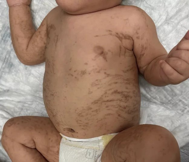
Clinical presentation of Incontinentia Pigmenti showing characteristic linear vesicular and hyperpigmented lesions along Blaschko’s lines. Image sourced from theCureusarticle“Incontinentia Pigmenti: A Rare Case of Survival of a Male Infant”by Riley Shin, Helen Chen & Michelle Tarbox, published 04 March 2025 (CC BY 4.0).
What are the Stages of IP?
IP has 4 distinct stages that can overlap.[6]
- Stage 1: Vesicular
This stage starts with erythema that develops into characteristic red, blister-like lesions. It is usually present at birth or starts in early infancy, affecting the scalp and extremities primarily. - Stage 2: Verrucous
Characterised by wart-like lesions, this is the phase where the blisters start to heal and develop raised crusts or scabs. - Stage 3: Hyperpigmentation
Usually appearing at 6-12 months of age, the slate-grey, blue, or brown skin lesions appear in an irregular, swirly pattern, often described as a “marble cake” appearance. These hyperpigmented lesions can occur in places other than the lesions of stages one and two. - Stage 4: Hypopigmentation
Also known as the “atrophic stage”, the skin in this stage changes to pale, scar-like lesions. These lesions are hairless, and the sweat glands are reduced.
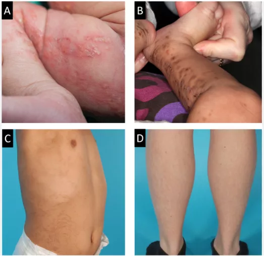
Figure showing the 4 stages of Incontinentia Pigmenti: (A) Stage 1: Vesicular. (B) Stage 2: Verrucular. (C) Stage 3: Hyperpigmentation. (D) Stage 4: Hypopigmentation. Image sourcedOraphenet Journal of Rare Diseases‘ article“Prevalence and clinical characteristics of incontinentia pigmenti: a nationwide population-based study”by Krogh Herlin, Sigrun Alba Johannesdottir Schmidt, Trine H. Mogensen & Mette Sommerlund (2024), licensed under CC BY 4.0.
How to Diagnose Incontinentia Pigmenti?
The diagnosis of IP is done based primarily on clinical findings, detailed history, and gene testing.
The clinical symptoms are divided into ‘major criteria’ and ‘minor criteria’.[7]
Major Criteria
The major criteria are the four distinct stages of the skin lesion of IP occurring from infancy to adulthood:
- Erythema, followed by the formation of blisters (vesicles) on the body, usually in a linear distribution. Erythema is most prominent before 6 months of age.
- Verrucous lesions follow the blisters. This is formed due to scab-like formation during the healing phase of the lesions.
- Hyperpigmented streaks usually occur in a swirly pattern on the trunk of the body.
- Pale, hairless, atrophic linear streaks.
The presence of at least one characteristic skin stage fulfills the major criterion.
Minor Criteria
These include involvement of other ectodermal structures:
- Teeth: Partial or complete absence of teeth. Small or abnormally shaped teeth
- Hair: Bald patches, woolly hair.
- Nails: Pitted, ridged, or curved nails
- Retina: Growth of new, abnormal blood vessels in the periphery of the retina, causing retinopathy.
- Family history: Consistent with X-linked inheritance or a history of multiple miscarriages.
The presence of any of the minor criteria supports the clinical diagnosis.
Gene Testing:
Detection of an IKBKG gene mutation alone is sufficient to confirm the diagnosis, even in the absence of full clinical staging. This molecular genetic testing is widely available and highly sensitive in detecting abnormalities.
Skin Biopsy:
If the gene testing does not give a substantial result, a skin biopsy of a blister can be done to confirm questionable findings.
Prognosis and Life Expectancy
The prognosis of IP depends widely on the extent of the disease and the severity of involvement of multiple organs. The skin lesions present in infancy and adolescence eventually heal and do not cause any trouble throughout life. However, the neurologic and ophthalmologic complications can lead to higher morbidity and mortality rates.[8]
If the disease affects the neurological system and causes neonatal seizures, the individual might have severe developmental and intellectual impairments in the future.
In case of eye involvement, they might suffer from vision impairment or blindness.
Thus, the prognosis of this disease depends on the severity of the systems involved at the time of birth or infancy. Individuals without early severe neurologic or ocular involvement generally have normal life expectancy and good long-term outcomes.[9]
Treatment for Incontinentia Pigmenti
There is no definitive treatment for IP, but the management is palliative and based on symptomatic care.
Routine visits to the dermatologist, ophthalmologist, and dentist can help aid in symptomatic relief and prevention of the progression of diseases of the skin or hair, eyes, and teeth, respectively.
- The skin lesions can be treated with topical steroids to reduce inflammation and antibiotics to prevent secondary infections in the blisters. However, please note that corticosteroids or antibiotics are prescribed only under medical supervision to reduce inflammation and prevent secondary infection.
- The ophthalmologist will carry out regular fundoscopy of the eye for early detection and prevention of retinal vascular abnormalities. In case of any abnormal developments, laser photocoagulation will be carried out to treat the symptoms.
- Assessment of neurological health will be done, including an MRI of the brain in infants with seizures, hypotonia, or developmental delay. For seizure control, anti-epileptic drugs will be given.
Take Away
Incontinentia Pigmenti is a rare genetic condition that most often affects the skin but can also involve the eyes, teeth, hair, and nervous system. Although the skin changes may look concerning, they usually improve with time and do not cause long-term problems. The most important part of care is early diagnosis and regular check-ups, especially for the eyes and nervous system, to prevent serious complications. With proper medical follow-up and support from a healthcare team, many people with Incontinentia Pigmenti grow up healthy and lead normal, active lives.
References
[1] Herlin LK, Schmidt SAJ, Mogensen TH, Sommerlund M. Prevalence and clinical characteristics of incontinentia pigmenti: a nationwide population-based study. Orphanet Journal of Rare Diseases. 2024;19(1).
[2] Incontinentia Pigmenti – Symptoms & Diagnosis | NFED. National Foundation for Ectodermal Dysplasias.
[3] Incontinentia pigmenti: MedlinePlus Genetics.
[4] Herlin LK, Schmidt SAJ, Mogensen TH, et al. Prevalence and clinical characteristics of incontinentia pigmenti: a nationwide population‑based study.Orphanet Journal of Rare Diseases. 2024;19:454. doi:10.1186/s13023‑024‑03480‑8.
[5] Scheuerle, Angela E., and Matilde Valeria Ursini. “Incontinentia Pigmenti.” PubMed, University of Washington, Seattle, 1993
[6] Incontinentia Pigmenti. NORD (National Organization for Rare Disorders). Published June 15, 2023.
[7] Landy SJ, Donnai D. Diagnostic criteria for incontinentia pigmenti. Journal of Medical Genetics. 1993;30(2):113‑121. Revised criteria include major clinical skin manifestations and minor systemic features, supported by genetic testing and family history.
[8] Shah KN. Incontinentia Pigmenti: Background, Pathophysiology, Etiology. Medscape.com. Published September 11, 2025. Accessed November 28, 2025.
[9] Incontinentia Pigmenti: What is it, Symptoms. Cleveland Clinic.