

What Is Inflammatory Myopathy?
Inflammatory myopathy is a term used to categorize a group of uncommon autoimmune diseases, characterized by chronic inflammation and skeletal muscle weakness. In contrast to the common muscle aches or aging, these diseases are caused by the immune system targeting healthy muscle tissue, which is gradually damaged and malfunctions.
Inflammatory myopathies may occur at any age; however, they are diagnosed more often in adults aged 40 to 60 years, and are not a normal part of aging. The resultant muscle weakness usually affects routine life, including climbing stairs, lifting, or even raising the arms, and in some cases, this may also affect the skin or lungs, among other body organs.
Inflammatory Myopathy vs. Myositis
Inflammatory myopathy is also referred to as myositis; the two terms are very close and are interchangeable. However, there is a subtle but significant difference between the two.
Myositis is a more general term that merely implies inflammation of the muscle. The causes of this inflammation may be either infections (such as viruses), direct trauma, particular medications, or autoimmune responses.
A more narrow form of myositis isInflammatory Myopathy. It refers specifically to chronic, idiopathic, immune-mediated muscle diseases and represents a subgroup of myositis rather than a completely separate entity. Under such circumstances, the immune system repeatedly attacks muscle tissue and causes progressive weakness.
In essence, all inflammatory myopathies are forms of myositis, but not all cases of myositis are classified as chronic autoimmune inflammatory myopathies. Thus, when used clinically and with patients, the term inflammatory myopathy is the one generally preferred to describe these particular, chronic autoimmune muscle diseases.
Types of Inflammatory Myopathy
The most significant autoimmune diseases, depending on which part of your body is involved, are:
Polymyositis (PM)
Polymyositis mainly attacks skeletal muscles on both sides of your body, especially those nearest to the trunk. It is characterised by the gradual weakening of muscles of the hips, thighs, shoulders, upper arms, and neck on either side of the body. The condition usually occurs in adults aged 30 to 60 years old, and it is more prevalent in women than in men.
It is important to note that true polymyositis is now considered rare, as many cases previously classified as polymyositis are reclassified as immune-mediated necrotizing myopathy or overlap myositis based on modern diagnostic criteria.
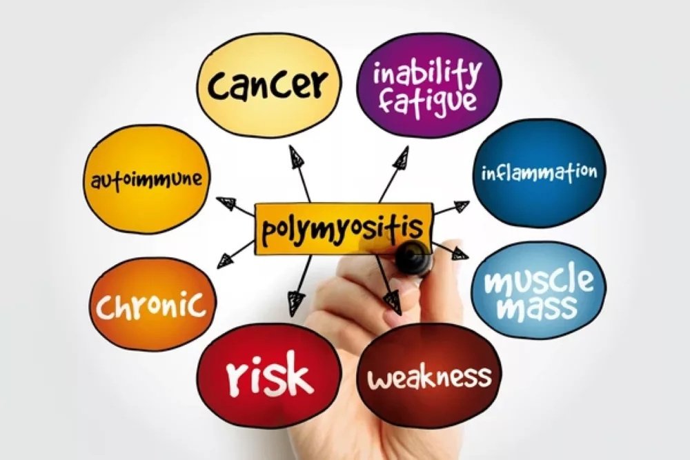
Conceptual illustration of polymyositis, an autoimmune inflammatory myopathy causing muscle weakness.
The affected individuals can experience:
- Problems with getting up a stair or out of a sitting position.
- Problem with lifting things over the head.
- Swallowing or speech problems.
- General weakness and fatigue.
Dermatomyositis (DM)
Dermatomyositis has many similarities with polymyositis, yet it also entails some unique skin alterations. These are periorbital blue-purple rash (heliotrope rash); erythematous rash on the face, anterior chest (V sign), shoulders (shawl sign), and lateral hips (Holster sign); and violaceous eruption on the knuckles (Gottron papules).
The pattern of weakness of the muscle resembles polymyositis in nature, except that proximal muscles are affected. Dermatomyositis may rarely be life-threatening, particularly in cases associated with malignancy, severe interstitial lung disease, or cardiac involvement, especially during the first year of disease. It presents itself in both children (juvenile dermatomyositis) and adults at a greater rate in women.
Inclusion Body Myositis (IBM)
Inclusion body myositis is a degenerative muscle disease. It normally occurs among individuals over the age of 50. IBM, in contrast to other inflammatory myopathies, is more prevalent in men and is likely to impact other groups of muscles.
Inclusion body myositis will make your muscles weak in your extremities (you have hands and legs below your knees). This implies that patients usually suffer:
- Difficulty in grasping things with the wrists and fingers
- The muscles of the front thigh are weak, leading to frequent tripping
- Problem with fine motor skills
- Swallowing difficulties
The most treatment-resistant type of inflammatory myopathy is usually IBM, and it develops more gradually compared to the other types.
Immune-Mediated Necrotizing Myopathy (IMNM)
This is a recently identified subtype also known as necrotizing autoimmune myopathy. Necrotizing myopathies that are mediated by the immune system may result in more impairing and rapidly progressive weakness than the other inflammatory myopathies, and are more inclined to produce difficulty in swallowing.
IMNM can be associated with:
- Use of Statins (cholesterol-lowering drugs)
- Cancer
- Connective tissue diseases
- Viral infections
Antisynthetase Syndrome (ASyS)
Antisynthetase syndrome can also cause various changes and symptoms, including inflammatory arthritis, fever, interstitial lung disease, thick and rough patches of skin on the fingers (mechanic’s hands), and Raynaud syndrome. It is characterized by the presence of specific autoantibodies and is a unique clinical entity that has great involvement of the lungs.
Overlap Myositis
Overlap myositis is when an idiopathic inflammatory myopathy develops in a person who has another systemic rheumatic disease, such as systemic lupus erythematosus or systemic sclerosis. Such patients have the symptoms of the inflammatory myopathy and other autoimmune disorders.
Causes of Inflammatory Myopathy
Most inflammatory myopathies are autoimmune diseases, in which the body’s defense mechanism, which is supposed to protect against infections and disease, targets its own muscle fibres, blood vessels, connective tissue, organs, or joints.
Autoimmune Mechanism
In most cases, it is not clear what exactly induces this autoimmune response. It is believed that idiopathic inflammatory myopathy is a result of genetic and environmental factors. Researchers have found that some genetic markers, especially changes made in the Human Leukocyte Antigen (HLA) complex, can predispose a person to these conditions.
Environmental Triggers
Inflammatory myopathy can be caused by the following environmental factors in genetically susceptible individuals:
Viral Infections: Myositis is also seen in some patients who have viral infections or soon after the infection, which may indicate that viruses can cause the autoimmune response.
Medications:
- Statins (anti-cholesterol drugs)
- Immuno checkpoint blocking drugs (anti-cancer drugs)
- D-penicillamine
- Hydralazine
- Procainamide
Cancer Association: It is known that some inflammatory myopathies, especially dermatomyositis and necrotizing myopathy, have a well-defined association with cancer. The cancer can cause an autoimmune reaction to common antigens in the tumor as well as the muscle tissue.
Immune Checkpoint Inhibitors: These are cancer immunotherapy medications that may induce severe cases of myositis through the uncontrolled activation of the immune system.
Risk Factors
Although any person can have inflammatory myopathy, some factors predispose a person to it:
- Age: Adults-Most of the time, it occurs between the ages of 40 and 60. Children-Most of the time, it occurs during the ages 5-15.
- Sex: There is a 2-3 times higher prevalence of polymyositis and dermatomyositis among women compared to men, whereas IBM is more prevalent among men.
- Ethnicity: The risk for people of Sub-Saharan African origin is greater.
- Family history: The presence of close relatives who have autoimmune diseases predisposes an individual.
Symptoms of Inflammatory Myopathy
The general symptoms of chronic inflammatory myopathy involve a slow and progressive muscle weakness, which begins with the proximal muscles, which are the nearest to the trunk of the body.
Primary Muscle Symptoms
The characteristic symptom is progressive weakness of the muscles, especially the following:
- Shoulders and upper arms
- Hips and thighs
- Neck muscles
This weakness will give practical problems:
- It becomes hard to walk up the stairs.
- Getting out of a sitting posture needs help.
- It is hard to lift anything higher than our shoulders.
- It is difficult to comb hair or to reach high shelves.
- There is an increased effort to get out of bed or a bathtub.
The other symptoms are tiredness after walking or standing, tripping or falling, or swallowing or breathing difficulty.
Muscle Pain and Tenderness
The muscle pain may not be common in patients with dermatomyositis, but it can occur, particularly with the involvement of the fascia. Certain patients have muscle tenderness to touch, although this is dependent on the subtype.
Skin Manifestations (in Dermatomyositis)
The skin abnormalities that are common in dermatomyositis are:
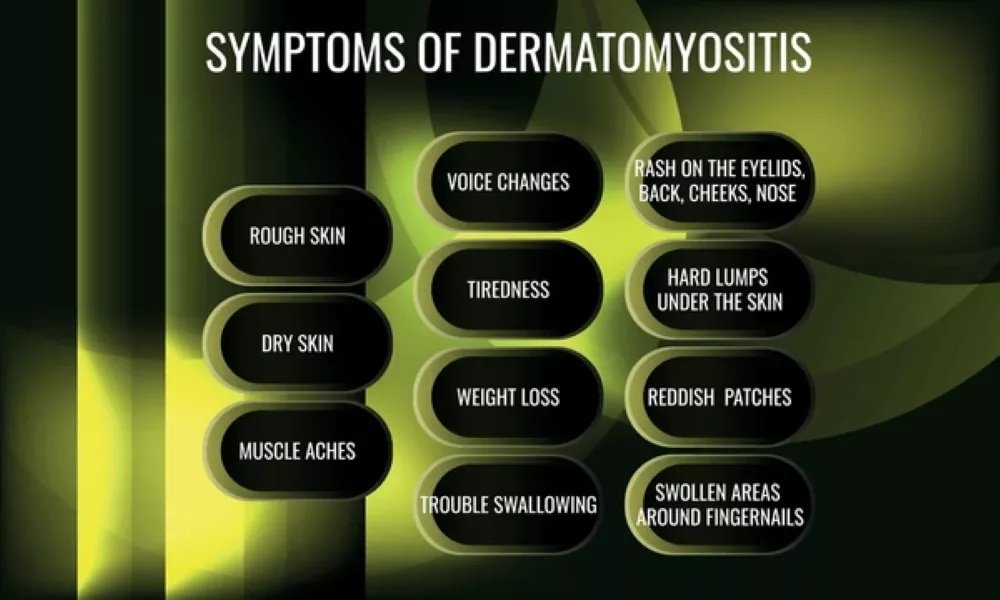
Infographic highlighting key symptoms of dermatomyositis, including muscle inflammation and skin changes.
- Heliotrope rash: This is a purple discoloration around the eyes.
- Gottron papules: These are bumps that are high, scaly on the knuckles, at the elbows, or on the knees.
- V-sign (A V-shaped rash on the chest).
- Shawl sign
- Photosensitive rashes
Extramuscular Manifestations
Inflammatory myopathies can affect other organs:
Lung Involvement: Interstitial lung disease is particularly common with antisynthetase syndrome that results in:
- Shortness of breath
- Chronic dry cough
- Decreased exercise tolerance
Heart Problems. These are not as common; still, they may consist of:
- Irregular heartbeats
- Heart muscle inflammation
- Pericarditis
Joint Problems. Arthritis can also be found particularly in antisynthetase syndrome and overlap myositis, which causes joint pain and stiffness.
Difficulties with swallowing. The weakness of the muscles of the throat causes dysphagia, which predisposes to choking and aspiration.
Raynaud’s phenomenon. Here, fingers and toes can become white or blue in the cold or in case of stress.
Inflammatory Myopathy Diagnosis
Clinical examination (especially of muscle weakness patterns), imaging and electromyography of the muscles, analysis of specific autoantibodies in the laboratory, and in most cases, histologic examination of biopsied muscle are all diagnostic tools.
Clinical Evaluation
An excellent medical history and physical examination by your doctor will entail examining:
- Distribution of muscle weakness
- Autoimmune diseases in the family.
- Past infections or medications
- Presence of skin changes
- Problem with certain movements.
- Onset and progression of symptoms
Laboratory Tests
Muscle Enzyme Levels: Your level may be high, indicating that you have an inflammatory myopathy. People with polymyositis and dermatomyositis normally have very high levels of creatine kinase, whereas people with inclusion body myositis have levels that are slightly elevated or even normal.
Other enzymes of muscle tested are:
- Aldolase
- Aspartate aminotransferase (AST)
- Alanine aminotransferase (ALT)
- Lactate dehydrogenase (LDH)
Autoantibody Testing: Autoantibodies allow for determining the subtypes and anticipate clinics:
- Anti-Jo-1 (antisynthetase syndrome)
- Anti-Mi-2 (dermatomyositis)
- Anti-SRP
- Anti-HMGCR
- Anti-TIF1-g
- Anti-MDA5
Additional Blood Tests:
- Antinuclear antibody (ANA)
- Inflammatory markers (ESR, CRP)
- Complete blood count
- Liver and kidney function tests
Electromyography (EMG)
Neurophysiological tests include studies of electroneurography or nerve conduction and needle electromyography (EMG) studies, and they allow the exploration of the peripheral nervous system function, including nerves, neuromuscular junctions, and muscle.
EMG is useful in the differentiation of myopathy and nerve diseases and may be useful in selecting the site of muscle biopsy. Typical findings include short-duration, low-amplitude, polyphasic motor unit potentials with early recruitment, along with spontaneous activity such as fibrillation potentials
Magnetic Resonance Imaging (MRI)
MRI of muscles helps:
- Determine sites of foci of active inflammation (muscle edema)
- Muscle damage and fat replacement
- Guide optimal biopsy sites
- Monitor treatment response
MRI implies applying specific sequences (T2 and STIR images) that highlight inflamed muscle tissue, which, in the scan, appears bright.
Muscle Biopsy
Muscle biopsy is the gold standard of diagnosis, although in classic cases with characteristic clinical, serologic, and imaging findings, a biopsy may sometimes be avoided. The test is conducted as an outpatient procedure by a doctor numbing a local anesthetic area on the part of the body and excising a small piece of muscle. This is examined under a microscope to reveal any trace of chronic inflammation or muscle fibre death that may indicate the presence of an inflammatory disease.
Differential Diagnosis
The physicians have to exclude other diseases that may resemble inflammatory myopathy:
| Condition | Key Distinguishing Features |
|---|---|
| Muscular Dystrophy | Genetic, progressive development, family history, and various findings of biopsy. |
| Toxic Myopathy | Recent exposure to drugs (steroids, statins) has a solution with drug withdrawal. |
| Metabolic Myopathy | Intolerance to exercise, metabolic abnormalities. |
| Endocrine Myopathy | Thyroid/ parathyroid diseases are present. |
| Motor Neuron Disease | Different EMG pattern, no muscle inflammation |
| Myasthenia Gravis | The weakness changes, positive antibody. |
Treatment of Inflammatory Myopathy
The purpose of treatment is to decrease inflammation, to regain the strength of the muscles, to cope with the complications, and to survive.
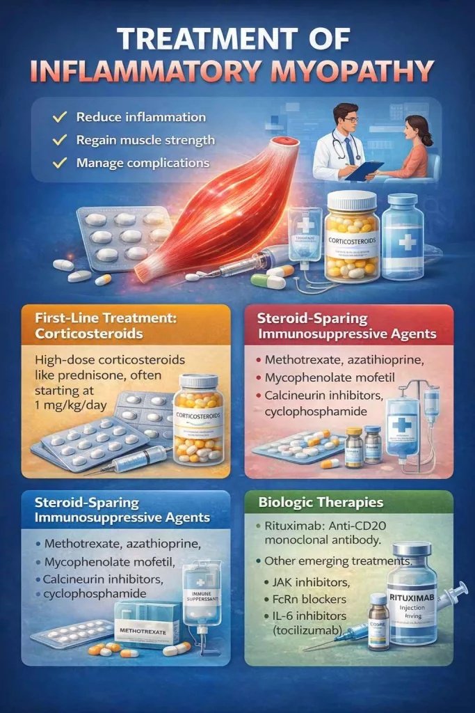
Overview of treatment strategies for inflammatory myopathy, from corticosteroids to advanced immunotherapies.
First-Line Treatment: Corticosteroids
Clinicians usually prescribe corticosteroids at a dose of 1 mg/kg/day, given as a single daily dose or in divided doses. In severe cases with marked weakness or organ involvement, they initiate treatment with intravenous methylprednisolone.
The treatment normally assumes the following:
- 4-8 weeks of high doses of corticosteroids till muscle enzymes are normal.
- Monthly reduction of dosage to the minimum.
- Attention to disease activity and steroid side effects.
Steroid-Sparing Immunosuppressive Agents
Methotrexate or azathioprine can be started alongside glucocorticoids, especially in patients who have a moderate or severe disease.
First-line steroid-sparing agents:
- Methotrexate: 15-25mg/week, works in most patients.
- Azathioprine: 2-3 mg/kg/day
The second-line agents to be used in refractory cases are:
- Mycophenolate mofetil: In lung disease
- Calcineurin inhibitors (Tacrolimus, Cyclosporine): Especially when it comes to the lungs.
- Cyclophosphamide: The final option: Use in case of serious, life-threatening disease.
Intravenous Immunoglobulin (IVIG)
The steroid and immunomodulating agents’ sparing effect was reported to be 40.9%. IVIG is particularly helpful for:
- Dermatomyositis
- Swallowing difficulties
- Patients are not able to tolerate other immunosuppressants
Biologic Therapies
Rituximab: Anti-CD20 Monoclonal antibody rituximab can be considered in the treatment of refractory cases. This B-cell-depleting treatment has proven successful in those patients who did not respond to traditional therapies.
Other emerging therapies:
- JAK inhibitors
- FcRN blockers
- IL-6 inhibitors (tocilizumab)
Treatment for Inclusion Body Myositis
Unfortunately, IBM does not usually respond well to immunosuppressive therapy. IBM has no standard course of treatment. The condition is mostly unresponsive to corticosteroids and immunosuppressive medications. Management focuses on:
- Physical therapy and exercise
- Assistive devices (cane, walkers, wheelchairs)
- Swallowing therapy for dysphagia
- Supportive care
Conclusion
Inflammatory myopathy is a complex of autoimmune conditions that has to be diagnosed and managed by experts and requires extensive management. Though these conditions may seriously influence the quality of life, the development of disease mechanisms, better diagnostic tools, and changing the treatment choices bring a ray of hope.
The importance of the early identification of symptoms, early diagnosis, and the prompt administration of proper treatment is essential to avoid irreversible muscle damage and severe complications. Through appropriate medical treatment, dietary changes, and constant observation, most patients with inflammatory myopathy will be able to have good control over the disease and lead fulfilling and productive lives.
It is important to keep in mind that, although the inflammatory myopathy is also a challenging condition, you can manage it, provided you are willing to apply the appropriate mix of medical knowledge, patient dedication, and holistic care.
References
[1] Dalakas MC. Inflammatory muscle diseases. N Engl J Med. 2015;372(18):1734-1747. doi:10.1056/NEJMra1402225
[2] Mammen AL. Autoimmune myopathies: autoantibodies, phenotypes and pathogenesis. Nat Rev Neurol. 2011;7(6):343-354. doi:10.1038/nrneurol.2011.63
[3] Tanimoto K, Nakano K, Kano S, et al. Classification criteria for polymyositis and dermatomyositis. J Rheumatol. 1995;22(4):668-674.
[4] Sontheimer RD. Cutaneous features of classic dermatomyositis and amyopathic dermatomyositis. Curr Opin Rheumatol. 1999;11(6):475-482. doi:10.1097/00002281-199911000-00006
[5] Needham M, Mastaglia FL. Inclusion body myositis: current pathogenetic concepts and diagnostic and therapeutic approaches. Lancet Neurol. 2007;6(7):620-631. doi:10.1016/S1474-4422(07)70171-0
[6] Mammen AL, Chung T, Christopher-Stine L, et al. Autoantibodies against 3-hydroxy-3-methylglutaryl-coenzyme A reductase in patients with statin-associated autoimmune myopathy. Arthritis Rheum. 2011;63(3):713-721. doi:10.1002/art.30156
[7] Connors GR, Christopher-Stine L, Oddis CV, Danoff SK. Interstitial lung disease with the idiopathic inflammatory myopathies: 2010;138(6):1464-1474. doi:10.1378/chest.10-0180
[8] Miller FW, Lamb JA, Schmidt J, Nagaraju K. Risk factors and disease mechanisms in myositis. Nat Rev Rheumatol. 2018;14(5):255-268. doi:10.1038/nrrheum.2018.48
[9] Smoyer-Tomic KE, Amato AA, Fernandes AW. Incidence and prevalence of idiopathic inflammatory myopathies among commercially insured, Medicare supplemental insured, and Medicaid enrolled populations. BMC Musculoskelet Disord. 2012;13:103. doi:10.1186/1471-2474-13-103
[10] Rider LG, Giannini EH, Harris-Love M, et al. Defining clinical improvement in adult and juvenile myositis. J Rheumatol. 2003;30(3):603-617.
[11] Fathi M, Lundberg IE. Interstitial lung disease in polymyositis and dermatomyositis. Curr Opin Rheumatol. 2005;17(6):701-706. doi:10.1097/01.bor.0000179948.65895.53
[12] Targoff IN, Miller FW, Medsger TA Jr, Oddis CV. Classification criteria for the idiopathic inflammatory myopathies. Curr Opin Rheumatol. 1997;9(6):527-535. doi:10.1097/00002281-199711000-00008
[13] Amato AA, Dumitru D. Acquired myopathies. In: Dumitru D, Amato AA, Zwarts MJ, eds. Electrodiagnostic Medicine. 2nd ed. Philadelphia: Hanley & Belfus; 2002:1258-1291.
[14] Joffe MM, Love LA, Leff RL, et al. Drug therapy of the idiopathic inflammatory myopathies: predictors of response to prednisone, azathioprine, and methotrexate and a comparison of their efficacy.
[15] Villarreal R, Linklater H, Rakheja D, Jorizzo J. Evaluation of treatment options for dermatomyositis. J Am Acad Dermatol. 2009;60(5):821-831. doi:10.1016/j.jaad.2008.11.883
[16] Dalakas MC, Illa I, Dambrosia JM, et al. A controlled trial of high-dose intravenous immune globulin infusions as treatment for dermatomyositis. N Engl J Med. 1993;329(27):1993-2000. doi:10.1056/NEJM199312303292704
[17] Oddis CV, Reed AM, Aggarwal R, et al. Rituximab in the treatment of refractory adult and juvenile dermatomyositis and adult polymyositis: a randomized, placebo-phase trial. Arthritis Rheum. 2013;65(2):314-324. doi:10.1002/art.37754