
 is characterized by the classical clinical tetrad of nail dysplasia, elbow deformities, hypoplasia or absence of...)
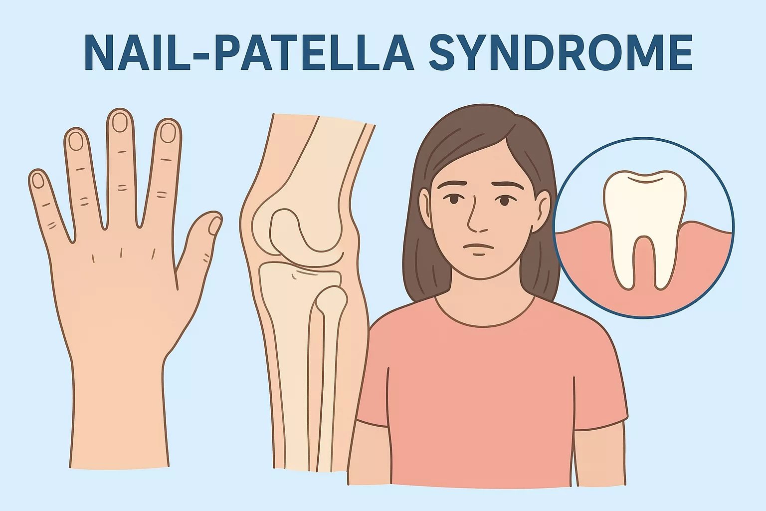
Nail patella syndrome (NPS) is characterized by the classical clinical tetrad of nail dysplasia, elbow deformities, hypoplasia or absence of patella, and presence of iliac horns. It is a rare, multisystemic, and autosomal dominant condition. Renal, neurological and ocular involvement can also occur in this condition. Dr E. M. Little first described the phenotype and hereditary nature of NPS.Nail patella syndrome revisited: 50 years after linkage. Ann Hum Genet. 2005 Jul;69(Pt 4):349-63.” style=”position:relative;color:#309b65;cursor:help;border-bottom:1px dotted #309b65;font-weight:bold”>[1]
The genetic basis of this condition can lead to significant variability in the clinical presentation of the patients. Diagnosis can be clinical through physical examination and radiological findings. Genetic testing can further aid in the confirmation of the diagnosis. It is common for families affected by NPS to remain undiagnosed for multiple generations. The prognosis of NPS is good, but complications can occur. The incidence of the syndrome is 1 in 50,000.[2]
Causes of Nail Patella Syndrome
Nail patella syndrome occurs due to a heterozygous loss-of-function mutation in the LMX1B gene on chromosome 9q34.1,[3] This gene encodes a transcription factor involved in multiple significant embryological developments. NPS is highly penetrant. However, there is inter- and intrafamilial variability that contributes to the condition’s variable clinical presentations.[4]
Clinical Presentation of Nail Patella Syndrome
Signs and symptoms of NPS are usually present at birth or during early childhood. They can vary in severity and range. Patients of NPS typically appear very lean. They have decreased muscle mass in the upper arms and legs. They find it challenging to obtain muscle mass and body weight. Another characteristic symptom is iliac horns. These are small horn-like outgrowths that develop on the bones of the pelvis.
Nail Abnormalities
The typical nail symptoms include the underdevelopment of finger and toenails.
Nails in NPS are usually:
- Hypoplastic, absent, or dystrophic nails
- Pitted
- Ridged horizontally
- Ridged longitudinally
- Separated into two halves by a longitudinal ridge or cleft of skin
- Thicker than usual
- Thinner than usual
- Discolored
- Limited to triangular lunulae[5]
Skeletal Abnormalities
Patients also develop skeletal abnormalities, including the patella and elbow. These abnormalities include:
- The arm bones that meet at the elbow can be underdeveloped or malformed.
- Excess skin webbing near the antecubital area (transition area between the anatomical arm and the forearm).
- Limited range of motion of the elbow.
- Inability to fully extend the forearm or supinate and pronate the forearm at the elbow joint.
- Patella can be absent, malformed and underdeveloped.
- Malformed, underdeveloped fibula and femur, which can lead to subluxation of the patella andbow leg deformities.
Ocular Symptoms
- NPS patients can present Lester’s sign (dark pigmentation around the iris that resembles a flower or clover leaf)[6]
- Open-angle Glaucoma (increased pressure in the eyes)
- Ocular hypertension
Renal Symptoms
NPS patients can present with kidney disorders that can lead to even renal failure. Kidney abnormalities due to NPS include:
- Microhematuria (microscopic traces ofblood in the urine)
- Proteinuria (high level of protein in the blood)
- Nephrotic syndrome, including edema and elevated creatinine
- Renal failure required dialysis or even kidney transplantation
Neurological symptoms
- Numbness
- Tingling
- Neuropathic pain
Diagnosis of Nail Patella Syndrome
Diagnosis of NPS starts from assessing the individual’s clinical presentation and family history of disease. The healthcare provider performs laboratory and imaging tests for further confirmation of the diagnosis.
Laboratory Studies
- Gene Sequencing: LMX1B sequencing is diagnostic in the majority of patients.
- Urinalysis: Performed to check for proteinuria without or with hematuria (an initial manifestation of NPS)
- Renal Biopsy: Used for the evaluation of progressive renal insufficiency, hypertension, and proteinuria in NPS.
- Light and Electron Microscopy: It usually reveals glomerulonephritis and basement membrane thickening. Electron microscopy detects the presence of collagen-like fibrils in the basement membrane, which cannot be observed with light microscopy.
Imaging Tests
Radiography plays an essential role in diagnosing most of the musculoskeletal characteristics associated with this condition. Imaging studies include CT scans, X-rays, and MRIs. These imaging tests demonstrate a small or absent patella and various morphological anomalies of the trochlea and condyles. They also indicate bilateral iliac horns, dysplasia, and subluxation.[7]
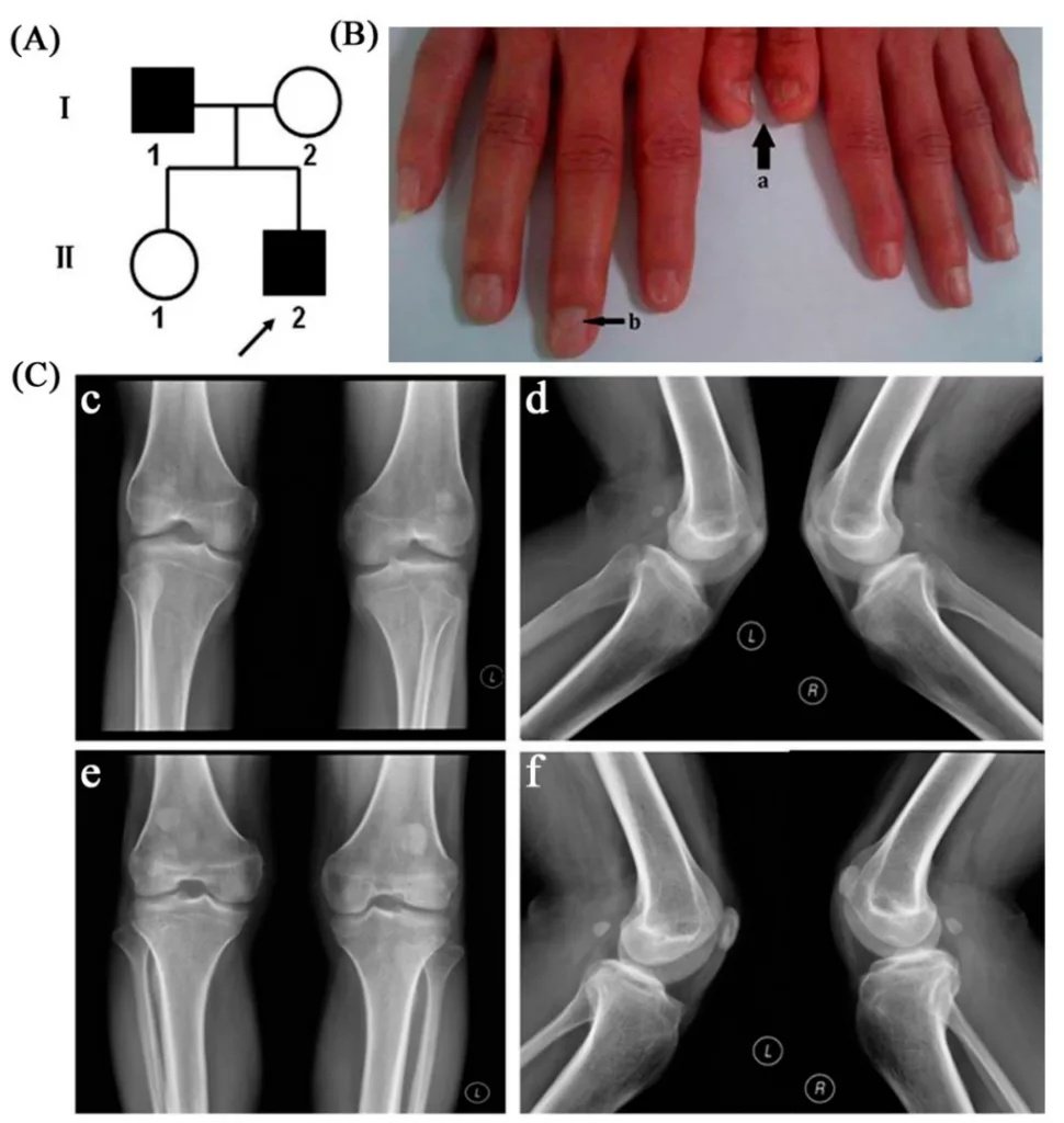
(A) Chinese pedigree with nail patella syndrome; patients are indicated by solid black, denoting the proband; (B) Clinical manifestation of the proband’s nails (a: short nail bed with longitudinal ridging;b: triangular lunula at the base of the nail); (C) Radiographic examination results of patients’ knee joint. The radiographs of the proband’s knee joint showed severe bilateral patellar dysplasia (c,d); The radiographs of the father showed bilateral hypoplastic, superiorly misplaced patellae (e,f). Image Courtesy: A Microdeletion of Chromosome 9q33.3 Encompasses the Entire LMX1B Gene in a Chinese Family with Nail Patella Syndrome et al.2014,doi.org/10.3390/ijms151120158, available via: https://www.mdpi.com/1422-0067/15/11/20158, CC BY 4.0.
Management and Treatment of Nail Patella Syndrome
Since there is no cure for NPS, treatment focuses on managing the symptoms.
Medical Treatment
Treatment of renal involvement focuses on slowing the progression of proteinuria. The two classes of drugs used for patients with genetic or acquired proteinuria are:
- Angiotensin receptor blockers
- Angiotensin-converting enzyme inhibitors[8]
Dialysis or transplantation may be required for end-stage renal disease.
For musculoskeletal abnormalities, treatment options include:
- Acetaminophen
- Analgesics such as non-steroidal anti-inflammatory drugs NSAIDS
- Physiotherapy, bracing, and splinting
As of now, treatment involves using eye drops containing a combination of beta-blockers and carbonic anhydrase inhibitors.
Surgical Management
Patellar pain and patellofemoral dislocation are the common symptoms of NPS. The healthcare provider tries to initially treat them with conservative measures. They switch to the surgical treatment option when they cannot achieve success via conservative treatment. There are multiple surgical approaches and techniques for the correction of patellar instability. Patellar realignment or synovial removal can improve symptoms.[9]
Due to a wide presentation of signs and symptoms, patients with NPS should be monitored routinely for new or worsening symptoms. Doctors monitor blood pressure for hypertension, the morning urine albumin to creatinine ratio for kidney disorders, and screening for glaucoma.
Differential Diagnosis and Prognosis
Several conditions can be mistaken for NPS due to its wide presentation and non-specific symptoms. However, the main differential diagnosis of NPS includes:
- Geniopatellar syndrome (lacks nail dysplasia but shares patella and genital anomalies)
- Patella aplasia-hypoplasia syndrome (present hypoplastic patellae)
- Radial and patellar aplasia or hypoplasia (present similar skeletal abnormalities)
- Meier-Gorlin syndrome (present patellar aplasia)
Patients with NPS have a normal lifespan. Conservative and surgical treatment exist for patients with patellofemoral symptoms. Nephropathy and glaucoma are the two potentially severe manifestations. But they are preventable and treatable with screening.
A Quick Review
Patients with NPS can develop multiple abnormalities ranging from mild to severe. This wide range of anomalies can make understanding the various clinical manifestations of the condition challenging. Genetic counselling should be offered to all patients with NPS and their families. If you or your partner has NPS and is planning to have a baby, you must visit your genetic counsellor. They will help you understand the chances of your baby inheriting this disorder, as there is no way to prevent it. A multidisciplinary approach is best to treat this condition.
References
[1] McIntosh I, Dunston JA, Liu L, Hoover-Fong JE, Sweeney E. Nail patella syndrome revisited: 50 years after linkage. Ann Hum Genet. 2005 Jul;69(Pt 4):349-63.
[2] Sweeney, E., Hoover-Fong, J. E., & McIntosh, I. (2020). Nail-patella syndrome.
[3] Lee, B. H., Cho, T. J., Choi, H. J., Kang, H. K., Lim, I. S., Park, Y. H., … & Cheong, H. I. (2009). Clinico-genetic study of nail-patella syndrome.Journal of Korean medical science,24(Suppl 1), S82-S86.
[4] Lovelace, P. D., & May, L. A. (2023). Nail-patella syndrome. InStatPearls [Internet]. StatPearls Publishing.
[5] Price, A., Cervantes, J., Lindsey, S., Aickara, D., & Hu, S. (2018). Nail-patella syndrome: clinical clues for making the diagnosis.Cutis,101(2), 126-129.
[6] Price, A., Cervantes, J., Lindsey, S., Aickara, D., & Hu, S. (2018). Nail-patella syndrome: clinical clues for making the diagnosis.Cutis,101(2), 126-129.
[7] Price, A., Cervantes, J., Lindsey, S., Aickara, D., & Hu, S. (2018). Nail-patella syndrome: clinical clues for making the diagnosis.Cutis,101(2), 126-129.
[8] Proesmans W, Van Dyck M, Devriendt K. Nail-patella syndrome, infantile nephrotic syndrome: complete remission with antiproteinuric treatment. Nephrol Dial Transplant. 2009 Apr;24(4):1335-8.
[9] Lippacher, S., Mueller-Rossberg, E., Reichel, H., & Nelitz, M. (2013). Correction of malformative patellar instability in patients with nail-patella syndrome: a case report and review of the literature.Orthopaedics & Traumatology: Surgery & Research,99(6), 749-754.