
 growth commonly present in the jaw. An odontoma is considered a type of odontogenic...)

Odontoma You Need An odontoma is a non-cancerous (benign) growth commonly present in the jaw. An odontoma is considered a type of odontogenic tumor but is more accurately classified as a hamartoma (a developmental malformation rather than a true neoplasm. It is also known as a dental hamartoma, which means it is a non-malignant anomaly composed of disorganized cells of tooth origin. Doctors identify them as benign calcified odontogenic tumors that have a prevalence of approximately 20–67% among all odontogenic tumors (making them the most common odontogenic tumor).[1]
Healthcare experts divide them into compound and complex types. Generally, these unusual growths are asymptomatic; hence, many patients are unaware of the disorder. In numerous cases, an odontoma diagnosis is made in routine radiographic analysis (X-rays). To prevent further dental complications, oral and maxillofacial surgeons treat the condition with surgical excision. Fortunately, there is a very low risk of recurrence.
What Is An Odontoma?: Odontoma You Need
An odontoma is a developmental anomaly composed of the very structures that make up a tooth, i.e., odontogenic origin. Many believe that, in reality, odontomas are hamartomas of aborted tooth formations. Just like a tooth, you will find variable amounts of tooth structures, i.e., enamel, dentin, and pulpal tissues. However, unlike a normal tooth, the structures are arranged in a disorganized pattern. Histopathological studies have shown that odontomas are made up of developed and well-differentiated enamel, dentin, cementum, and pulp tissues.[2] Enamel and dentin make up the crown portion of your tooth, while cementum is the protective covering of the tooth root. Odontomas constitute about 22% of all the odontogenic tumors of the jaw.
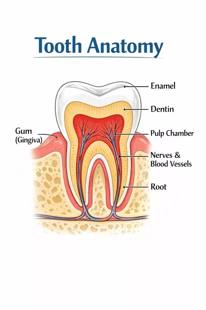
Different structures of a normal tooth are present in an odontoma as well.
Location
Odontomas are frequently found within the jawbone. The intrabone (intraosseous) lesions can grow in any region of the jaw. However, the following locations are preferred by these tumors:
- Anterior (front) maxilla, i.e., between the upper incisor and canine region.
- Posterior (back) mandible, i.e., between the premolar and molar area.
Dentists have also found that most odontomas arise on the right side of the jaw. Odontomas can appear as or be associated with supernumerary teeth. They are commonly associated with impacted or unerupted teeth and may interfere with normal eruption.
Types Of Odontomas
Based on the differences in appearance, organization of cells, and preferred locations, dental odontoma is classified into two types, i.e., compound and complex odontomas.
Compound Odontoma
This is the more common type of odontoma that generally grows slowly. According to one study, compound odontomas constitute 71.4% of all odontomas. The preferred site for a compound odontoma is the front side of your upper jaw (anterior maxilla).[3] This dental deformity is typically seen in young adults (below 20 years of age) and tends to have a slight preference for men.
This type of odontoma is often associated with unerupted or impacted teeth and may be found between adjacent teeth. Compound odontomas exist as multiple small tooth-like structures. The organization of cells/tissues in a compound type is very similar to a normal tooth. Therefore, on radiograph, they appear as radio-opaque (white) masses that resemble normal tooth structures. The well-defined masses are surrounded by a thin radiolucent band (black area).
These miniature teeth are asymptomatic. However, they can impede the normal eruption of a tooth.[4] They can also cause displacement of tooth roots.
Complex Odontoma
This type is characterized by a haphazard distribution of odontogenic layers. Unlike a compound odontoma, the orientation of enamel, dentin, and cementum in a complex odontoma does not resemble a normal tooth. Thus, on the radiograph, you will find dense, opaque masses surrounded by a darker line. Complex odontomas mostly occur on the posterior (back) region of the lower jaw (mandible). In most cases, they are asymptomatic but can cause problems when they grow to large sizes. Complications arising from large complex odontomas include evident swelling, delayed eruption of teeth, and displacement of erupted teeth.
A rare type of odontoma is the ameloblastic fibro-odontoma. It is usually diagnosed in the early stages of life (below 20 years of age). Like other types, this type also causes displacement of teeth and hindrance in the eruption of teeth.[5]
Odontoma Symptoms
In the vast majority of cases, odontomas do not cause any symptoms. This is why most diagnoses are made during routine checkups. In rare instances, an odontoma can cause pain and frequent infections. But these symptoms are only seen in severe cases.[6] The tooth-like growth can rarely cause neuralgic pain if the tumor compresses nearby nerves. Moreover, huge erupting odontomas can potentially cause infection (acute or chronic) of the paranasal sinuses.
Complications Associated With Odontomas
Large-sized odontomas are linked to the following complications:
Swelling
Large odontomas (especially erupted odontomas) can present with evident swelling in the oral cavity. Patients can notice severe swelling of the soft tissue in the floor of the mouth. In rare cases, excessive swelling induced by an odontoma can interfere with your breathing.[7] Expansion of the cortical bone is not common, but it can still be a finding.
Tooth Displacement
Large-sized odontomas present in the roots of an erupted tooth can displace the teeth. They are linked to malpositioning of the adjacent teeth. Studies show that large odontomas in the posterior region of the mandible can displace the adjacent molars. They may even alter the normal root morphology.[8]
Eruption Disturbance
Odontogenic tumors are frequently associated with issues in tooth eruption. Delayed eruption is seen in both deciduous and permanent teeth. A meta-analysis evaluating aspects of different odontoma types concluded that the most significant feature associated with odontoma is alteration in tooth eruption.[9] Delays in eruption (especially of permanent molars) are seen in 37-87% of cases.[10]
Odontoma Causes
The exact cause of odontoma is unknown. However, many expert researchers believe that several factors can cause developmental disturbances, which can eventually lead to tumor formation.
Genetics
Odontomas have been linked to different genetic syndromes like Gardner syndrome and otodental syndrome, etc. Thus, many researchers believe that genetics has a role in the occurrence of odontogenic tumors.[11]
Trauma
Local trauma endured during primary dentition can lay the foundation for an odontoma. In some pediatric cases, parents inform the doctors of trauma to the child in the exact same region where the odontoma has developed.[12]
Trauma to the face, head, and neck region can potentially disturb normal tooth development and cause rare anomalies (odontoma-like malformations).[13]
Infections
Past infections have been proposed as a contributing factor, although the evidence is limited. This is why doctors take the history of trauma and infection from people with odontomas.
Odontoma Diagnosis
Doctors identify odontomas with the help of radiographic images. Conventional dental radiographs, like panoramic X-rays, are considered primary tools in identifying the tooth-like anomalies in bone. However, modern times doctors rely on Cone Beam Computed Tomography (CBCT) as it provides a clearer 3D picture of the orodental structures.

A panoramic X-ray of a patient shows an odontoma in the right upper canine region of the patient. The lesion has displaced the root of the canine.
On a radiograph, odontomas appear as well-defined lesions that are radiopaque (white). The main well-circumscribed lesion is surrounded by a thin band of fibrous capsule, which is radiolucent (black). Most odontomas range in size from 1 to 2 cm in diameter. However, giant odontomas can exceed 3 cm.
Differential Diagnosis
Different pathologies have presentations similar to an odontoma. Some of these pathologies are cancerous growths that require early diagnosis and treatment. Therefore, it is important to differentiate between the conditions.
Osteoma Vs Odontoma
Both conditions are asymptomatic, slow-growing, and benign in nature. However, they both differ in the composition of the structures. While odontomas appear as tooth-like structures composed of enamel, dentin, pulp, and cementum, osteomas are bony, connective tissue tumors. Moreover, most odontomas arise in the anterior maxilla and posterior mandible region. On the other hand, the preferential site for osteomas is the ramus or condyle of the mandible.
Odontoma Treatment
Surgical excision of the slow-growing tumor is the go-to treatment modality for an odontoma. Unlike aggressive tumors such as ameloblastoma, odontomas are usually well-encapsulated and easily managed by surgical excision. As these anomalies are mostly diagnosed at young ages, doctors prefer performing odontoma surgery as soon as possible. This is done to prevent associated complications. In several cases, odontoma management requires an interdisciplinary approach. Usually, a pediatric dentist, oral/maxillofacial surgeon, and orthodontist collaborate to remove the anomalous growth and fix any teeth displacement, impaction of neighboring teeth caused by the odontoma.[14]
The process of removal is simple and conservative. There is minimal postoperative pain and complications. Complete healing of the bone takes six to twelve months.
Final Word
An odontoma is a benign tumor that grows slowly in your jawbones. It is a dental hamartoma, which means that the tumor is made up of tooth cells/tissues, i.e., enamel, dentin, pulp, and cementum, etc.
There exist two types of odontomas. Compound odontomas have tooth layers arranged in an orderly pattern, which makes them look like tooth-like structures on radiographs. These odontomas preferentially grow on the anterior maxilla. On the other hand, complex odontomas have enamel and dentin arranged in a disorderly manner and arise in the posterior mandible. The tumors are asymptomatic, but large odontomas can cause complications like displacement of roots, swelling in the oral cavity, and hindrance in the eruption of primary and secondary dentition.
The exact cause of odontoma formation is unknown. However, many believe that genetics, past trauma, and infections can disrupt the normal tooth development and lead to the development of benign calcified tumors. Doctors diagnose them during routine checkups. These tumors are seen as dark (radiolucent) masses on X-rays and CBCT scans. Oral surgeons treat the tumors by surgically excising them. There are very low chances of recurrence. Sometimes, orthodontic treatment is required to fix the misaligned/displaced roots (caused by an odontoma).
References
[1] Satish, V., Prabhadevi, M. C., & Sharma, R. (2011). Odontome: a brief overview.International journal of clinical pediatric dentistry,4(3), 177.
[2] da Costa Jerônimo, E. O., Gomes, M. S. J., Vicente, E. C., & Tempest, L. M. (2023). ODONTOMA-DIAGNOSTIC CRITERIA AND TREATMENT: LITERATURE REVIEW.Revista Contemporânea,3(11), 24449-24460.
[3] DeColibus, K. A., Rasner, D. S., Okhuaihesuyi, O., & Owosho, A. A. (2023). Clinicoradiopathologic analysis of odontomas: a retrospective study of 242 cases.Dentistry Journal,11(11), 253.
[4] Onda, T., Hayashi, K., Katakura, A., & Takano, M. (2022). Compound odontoma obstructing the eruption of a mandibular premolar.Oxford Medical Case Reports,2022(9), omac102.
[5] Kumar, L. S., Manuel, S., Khalam, S. A., Venugopal, K., Sivakumar, T. T., & Issac, J. (2014). Ameloblastic fibro-odontoma.International journal of surgery case reports,5(12), 1142-1144.
[6] Sánchez, O. H., Berrocal, M. L., & González, J. M. (2008). Metaanalysis of the epidemiology and clinical manifestations of odontomas.Med Oral Patol Oral Cir Bucal,13(11), E730-4.
[7] Jhamb, A. V., Dolas, R., Pandilwar, P., Jhamb, P. A., & Mohanty, S. (2012). Odontoma: Case series and report of cases complicated by infection and multiple denticles.Open J Stomatol,2, 44-9.
[8] Johar, H. F., Erlina, Y., & Muttaqin, G. F. (2025). Multidisciplinary Approach to Complex Odontoma in Growing Individuals: Two Clinical Cases.Annals of Orthodontics and Periodontics Specialty,5, 63-74.
[9] Sánchez, O. H., Berrocal, M. L., & González, J. M. (2008). Metaanalysis of the epidemiology and clinical manifestations of odontomas.Med Oral Patol Oral Cir Bucal,13(11), E730-4.
[10] Ohtawa, Y., Ichinohe, S., Kimura, E., & Hashimoto, S. (2013). Erupted complex odontoma delayed eruption of permanent molar.The Bulletin of Tokyo Dental College,54(4), 251-257.
[11] Schuch, L. F., Silveira, F. M., Pereira-Prado, V., Sicco, E., Pandiar, D., Villarroel-Dorrego, M., & Bologna-Molina, R. (2024). Clinicopathological and molecular insights into odontogenic tumors associated with syndromes: A comprehensive review.World Journal of Experimental Medicine,14(4), 98005.
[12] Santana, J. S., Delbem, A. C. B., Nagata, M. E., Pessan, J. P., de Morais, L. A., Cavazana, T. P., … & Hosida, T. Y. (2024). Odontoma como Causa de Retenção Prolongada de Dente Primário: Relato de Caso Clínico.ARCHIVES OF HEALTH INVESTIGATION,13(9), 2897-2901.
[13] Silva, D. R., & Shahinian, A. L. (2022). Odontoma malformation and disturbances of eruption subsequent to traumatic dental injuries: A literature review and a case report.Dental traumatology,38(2), 98-104.
[14] Muczkowska, N., Czochrowska, E., Masłowska, K., Wojtowicz, A., & Popowski, W. (2025). Combined surgical and orthodontic treatment of complex odontoma in growing patients: presentation of two cases.Dentistry Journal,13(2), 82.