
 is a rare condition characterized by...)

Osteonecrosis Jaw Symptoms Jaw osteonecrosis, avascular necrosis, or osteonecrosis of the jaw (ONJ) is a rare condition characterized by destruction and death of the jawbone, mostly caused by bisphosphonate therapy (for cancer/osteoporosis management). Rarely, it can also arise after a dental surgical procedure like extraction or implant placement, etc. However, it can also occur in healthy individuals. The incidence of ONJ in patients receiving bisphosphonate therapy for metastatic cancer was found to be 2.08%.[1]
Patients with bone disorders like osteoporosis are at a greater risk of the disease. A meta-analysis found the prevalence of medication-related osteonecrosis of the jaw in osteoporotic patients following a dental extraction to be 1.7%.[2] The exposed jawbone in the oral cavity usually presents with pain, soreness, and tooth mobility, etc. In mild-to-moderate cases, doctors opt for conservative therapies, but in severe cases, the dead jawbone can’t heal itself; therefore, doctors treat it by removing the necrotic (dead) bone.
What Is Osteonecrosis Of The Jaw?: Osteonecrosis Jaw Symptoms
“Osteo” means bone, and “necrosis” means cell death. Therefore, this rare yet serious condition means that bone cells present in your jawbone die. It may present with exposed necrotic bone or, in some cases, without visible bone exposure (non-exposed variant). This cell death leads to poking of the bone through your gums, which consequently leads to multiple symptoms. To better diagnose and treat the condition, doctors divide ONJ into 4 stages, which are mentioned below:
Stages Of Osteonecrosis Of The Jaw
Based on the severity of the disease, osteonecrosis is classified into:
Stage 0:
The condition is termed stage 0 if the patient is on anti-resorptive medicines (bisphosphonates) and experiences non-specific jaw pain. There is no visible exposed bone, but patients may have symptoms such as dull pain or radiographic changes without clear clinical findings.
Stage 1:
Dead bone is exposed, but there is little to no pain experienced by the patient.
Stage 2:
Symptoms appear along with bone exposure. Patients notice gum swelling and pain. Moreover, there are evident signs of infection.
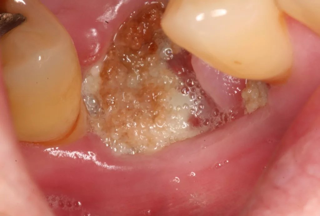
Exposed dead bone can be seen. “Medication Associated Osteonecrosis of the jaw Stage 2” Image courtesy of Coronation Dental Specialty Group, CC BY-SA 4.0, viaWikimedia Commons.
Stage 3:
The disease has spread beyond the alveolar bone (bone adjacent to your teeth). The symptoms are the same as stage 2, but now necrosis is seen in the sinuses and other parts of the facial bones, too.
Symptoms Of Osteonecrosis Of The Jaw
Exposed bone is known to cause a great deal of pain. Therefore, jaw necrosis can cause significant debilitation. Patients suffering from this bone disorder experience the following symptoms:
Pain
Usually, pain is one of the first signs of exposed bone in osteonecrosis of the jaw. Sufferers report pain in the jaw and the oral cavity. Initially, the condition may present with a dull ache in the mandible. Painful gums (mucous membranes) are often accompanied by other issues, like swollen gums and frequent oral ulcerations.[3] Pain arises due to the death of bone cells and consequent fluid buildup (edema) and swelling (inflammation).
Tooth Mobility
The loss of supportive bone ultimately results in loosening of the teeth. Patients notice mobility in their teeth. Clinical studies conclude that jaw pain, ulcers, and loose teeth with signs of infection are salient features of medication-related osteonecrosis.[4]
ONJ patients do not experience generalized tooth mobility. Loosening of the teeth is only seen in the necrotic bone region.[5] The combination of these symptoms makes eating difficult and leads to reduced quality of life.
Mouth Ulcers/Sores
Most of the time, there are chronic, non-healing ulcers. The sores are painful and cause difficulties with chewing, speaking, and swallowing. The presence of persistent ulcers (existing for more than 8 weeks) with bone exposure points towards ONJ. Ulcers are commonly found in areas of exposed bone and can occur on the upper (maxilla) or lower (mandible) jaw.
Gum Swelling And Pus Drainage
Swollen gums are seen in the majority of cases. This is because the dead (and exposed) bone known as the sequestrum acts like a foreign body that triggers chronic inflammation. This leads to soft tissue irritation, infection, and inflammation of the gums. Moreover, secondary infection causes discharge of purulent pus from the exposed bone. Foul-smelling pus is a key indicator of secondary jaw infection.
Causes Of Osteonecrosis Of The Jaw
In most of the cases, the main reason for jaw cell death is the result of a compromise in the blood supply to the bone cells. Slowing down or stoppage of blood flow to the hard tissues deprives the structures of nutrition. Therefore, experts term osteonecrosis as a type of avascular (lack of vascular supply) necrosis. In addition to reduced blood supply, other mechanisms include suppressed bone remodeling, infection, inflammation, and impaired angiogenesis. Based on the different underlying causes leading to the bone necrosis, osteonecrosis is divided into different types.
Types Of Osteonecrosis
The different conditions that ultimately lead to painful bone conditions in the jaw include:
Medication Related Osteonecrosis (MRONJ):
This type of ONJ usually develops when microbial contamination and local trauma occur in patients taking certain types of drugs. Antiresorptive and antiangiogenic drugs are linked to MRONJ.
These drugs are generally prescribed to osteoporosis patients. Bisphosphonates (first-line antiresorptive drugs) are given to osteoporosis and cancer patients (and other bone conditions) to strengthen bones and reduce fracture risks. Antiresorptive medicine like denosumab works by slowing down the process of bone breakdown, and antiangiogenic drugs prevent the formation of new blood vessels (which starve the tumors of nutrients, consequently leading to their death). Bisphosphonate-related osteonecrosis of the jaw (BRONJ) is a major concern in patients taking the drug. The condition results from suppressed bone remodeling, reduced healing capacity, and increased susceptibility to infection.[6]
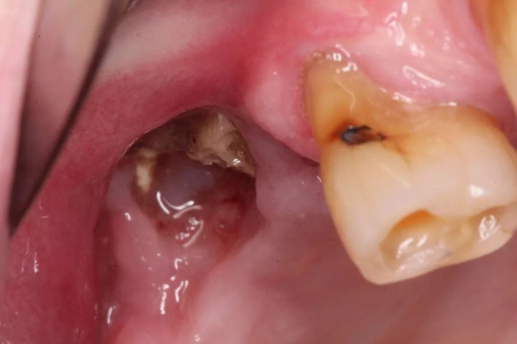
Exposed necrotic bone following upper 1st molar extraction in a patient on denosumab (anti-resorptive medicine). Image courtesy “MRONJ in a patient on denosumab upper right molar” byIan Frust, made available by Wikimedia Commons under CC BY-SA 4.0.
So, the question arises, why do bisphosphonates cause osteonecrosis of the jaw?
The over-suppression of bone metabolism/remodeling by bisphosphonates also causes the prevention of microdamage repair in the bone cells. This eventually results in the formation of dead bone (sequestrum). Microdamages occur when the bone is exposed to trauma (like tooth extraction) or infection.[7]
Traumatic Osteonecrosis:
Bone necrosis after direct trauma is not very frequently seen, but still poses a debilitation. Traumatic ONJ can be brought about by chemical, mechanical, or thermal trauma/injury to the bone. Impact trauma to the maxillofacial region sustained during falls, brawls, and traffic accidents can trigger cell death in the jaw bones. This can be attributed to impaired arterial supply to the bone following traumatic injuries.[8]
Rarely, leaking of pulp devitalizing materials like sodium hypochlorite beyond the root canal system (into the surrounding bone) can trigger osteonecrosis by chemical trauma.
Non-Traumatic Osteonecrosis:
This type of bone death sequelae is linked to cancers, acquired and congenital pathologies. Diabetes mellitus compromises your ability to heal vascular breakage; thus, denture trauma can result in acquired non-traumatic ONJ in such patients. Moreover, oral metastatic (cancerous) lesionsimpact the blood supply of the bones.
Infection of the bone, i.e., osteomyelitis, can atypically present with exposed necrotic bone in patients. Infections like osteomyelitis may mimic or coexist with ONJ. Other infections that can contribute to the cause include TB, syphilis, shingles, and actinomycosis. Moreover, intranasal use of narcotics like cocaine is known to cause soft and hard tissue destruction. Prolonged usage can eventually result in small areas of necrosis and sequestration (poking of dead bone cells). All these irritating events lead to bone hypoxia and bone resorption.[9]
Spontaneous Osteonecrosis:
It arises without any apparent cause and is termed idiopathic (without an identifiable cause) ONJ. Spontaneous osteonecrosis usually starts with ulceration. This condition, i.e., oral ulceration with bone sequestration, is rare, and clinical data about it are scarce.
Osteoradionecrosis (ORN) is a severe type of jawbone cell death that is induced by exposure to radiation therapy (for cancer treatment). Some clinicians consider ORN a type of ONJ, while others consider it a separate entity.[10]
Risk Factors
The bone disorder usually arises when there is a combination of different factors. Based on the high prevalence of ONJ in certain groups, experts identify the following conditions/habits as risk factors for osteonecrosis:
- Cancer treatment (chemotherapy)
- Old age (above 65 years)
- Facial trauma or fracture
- Diabetes mellitus
- Cigarette smoking
- Dentures(especially ill-fitting ones)
- Immunocompromise (due to long-term corticosteroid usage)
- Neoplasm (oral malignancy)
How Is Osteonecrosis Of The Jaw Diagnosed?
The primary step in diagnosing jaw osteonecrosis is history taking. Your dentist will take a full account of the symptoms and any associated previous activities (dental extraction, trauma, etc.). Due to the high prevalence of MRONJ, the drug history of IV bisphosphonates is crucial. A dentist physically examines the mouth to see areas of necrotic and exposed bone. A key diagnostic criterion is exposed bone (or bone that can be probed) persisting for more than 8 weeks in a patient with relevant drug history and no history of radiation therapy to the jaws.
Imaging Scans
At times, dentists take help from imaging scans. Dental imaging, like X-rays or MRI scans, can aid in detecting the extent of bone destruction.
Differential Diagnosis
ONJ has presentations similar to the following conditions:
Osteomyelitis vs Osteonecrosis Of The Jaw
While both are painful conditions that cause destruction of the jawbone, osteomyelitis is the result of bacterial (mostly) bone marrow infection, and ONJ is bone cell death induced mainly by bisphosphonates. Systemic features like fever are more common in osteomyelitis but may also occur in advanced ONJ with secondary infection.
Treatment For Osteonecrosis Of The Jaw
The therapeutic options for osteonecrosis depend on the severity of the disease. Current guidelines favor conservative management in early stages and reserve surgery for advanced disease. In general, a team of clinicians, i.e., oral surgeons, oncologists, and rheumatologists, treats the condition (depending on the underlying cause). Experts opt for treatment options according to the stage of ONJ. All treating doctors prefer adopting a conservative approach over surgery.

In stage 0, doctors keep the patient under observation. They may prescribe prophylactic antibiotics and antiseptic mouthwashes to prevent an infection (which plays a role in the occurrence of the disease). However, management focuses on pain control and monitoring.
Stage 1
At this stage, your healthcare provider will perform a procedure called debridement. In this conservative surgical approach, the surgeon scrapes away the dead and exposed bone cells with the help of bone saws and rongeurs. The patient is advised to use oral antibiotics with antiseptic mouthwashes (containing chlorhexidine). This treatment type prevents further bone loss and allows the gums to heal.
Stages 2 and 3
For advanced stages with symptoms, doctors perform sequestromy. In this procedure, doctors surgically remove the necrotic (dead) jawbone. Any affected teeth and a small amount of healthy tissue surrounding the dead bone are also removed to ensure a better prognosis. In case of disease progression into the sinus, patients may need to undergo sinus surgery as well.
Nowadays, doctors are using regenerative therapy with surgery to improve outcomes. Leukocyte-rich and platelet-rich fibrin (L-PRF) therapy is being used as an adjunctive therapy to surgical debridement in stages 2 and 3 for enhanced results.[11]
At times, sequestromies are paired with ridge modeling and reconstruction procedures to rebuild soft and hard tissues. After surgery, patients are prescribed mouth rinses, painkillers, and systemic (oral) antibiotics.
Stem-Cell Therapy
Due to the increasing cases of MRONJ, health professionals are looking for efficient and novel therapeutic ways of management. Stem cell therapy has shown some good results. However, exosome therapy has proven to be a more potent therapy.[12] It is an advanced regenerative treatment in which tiny vesicles (derived from stem cells) are used to deliver growth factors to the affected site. It is important to note that stem cell and exosome therapies are currently experimental and lack sufficient clinical evidence for routine use.
Osteonecrosis Of The Jaw Prognosis
The prognosis of surgical management is generally good. Early-stage disease has a better outcome with conservative management, while advanced stages may require surgical intervention. Reported cure rates vary widely depending on study population and treatment approach. According to a paper by the American Association of Oral and Maxillofacial Surgeons, the overall cure rate of the disease is 45.7%. Conservative treatment has a poor prognosis, but the overall cure rates were:[13]
- 25.8% at 12 months
- 50.8% at 36 months
- 72.4% at 60 months
How Can I Prevent ONJ?
A key preventive strategy is dental evaluation and necessary treatment before starting antiresorptive or antiangiogenic therapy.
Other strategies include:
- Using atraumatic surgical techniques when extraction is necessary
- Maintaining good oral hygiene
- Avoiding invasive dental procedures during therapy when possible
- In case of an invasve treatment like extraction, several strategies have been adopted by expert clinicians to prevent the onset of ONJ. Primary closure involves sealing the extraction site with a gum (mucosal) flap. In an epi-periosteal flap, a tissue flap without the periosteum (a membrane that directly covers your bone) is used to cover the wound. These techniques have good and similar results in preventing osteonecrosis of the jaw.[14]
Final Word
Osteonecrosis of the jaw is a painful condition in which the jawbone dies and pokes out of your gums. Dead bone (sequestrum) results from compromise in the blood flow to the hard tissues. Doctors classify it into stages 0 to 3. There are no symptoms in stage 0, no pain despite bone exposure in stage 1. Pain is and gum swelling is present in stage 2, while necrosis extends into the sinuses in stage 3.
Patients experience pain, gum swelling/pus discharge, frequent oral ulcers, and tooth mobility, etc. Osteonecrosis of the jaw is caused mainly by IV use of antiresorptive medicines (like bisphosphonates and denosumab) in osteoporosis/cancer patients. Moreover, mechanical, chemical, or thermal trauma can trigger ONJ. Non-traumatic ONJ is the outcome of neoplasms (cancers) and infections, while spontaneous ONJ arises due to unknown (idiopathic) causes.
Doctors diagnose the condition as ONJ if the bone exposure exists for more than 8 weeks. For mild (stage 1) cases, doctors prescribe oral antibiotics and mouthwashes, while for moderate cases, surgeons perform debridement of the bone. For stages 2 and 3, surgeons remove the dead bone (sequestromy) along with any mobile teeth and reconstruct/remodel the bone. Modern regenerative therapy (exosome and platelet-rich fibrin) has shown promising results in improving symptoms.
References
[1] Zhang, C., Shen, G., Li, H., Xin, Y., Shi, M., Zheng, Y., … & Zhao, J. (2024). Incidence rate of osteonecrosis of jaw after cancer treated with bisphosphonates and denosumab: A systematic review and meta‑analysis.Special Care in Dentistry,44(2), 530-541.
[2] Kostares, E., Kostare, G., Kostares, M., Pitsigavdaki, F., Perisanidis, C., & Kantzanou, M. (2025). Prevalence of Osteonecrosis of the Jaw Following Tooth Extraction in Patients with Osteoporosis: A Systematic Review and Meta-Analysis.Journal of Clinical Medicine,14(17), 5988.
[3] Sharma, S., Shankar, R., Kiran, B. S. R., Breh, R., Sarangi, S., & Upadhyay, A. K. (2023). A narrative review of osteonecrosis of the jaw: what a clinician should know.Cureus,15(12), e51183.
[4] Kuehn, S., Scariot, R., & Elsalanty, M. (2023). Medication-related osteonecrosis: why the jawbone?.Dentistry Journal,11(5), 109.
[5] Xie, R., Wang, W., Bian, L., Qian, Y., Li, J., & Zhang, H. (2024). Comparative clinical study of phosphorous necrosis and medical-related osteonecrosis of the jaws.Clinical Oral Investigations,28(2), 147.
[6] Srivichit, B., Thonusin, C., Chattipakorn, N., & Chattipakorn, S. C. (2022). Impacts of bisphosphonates on the bone and its surrounding tissues: mechanistic insights into medication-related osteonecrosis of the jaw.Archives of Toxicology,96(5), 1227-1255.
[7] Gupta, M., & Gupta, N. (2018). Bisphosphonate related jaw osteonecrosis.
[8] Alalawi, W. A., & Almajed, E. (2018). Unilateral hard palate necrosis after ascending palatine artery embolization.Journal of Craniofacial Surgery,29(5), e437-e438.
[9] Cheng, E. Y., & Mirzaei, A. (2025). Classical non-traumatic osteonecrosis versus osteonecrosis of the jaw: Distinct manifestations of a shared pathophysiological spectrum.Journal of Orthopaedics.
[10] Kün-Darbois, J. D., & Fauvel, F. (2021). Medication-related osteonecrosis and osteoradionecrosis of the jaws: Update and current management.Morphologie,105(349), 170-187.
[11] Yalcin-Ülker, G. M., Duygu, G., Tanan, G., Cakir, M., & Meral, D. G. (2023). Use of leukocyte-rich and platelet-rich fibrin (L-PRF) adjunct to surgical debridement in the treatment of stage 2 and 3 medication-related osteonecrosis of the jaw.Journal of Craniofacial Surgery,34(3), 1039-1044.
[12] Mazreku, M., Danišovič, L. U., Klein, M., & Kleinová, M. (2025). Recent stem-cell-based and stem-cell-free possibilities for the therapeutic management of the osteonecrosis of the jaw.Biomolecules,15(4), 595.
[13] Kaibuchi, N., Hoshi, K., Yamazaki, A., Miyamoto-Sangu, N., Akagi, Y., & Okamoto, T. (2021). The progress of medication-related osteonecrosis of the jaw with conservative initial treatment: A 12-year retrospective study of 129 patients.Bone Reports,14, 101072.
[14] Ribeiro, L. N., Severino‐Lazo, R., Vasconcelos, B. C. D. E., de Moraes, S. L. D., & Carvalho, M. D. V. (2025). Preventive Treatments for Osteonecrosis of the Jaw: A Systematic Review and Network Meta‐Analysis.Oral Diseases.