

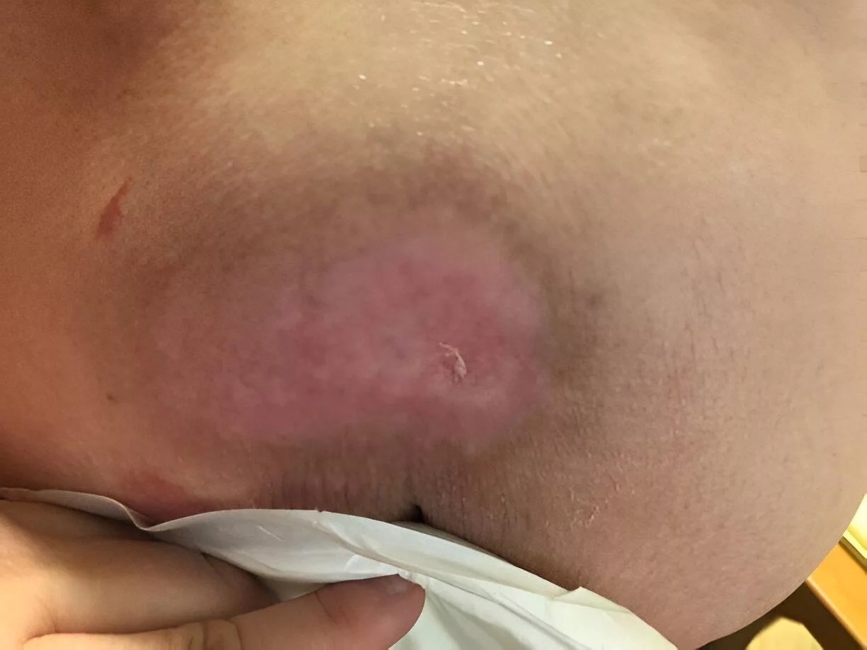
Pressure Ulcers Risks Pressure ulcers, also known as pressure injuries, bedsores, or decubitus ulcers, are localized skin and soft tissue injuries that occur due to prolonged pressure and shear exerted over the bony prominences. While they are most commonly associated with immobility, they can also develop in patients with medical devices such as casts, braces, or oxygen tubing that exert continuous pressure on the skin. Pressure ulcers can vary in their characteristics and severity. More than 70% occur at high-risk sites such as the sacrum, heels, and ischial tuberosities, but they can also develop at the trochanter, occiput, scapula, lateral malleolus, elbow, shoulder, and ear. This condition demands prompt treatment to avoid potential lethal complications.
Although it is a preventable condition in most cases, it continues to pose a major health burden by affecting more than 3 million individuals annually in the United States. There is an increased national attention to the prevalence of this condition, which has largely remained unchanged, accompanied by a continuous increase in the cost of its care.[1]
Stages of Pressure Ulcers: Pressure Ulcers Risks
There are different methods to stage the pressure ulcers. Kottner and colleagues reviewed all the most common global classifications in 2020. The most widely accepted classification system is the National Pressure Injury Advisory Panel (NPIAP) in the United States.[2]The stages of pressure ulcers according to NPIAP are as follows:
Stage I:
Stage I is the mildest one. It only affects the upper layer of your skin. In this stage, the wound has not opened yet. The skin may look pink or red, but it can be hard for people with darker skin colors to see the color change. Your skin may feel tender to the touch, warmer, or cooler than normal.
Stage II:
In the second stage, the sore area breaks through the top layer of skin and some of the layer below the skin. This breakdown creates a shallow, pink or red base open wound. You can also see skin abrasions or blisters.
Stage III:
The notable wound extends to the subcutaneous tissue. The wound has greater depth, often with drainage or slough, but bone, tendon, and muscle are not exposed.
Stage IV:
It is the most serious stage of pressure ulcers. The wound penetrates below the subcutaneous fat into your deep tissues, including tendons, muscles and ligaments. In some of the most severe cases, the wounds can extend down to the cartilage and bones. Undermining and sinus tracts may also be present.
Unstageable:
The depth is unknown, as the depth of the wound or sore is confusing. They are hard to diagnose because the bottom of the sore is covered with escher (hard plaque that’s black or brown) and slough (debris that appears green, yellow, or brown).
Deep Tissue Injury:
It is another category mentioned in the NPIAP guidelines, and this injury occurs with prolonged pressure and shear forces at the interface of bone and muscles. It results in non-intact or intact skin with deep red, purple, or maroon, persistent discolouration. However, it can not be used to describe the neuropathic, traumatic, or dermatological injuries.
Causes & Risk Factors for Pressure Ulcers
The development of the condition is a complex and multifactorial process. Both internal and external factors contribute to the formation of these ulcers. The intrinsic factors (patient-related) that can speed up the process of tissue damage include:
- Anemia
- Malnutrition
- Endothelial dysfunction
- Older age and frailty
- Cognitive impairment
The extrinsic factors that can speed up the process of tissue deformation and ischemia are:
- Prolonged pressure
- Shear force
- Friction
- Moisture
The other risk factors identified by the researchers include:The patients with the following conditions can exhibit a predisposition to decubitus ulcers:
- Prolonged anesthesia
- Neurological disorders
- Dehydration
- Hypotension
- Malnutrition
- Cardiovascular disorders
- Surgical patients
The dysfunction of the neurological mechanisms responsible for regulating local blood flow can lead to the formation of pressure ulcers. Immobility in bedridden patients undergoing surgery for two hours is enough to form the foundation of these ulcers.[3] Prolonged pressure on the tissues can also cause occlusion in the capillary bed and reduce the levels of oxygen in that area. After some time, ischemic tissues begin to accumulate toxic metabolites, and subsequently, tissue necrosis and ulceration occur.
Pathophysiology of the Pressure Ulcers
The tissues can withstand an abnormal amount of external pressure. However, constant pressure exerted over a prolonged time can cause the abnormality. An external pressure that exceeds the arterial capillary pressure of 32 mm Hg can impede the flow of blood. Additionally, pressure that is greater than the venous capillary closing pressure of 8 – 12 mm Hg can impair the return of the venous blood. Prolonged pressure above these values causes ischemia and necrosis.
Moisture can lead to or worsen existing ulcers through tissue breakdown and maceration. Friction caused by skin rubbing can lead to the development of ulcers by contributing to breaks in the superficial layers of the skin. Researchers have also found a link between the pathophysiology of COVID-19 and the development of pressure ulcers. The pro-inflammatory cytokines interleukin-6 and tumor necrosis factor-alpha (found abundantly in COVID-19 patients) can contribute to the onset and maintenance of inflammation that occurs after necrosis and the formation of pressure ulcers.[4]
Signs & Symptoms of Pressure Ulcers
Signs and symptoms may vary according to the stage of the condition. As the affected area has no tears or surface breaks at the first stage, the symptoms can include:
- The affected site appears red in lighter skin tones and purple, blue, or darker in people with darker skin tones.
- It has a higher temperature than the surrounding normal tissues.
- Firmer than surrounding tissues
- Sore to touch
- It can cause a mild itching or burning sensation.
In stage II, pressure ulcers may appear as:
- Pain
- A crater-like or shallow wound or blister that may burst
- Drainage or pus may begin to appear, signaling early tissue damage.
- Sore, swollen, or red tissue around the ulcer
In the third stage, the symptoms can progress into:
- The wound deepens into a crater-like lesion
- Discolored drainage
- Foul odor
You can notice the following symptoms in stage IV of this condition:
- Extreme pain
- Foul smell and pus are signs of infection
- Dead tissue turns black, forming a hard, dry covering known as eschar
- Large ulcers may expose muscle, tendon, or bone
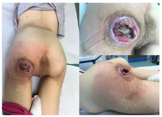
The patient with pressure ulcers in ischial region. Image Courtesy: Reconstructive Options for Pressure Ulcers in Pediatric Patients by Krakowczyk et al. 2024, doi.org/10.3390/children11060691, available via https://www.mdpi.com/2227-9067/11/6/691 CC BY 4.0.
Diagnosis of Pressure Ulcers
Pressure injuries are diagnosed primarily via visual inspection of the skin and its surrounding tissues.
History & Physical Examination:
At first, the healthcare provider carefully examines the affected area, size, depth and discharge from the sore. They can also stage the ulcers depending on their appearance. A thorough history and overall assessment of the patient are necessary to make a diagnosis. The clinicians gather the following information:
- Discharge or foul odor from the ulcer site.
- Duration of the hospital stay.
- Duration of being bedridden or immobile.
- Assessment of the associated medical causes that can cause the pressure injury (stroke, road accident, etc.)
- Natural history of the injury and the site at which it was first developed.
- A brief history of any systemic disease, such as diabetes or peripheral vascular disease.
- Patients’ nutritional status through examination and diet history.
Additional Assessment Options:
- A healthcare provider may perform a blood test, such as blood cultures, to check for the infection.
- In some cases, a tissue sample can be taken to determine the extent of the tissue damage. A biopsy can also rule out other conditions.
- X-rays or MRIs can also be used in some cases to assess the depth of the ulcer and rule out bone infection (osteomyelitis).
Risk Assessment:
While assessing the characteristics of the wound, it is essential to keep the risk assessment instruments in mind to lower the risk of the incidence of pressure injury. There are three risk assessment scales:
- Braden Scale
- Norton Scale
- Waterlow scale
The most widely adopted is the Braden Scale, which evaluates:
- Sensory perception
- Moisture
- Activity
- Mobility
- Nutrition
- Friction and shear
The scores that indicate the risk are:
- Nine or below: very high risk
- 10 – 12: high risk
- 13 – 14: moderate risk
- 15 – 18: mild risk[5]
Management & Treatment
Management and treatment of pressure can be non-surgical and surgical. Depending on the severity, the pressure ulcer can take weeks or months to heal. Stage one and two pressure ulcers are easy to manage and treat.
Non-Surgical Management:
Pressure ulcers in stages one and two are manageable and treatable without the need for surgery.
Stage I & Stage II
- The first step in managing stage I and stage II ulcers is to remove pressure from the area, as any excess pressure can cause the ulcer to break through the skin surface.
- Keep the affected area clean and dry to reduce tissue damage. If you are bedridden, change your position or add blankets or a pillow for extra padding.
- Additionally, staying hydrated and eating diet rich in protein, vitamins, and minerals support tissue repair.[6]
Stage III & IV
- Stage III and IV ulcers usually need medical intervention. If abscesses or necrotic tissue are present, clinicians perform debridement (removal of devitalized tissue), which may be surgical, enzymatic, autolytic, or mechanical depending on the wound.
- Antibiotics are prescribed only if there are clear signs of infection such as cellulitis, osteomyelitis, or systemic sepsis.
- Dressings are chosen based on wound depth, presence of infection, and exudate levels. According to NPIAP recommendations, silicone foam dressings are commonly preferred. Other dressing options include hydrocolloid, hydrogel, alginate, or negative-pressure wound therapy (vacuum-assisted closure) for large or exudative wounds.
- Topical antiseptics like hydrogen peroxide or iodine are generally avoided as they may damage healthy tissue, but antimicrobial dressings (such as silver-based) can be used in selected infected wounds.
Surgical Management:
Surgical management is intended to fill the dead space and offer durable skin through flap reconstruction. The surgical treatment options for pressure ulcers include:
Surgical Debridement
During surgical debridement, doctors carefully remove the non-viable tissue using surgical instruments like electrocautery or a scalpel. It can also involve removing the affected bone in some cases. It creates a clean wound bed with healthy tissue to encourage healing.
Skin Grafting
Skin grafting involves transplanting healthy skin from one area of the body to the ulcer site. After grafting the skin onto the site, it is secured with sutures, staples, or a special dressing. It accelerates the healing process, improving both function and appearance. Skin grafts for pressure ulcers require careful consideration of factors such as the patient’s overall health, vascularity of the wound bed, and depth of the ulcers, which can impact the healing.[7]
Prevention of Pressure Ulcers
The preventive strategies that can lower the risk of pressure ulcers include:
- Changing positions after some time is necessary if you are bedridden or in a wheelchair
- Eat a nutritious diet and stay hydrated.
- Check your skin regularly for changes in color and sensations
- Keep your skin dry and clean.
- Apply a moisture barrier cream to protect our skin
- Participate in physical therapies.
- Use mattresses or foam cushions that can ease pressure on your skin[8]
- Change and wash your bedsheets, clothes and undergarments on a regular basis.
- Quit smoking, as nicotine can slow wound healing.
Differential Diagnosis
The differential diagnosis of pressure ulcers includes the following conditions:
- Venous ulcers
- Diabetic ulcers
- Dermatitis
- Osteomyelitis (infectious disease of the bone marrow)
- Pyoderma gangrenosum (inflammatory skin condition)[9]
Prognosis of Pressure Ulcers
The prognosis varies depending on the anatomic location, stage of injury and treatment regimen. Factors that can affect the prognosis are stage and size of pressure ulcer, and advancing age.[10] Early-stage pressure ulcers have a good prognosis with appropriate care. The later stages are difficult to heal and can lead to severe complications like infection.
Complications
Complications often develop with pressure ulcers.
- There is a risk of potentially life-threatening bacterial infections, such as septicemia and cellulitis. The patient can also develop sepsis or require an amputation.
- Some patients can also develop sinus tracts.
- Bacteria in your bloodstream can cause endocarditis or meningitis, osteomyelitis or septic arthritis.
- If there is inadequate post-operative care, then complications secondary to the reconstructive surgery (abscess formation, wound dehiscence, seroma or hemotoma formation) can also occur.
- The wounds are catabolic, and they can cause severe fluid and protein loss. They can result in malnutrition or hyponatremia. Due to a draining ulcer, the body loses about 50 grams of protein.
Final Remarks
Pressure injuries occur when there is prolonged pressure on specific body areas that are more susceptible to friction and shear force injuries. They can vary in size, depth, and chronology. Additionally, the staging system helps interprofessional teams make the appropriate interventions. Consulting a clinician can aid in guiding the treatment and yield better results.
References
[1] Mervis, J.S. and T.J. Phillips, Pressure ulcers: pathophysiology, epidemiology, risk factors, and presentation. Journal of the American Academy of Dermatology, 2019. 81(4): p. 881-890.
[2] Kottner, J., et al., Pressure ulcer/injury classification today: An international perspective. Journal of Tissue Viability, 2020. 29(3): p. 197-203.
[3] Bansal, C., et al., Decubitus ulcers: a review of the literature. International journal of dermatology, 2005. 44(10): p. 805-810.
[4] Gefen, A. and K. Ousey, COVID-19: pressure ulcers, pain and the cytokine storm. Journal of wound care, 2020. 29(10): p. 540-542.
[5] Zaidi, S.R.H. and S. Sharma, Pressure ulcer, in StatPearls [Internet]. 2024, StatPearls Publishing.
[6] BoykoTatiana, V., T. LongakerMichael, and P. YangGeorge, Review of the current management of pressure ulcers. Advances in wound care, 2018.
[7] Ricci, J.A., L.R. Bayer, and D.P. Orgill, Evidence-based medicine: the evaluation and treatment of pressure injuries. Plastic and reconstructive surgery, 2017. 139(1): p. 275e-286e.
[8] Teasell, R. and D.K. Dittmer, Complications of immobilization and bed rest. Part 2: Other complications. Canadian Family Physician, 1993. 39: p. 1440.
[9] Okhovati, S., M. Esmaeili, and E. Shariat, Effect of intensive care unit nurses’ empowerment program on ability in visual differential diagnosis of pressure ulcer classification. Critical care nursing quarterly, 2019. 42(1): p. 89-95.
[10] Takahashi, P.Y., Pressure ulcers and prognosis: candid conversations about healing and death. Geriatrics (Basel, Switzerland), 2008. 63(11): p. 6-9.