
 is a rare lung disorder that causes the accumulation of surfactant within the alveoli. Surfactant is a fatty...)
What is Pulmonary Alveolar Proteinosis?
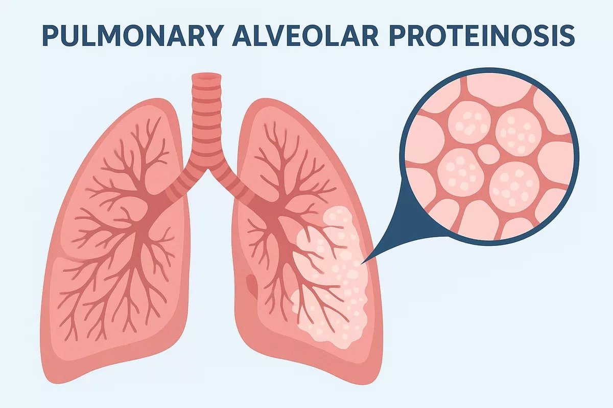
Pulmonary alveolar proteinosis (PAP) is a rare lung disorder that causes the accumulation of surfactant within the alveoli. Surfactant is a fatty and protein-rich material. Samuel H. Rosen et al. first described PAP in 1958. It develops due to impaired surfactant clearance, which may result from autoimmune antibodies, genetic mutations, or toxic exposures. PAP can be classified into three types: autoimmune (previously called idiopathic), secondary, and congenital.
The prevalence ranges from 3.7 to 40 cases per million. However, it can vary from country to country. The incidence is approximately 0.2 cases per million. Among them, approximately 90% of adult cases are autoimmune PAP. Males are more commonly affected than females at a 2:1 ratio. Congenital PAP is the rarest form and results from genetic mutations affecting surfactant production or clearance.
Etiology and Pathophysiology of PAP
The exact cause of PAP depends on its subtype. In all forms, the central problem is the accumulation of surfactant within the alveoli due to defective clearance by alveolar macrophages. The alveoli have thin walls that allow oxygen to pass from your lungs to your blood. The walls have a thin layer of surfactant that prevents collapse and maintains normal breathing function. Under normal circumstances, alveolar macrophages continuously remove excess surfactant, keeping the air sacs clear. However, in PAP, this clearance mechanism fails. The macrophages are either functionally impaired or reduced in number, causing surfactant to build up and block oxygen diffusion into the bloodstream.
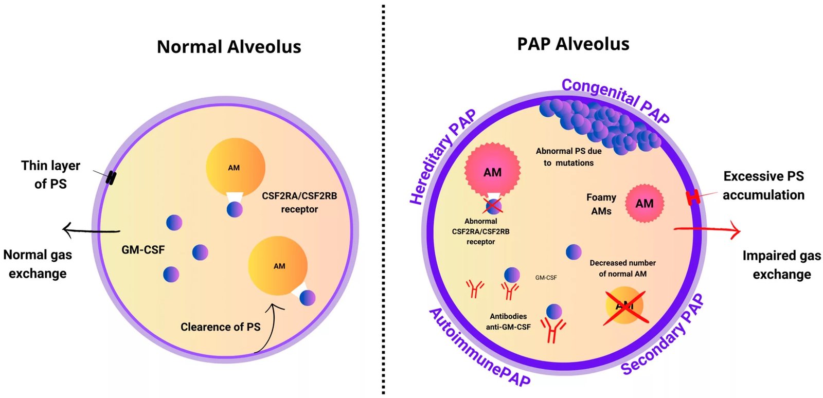
Comparison between a normal alveolus and a pulmonary alveolar proteinosis (PAP) alveolus. In PAP, surfactant accumulates due to defective macrophage function or GM-CSF signaling abnormalities, leading to impaired gas exchange. The diagram also illustrates autoimmune, hereditary, and secondary mechanisms of PAP pathogenesis.. Image Courtesy: A Comprehensive Outlook on Pulmonary Alveolar Proteinosis—A Review by Wołoszczak et al. 2024,doi.org/10.3390/ijms25137092, available via: https://www.mdpi.com/1422-0067/25/13/7092, CC BY 4.0.
Autoimmune PAP
Autoimmune PAP is the most common form and results from autoantibodies against GM-CSF. This antibody neutralizes GM-CSF signaling, which is essential for alveolar macrophage function. Cigarette smoking and infections may trigger or worsen the autoimmune process, although they are not the direct cause.
Secondary PAP
Secondary PAP can result from any disease that reduces the functionally effective population of alveolar macrophages. These disorders include:
- Myelodysplastic syndrome
- Chronic myelogenous leukemia
- Acute myeloid leukemia
- Acute lymphoid leukemia
- Myeloma
- Lymphoma
- Lung cancer
- Glioblastoma
- Mesothelioma
It is also linked with exposure to environmental and occupational dusts or fumes that damage alveolar macrophages, such as:
- Dust
- Talc
- Silica
- Aluminum
- Kaolin
- Indium
- Cellulose
- Titanium
- Cement
- Fiberglass
- Nickel
- Quartz
- Cereal dust
- Expoxy
- Polyvinyl chloride
- Paint
- Copper
- Welding
- Zirconium
- Smoke
Congenital PAP
Congenital PAP is caused by inherited mutations that affect surfactant metabolism or GM-CSF receptor function. These may include mutations in GM-CSF receptor alpha or beta subunits (CSF2RA, CSF2RB), surfactant proteins B and C (SFTPB, SFTPC), ATP-binding cassette 3 (ABCA3), and NKX2-1. It typically presents in newborns or infants and often has a poor prognosis without early intervention.
Signs and Symptoms of Primary Alveolar Proteinosis
Symptoms of PAP include:
- Shortness of breath (dyspnea)
- Chest pain
- Cyanosis
- Cough (Often dry with minimal sputum)
- Hemoptysis (rare)
- Fever
- Fatigue
- Malaise
- Weight loss
- Clubbed fingers
- Recurrent lung infections
Diagnosis of Pulmonary Alveolar Proteinosis
Diagnosis requires a combination of clinical assessment, imaging, laboratory testing, and sometimes bronchoscopy.
Physical Examination and History
Your healthcare provider will listen to your lungs for abnormal sounds and ask about smoking, occupational exposures, infections, or underlying hematologic diseases. Physical findings can be non-specific.
Radiography
X-rays in Pulmonary alveolar proteinosis show “batwing” distribution, while high-resolution CT (HRCT) demonstrates ground-glass opacities with interlobular septal thickening, known as the “crazy-paving” pattern.
Biomarkers
Clinicians can order a serum biomarker test for granulocyte–macrophage colony-stimulating factor GM-CSF autoantibodies. It can be done via the Enzyme-Linked Immunosorbent Assay (ELISA). An anti-GM-CSF antibody level greater than 2.8 μg/mL or above is considered abnormal and indicative of PAP.
Bronchoscopy and Bronchoalveolar Lavage (BAL)
Bronchoscopy with BAL is considered the gold standard for diagnosis. The lavage fluid typically appears milky and opaque, and microscopic examination reveals periodic acid–Schiff (PAS)-positive material, confirming the diagnosis..
Pulmonary Function Test
A pulmonary function test measures how well your lungs are doing their job. It is not specific or necessary for PAP. However, an increase in the alveolar-arterial oxygen gradient and a significant reduction in diffusion capacity are the most common findings that indicate PAP in patients.
Lung Biopsy
Biopsies can be helpful, but are not necessary for diagnosing the PAP. Lung biopsy uses surgery to remove tissues from your lungs for examination. Histology shows alveoli filled with eosinophilic, PAS-positive lipoproteinaceous material.
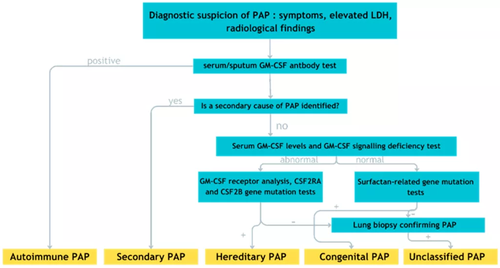
Algorithm for primary alveolar proteinosis diagnosis. The plus (+) sign indicates a positive test result, and the minus (−) sign indicates a negative test result. Image Courtesy: A Comprehensive Outlook on Pulmonary Alveolar Proteinosis—A Review by Wołoszczak et al. 2024,doi.org/10.3390/ijms25137092, available via: https://www.mdpi.com/ijms/ijms-25-07092/article_deploy/html/images/ijms-25-07092-g002.png, CC BY 4.0.
Pulmonary Alveolar Proteinosis Treatment
The most common treatment for PAP is whole-lung lavage (WLL).
Whole Lung Lavage
WLL remains the standard and most effective treatment for symptomatic PAP. Also known as “lung washing,” the procedure is done under general anesthesia using a double-lumen endotracheal tube to isolate each lung. Warm saline is repeatedly instilled and drained until the lavage fluid becomes clear. Each lung is usually washed separately, with procedures spaced weeks apart.
The procedure takes 2–6 hours per lung and provides significant improvement in oxygenation and symptoms. Many patients require repeat lavages over time, depending on disease progression. The following are the steps of WWL:
- After general anaesthesia, the healthcare provider inserts a double-lumen endotracheal tube to isolate one lung. A bronchoscope is sometimes used to confirm proper placement.
- One lung is ventilated with oxygen while the other lung is filled with warm sterile saline solution in repeated cycles.
- The saline is gently instilled, agitated through chest percussion or manual vibrations to help loosen the accumulated surfactant, and then drained out by gravity or gentle suction. The process is repeated several times until the returning fluid becomes clear. The same procedure is later performed on the other lung, usually in a separate session.
GM-CSF Therapy
Recombinant GM-CSF therapy, administered either by inhalation (nebulizer) or subcutaneous injection, helps restore macrophage function. It can improve oxygen levels and reduce the need for repeated WLL. This therapy is particularly beneficial in autoimmune and some congenital forms but is less effective in secondary PAP.
Supportive Treatment Options
Supportive therapies include:
- Bronchodilators: They are medications that relax the muscles around your airways to help you breathe more easily.
- Oxygen Therapy: Supplemental oxygen through a face mask or small tubes can aid in overcoming the breathing difficulties.
- Plasmapheresis: Plasma exchange and plasmapheresis replace your plasma with healthy donor plasma. It prevents your immune system from attacking healthy cells in your alveoli.
- Rituximab therapy: Can be considered in patients unresponsive to WLL or GM-CSF therapy, as it reduces B-cell production of GM-CSF antibodies.
- Lung Transplant: People with severe PAP and lung damage eventually need lung transplant surgery. It replaces one or both of their lungs with healthy lungs from a donor.
Differential Diagnoses of Pulmonary Alveolar Proteinosis
The differential diagnoses of PAP include the disorders that share restrictive physiology and diffuse interstitial changes on CT scan. These conditions include:
- Hypersensitivity pneumonitis
- Cardiogenic pulmonary edema
- Pneumocystis jiroveci pneumonia (PJP)
- Non-small cell lung cancer (NSCLC)
- Small cell lung cancer (SCLC)
- Sarcoidosis
- Respiratory distress syndrome
- Restrictive lung disease
- Interstitial pulmonary fibrosis
Prognosis of PAP
The prognosis is unpredictable as the disease course varies. Autoimmune PAP generally has an excellent outlook, with a five-year survival rate of over 90% when treated with WLL. Secondary PAP has a poorer prognosis, mainly due to underlying diseases such as hematologic malignancies or chronic infections. Congenital PAP often carries the worst prognosis if untreated.
Pulmonary Alveolar Proteinosis Complications
People with PAP are at an increased risk of developing an opportunistic infection. Lungs are the most common site of infection. Mycobacterium Tuberculosis and Nocardia are the most commonly reported opportunistic infections observed in PAP. Fungal infections are:
- Blastomyces
- Cryptococcus
- Aspergillus
- Histoplasma
Other infections are:
- Streptomyces
- Mucorales
- Coccidiodes
- Acinetobacter
Pulmonary fibrosis and chronic respiratory failure may develop in untreated or severe cases.
Final Remarks
PAP is a rare lung disease that clogs the air sacs in your lungs and can cause breathing problems. It presents a range of symptoms. Early diagnosis, timely whole-lung lavage, and appropriate follow-up are crucial for long-term management. Seek immediate treatment if you have this condition. A multidisciplinary team of healthcare providers can collaborate to enhance your quality of life.
References
[1] Rosen, S. H., Castleman, B., Liebow, A. A., Enzinger, F. M., & Hunt, R. T. (1958). Pulmonary alveolar proteinosis.New England Journal of Medicine,258(23), 1123-1142.
[2] Carrington, J. M., & Hershberger, D. M. (2018). Pulmonary alveolar proteinosis.
[3] Borie, R., Danel, C., Debray, M. P., Taille, C., Dombret, M. C., Aubier, M., … & Crestani, B. (2011).Pulmonary alveolar proteinosis. Eur Respir Rev 20: 98–107.
[4] McElvaney, O. J., Horan, D., Franciosi, A. N., Gunaratnam, C., & McElvaney, N. G. (2018). Pulmonary alveolar proteinosis. QJM: An International Journal of Medicine, 111(3), 185-186.
[5] Nishimura, M., Yamaguchi, E., Takahashi, A., Asai, N., Katsuda, E., Ohta, T., … & Baba, K. (2018). Clinical significance of serum anti-GM-CSF autoantibody levels in autoimmune pulmonary alveolar proteinosis.Biomarkers in Medicine,12(2), 151-159.
[6] Kamboj, A., Lause, M., & Duggirala, V. (2018). Severe pulmonary alveolar proteinosis in a young adult. The American Journal of Medicine, 131(5), e199-e200.
[7] Campo, I., Luisetti, M., Griese, M., Trapnell, B. C., Bonella, F., Grutters, J., … & Rodi, G. (2016). Whole lung lavage therapy for pulmonary alveolar proteinosis: a global survey of current practices and procedures. Orphanet journal of rare diseases, 11(1), 115.
[8] Bird, D., Evans, J., & Pahoff, C. (2022). Rituximab rescue therapy for autoimmune pulmonary alveolar proteinosis.Respiratory Medicine Case Reports,37, 101637. https://doi.org/10.1016/j.rmcr.2022.101637
[9] Carrington, J. M., & Hershberger, D. M. (2018). Pulmonary alveolar proteinosis.
[10] Punatar, A. D., Kusne, S., Blair, J. E., Seville, M. T., & Vikram, H. R. (2012). Opportunistic infections in patients with pulmonary alveolar proteinosis. Journal of Infection, 65(2), 173-179.