

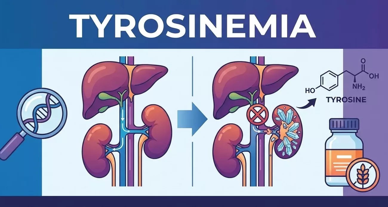
tyrosinemia metabolism disorder Tyrosinemia is a genetic disorder characterized by an error of metabolism in which the body cannot effectively break down the amino acid tyrosine, leading to the buildup of toxic substances in the liver and kidneys. The amino acid tyrosine is derived from the liberation of tyrosine from hydrolysis of dietary or tissue protein or from the hydroxylation of the essential amino acid phenylalanine.
There are three types of tyrosinemia based on their clinical presentation and genetic causes. All three hereditary types are autosomal recessive, whereas transient tyrosinemia is not genetic. If left untreated, the buildup of tyrosine and its byproducts in tissues and organs can lead to severe health complications. Newborns can temporarily have elevated tyrosine levels (transient tyrosinemia). It may be because of a vitamin C deficiency, high protein intake or an undeveloped liver due to preterm births. Tyrosinemia is estimated to occur at a rate of 1/100,000 to 1/120,000 worldwide, with significantly higher prevalence in certain populations, such as Quebec, Canada.
Types of Tyrosinemia: tyrosinemia metabolism disorder
The following are the types of tyrosinemia with their clinical presentations:
Transient Tyrosinemia
This type usually results from delayed enzyme maturation in the tyrosine catabolic pathway. Transient tyrosinemia is a benign condition that spontaneously resolves with no sequelae. Additionally, it is not an inborn error of metabolism because its cause is not a genetic mutation.
Hereditary Infantile Tyrosinemia or Tyrosinemia I
It is the most severe form of this disorder. It usually begins in the first few months of life. Infants with this condition do not gain weight or grow at an expected rate. Eating high-protein foods can lead to vomiting and diarrhea. Affected infants can have:
- Yellowing of the skin and whites of the eyes (jaundice)
- Increased tendency to bleed, particularly nosebleeds
- Cabbage-like odor
Tyrosinemia type I stems from fumarylacetoacetate hydrolase (FAH) deficiency. It causes the buildup of toxic metabolites that damage the liver, kidneys, and nerves. These include fumarylacetoacetate (FAA), maleylacetoacetate (MAA), and succinylacetone (SUA). Succinylacetone is the key diagnostic biomarker detectable in blood and urine.
Type I presents in two forms: acute hepatic failure and chronic progressive disease. Some patients can also experience rickets or porphyria-like neurologic crises that can change their mental state. The affected children can have reduced sensation in the arms and legs, respiratory failure, and abdominal pain. Without treatment, most children do not survive beyond the first decade of life due to liver failure or hepatocellular carcinoma. Early diagnosis and treatment allow many affected individuals to survive into adulthood.
Richner-Hanhart Syndrome or Tyrosinemia II
Tyrosinemia II begins in early childhood. It affects the child’s eyes, skin, and mental development. The clinical presentation of type II differs from that of type I. This presentation includes the:
- Painful keratitis
- Photophobia (abnormal sensitivity to light)
- Palmoplantar hyperkeratosis (thick and painful skin of the palms of the hands and the soles of the feet)
- Herpetiform corneal ulcers (distinctive, dendritic sores on the cornea)
- Variable intellectual disability or developmental delay.
Liver problems do not occur in this type.
Tyrosinemia III
Type III is the rare one. Many patients are asymptomatic; the following symptoms occur only in some cases.
- Seizures
- Intellectual disability
- Intermittent ataxia (a rare genetic neurological disorder that causes temporary episodes of poor balance and coordination).
Like type II, significant liver involvement does not occur in type III tyrosinemia.
Causes of Tyrosinemia
Tyrosinemias occur due to dysfunction of various genes in the tyrosine catabolic pathway and phenylalanine metabolism. It is inherited in an autosomal recessive pattern. Both copies of the gene in each cell must have a variant to cause this condition. Parents of an autosomal recessive condition are carriers of the mutated gene and do not manifest any signs or symptoms of the condition.
- Type I tyrosinemia is caused by a mutation in the FAH gene that encodes the fumarylacetoacetate hydrolase. The mutation leads to FAH deficiency, which results in succinylacetone accumulation (a diagnostic marker) and liver and kidney damage.
- Type II tyrosinemia results from a mutation in the TAT gene that encodes the enzyme tyrosine aminotransferase. The accumulation of tyrosine leads to dermatologic and ophthalmologic manifestations.
- A mutation in the HPD gene causes tyrosinemia type III. The HPD gene codes for 4-hydroxyphenylpyruvate dioxygenase. It is the least common of the three conditions.
Symptoms of Tyrosinemia
The symptoms of tyrosinemia are listed in Table 1 below.
Table 1: Clinical Manifestations of the Different Types of Tyrosinemia
| Types | Symptoms |
|---|---|
| Tyrosinemia Type I | Renal, liver and neurological disorders |
| Tyrosinemia Type II | Developmental delay, Corneal thickening, Hyperkeratosis of soles and palms |
| Tyrosinemia Type III | Asymptomatic to severe mental retardation and neurological anomalies |
Diagnosis of Tyrosinemia
Diagnosis of tyrosinemia is established through a combination of newborn screening, biochemical assays, clinical assessment, and genetic confirmation.
Physical Examination and History
The acute onset of the condition is dramatic with jaundice, hepatomegaly, and distinctive cabbage-like odor, purpuric lesions, melena, and epistaxis. Infants with the chronic form develop polyneuropathy and painful abdominal crises. Survivors can have hepatic nodules and cirrhosis. As the disease is autosomal recessive, the family pedigree typically does not reveal previously affected individuals.
Newborn Screening
Primary detection is performed by tandem mass spectrometry on dried blood spots. Succinylacetone is pathognomonic for tyrosinemia type I, while elevated tyrosine levels are nonspecific and may occur in all types or secondary conditions. Confirmatory steps include repeated SA, methionine, tyrosine levels, urine organic acids, liver function tests, and alpha-fetoprotein (AFP), which is used for hepatocellular carcinoma surveillance rather than diagnosis.
Biochemical Confirmation
Type I shows:
- Blood or urine SA
- Elevated tyrosine and methionine (not diagnostic alone)
- Renal tubular dysfunction (phosphaturia, glucosuria)
- Hepatomegaly or coagulopathy
AFP is extremely high in untreated Type I and is used for monitoring liver cancer risk.
Tyrosinemia Type II features:
- Plasma tyrosine >500-1000 µmol/L
- Urinary metabolites
- No SA
Type III presents:
- Milder tyrosine elevations (>500 µmol/L)
- No SA
- No organ failure
Genetic and Enzymatic Testing
The definitive diagnosis of the condition relies on sequencing, often with reduced enzyme activity in liver biopsy or fibroblasts:
- FAH gene for type I
- TAT for type II
- HPD for Type III
Prenatal testing uses amniocentesis for mutations or SA if a family history exists.
Management and Treatment of Tyrosinemia
Treatment varies by type. Lifelong patient monitoring can help prevent complications such as hepatocellular carcinoma.
Treatment of Type I Tyrosinemia
Doctors recommend nitisinone (NTBC), which inhibits 4-HPD, blocks succinylacetone production, and stabilizes liver and renal function. They recommend a combination of a low-tyrosine or low-phenylalanine diet, plus vitamin D, mineral supplements, and carnitine. Early NTBC therapy markedly reduces the risk of hepatocellular carcinoma. Liver transplantation is reserved for NTBC failure or established malignancy.
Treatment of Type II Tyrosinemia
A strict low-phenylalanine or tyrosine diet prevents skin or ocular lesions and neurodevelopmental issues. Diet alone is usually curative in type II, and ocular lesions resolve within days. NTBC is ineffective in type II due to upstream TAT deficiency. Vitamin supplements address deficiencies in vitamins E, D, and K, as well as in minerals. Supportive care includes topical keratolytic therapy and emollients.
Treatment of Type III Tyrosinemia
Doctors recommend a lifelong restricted diet with formulas or substitutes that suffice for mild symptoms. Phenylalanine supplementation is individualized and not universally required. There is no specific pharmacologic therapy. Doctors regularly monitor growth, neurodevelopment, and plasma tyrosine levels.
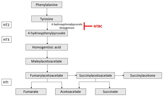
The tyrosine metabolism pathway, including Hereditary Tyrosinemia Type 1, 2, and 3, is associated with different steps within the pathway and the point at which NTBC interference occurs. Image Courtesy: Progress in Gene Therapy for Hereditary Tyrosinemia Type 1 by Thomas et al, 2025,doi.org/10.3390/pharmaceutics17030387, available via: https://www.mdpi.com/1999-4923/17/3/387, CC BY 4.0.
Prognosis of Tyrosinemia
It varies significantly by type, with Type I being the most severe.
Prognosis of Type I
Without treatment, children with tyrosinemia type I rarely survive past age 10. It may be due to liver failure or hepatocellular carcinoma. Early nitisinone therapy combined with dietary management dramatically improves outcomes. These improved outcomes allow many infants to reach adulthood. Complications of Type I may include liver cirrhosis, kidney failure, rickets, acute neurologic crises, and high hepatocellular carcinoma risk.
Prognosis of Type II
Tyrosinemia type II carries a good prognosis. Early dietary restriction of tyrosine and phenylalanine can effectively manage symptoms like corneal ulcers and palmoplantar hyperkeratosis. Intellectual disability affects about half of cases but remains manageable with support, and lifespan is typically normal with no severe complications.
Prognosis of Type III
Tyrosinemia type III often is mild or asymptomatic, with no impact on life expectancy. A tyrosine or phenylalanine-restricted diet is recommended. Mild to moderate intellectual disability or ataxia may persist.
A Quick Review
Tyrosinemia is a rare metabolic disorder caused by defects in tyrosine metabolism, leading to the accumulation of harmful byproducts in the body. The clinical severity varies by type, with tyrosinemia type I being the most serious. After diagnosis, a special diet and, sometimes, medications can help children manage symptoms.
References
[1] Russo, P. A., Mitchell, G. A., & Tanguay, R. M. (2001). Tyrosinemia: a review.Pediatric and Developmental Pathology,4(3), 212-221.
[2] Grompe, M. (2001). The pathophysiology and treatment of hereditary tyrosinemia type 1. InSeminars in liver disease(Vol. 21, No. 04, pp. 563-572). Copyright© 2001 by Thieme Medical Publishers, Inc., 333 Seventh Avenue, New York, NY 10001, USA. Tel.:+ 1 (212) 584-4662.
[3] King, L. S., Trahms, C., & Scott, C. R. (2017). Tyrosinemia type I.GeneReviews®[Internet].
[4] Locatelli, F., Puzenat, E., Arnoux, J. B., Blanc, D., & Aubin, F. (2017). Richner-Hanhart syndrome (tyrosinemia type II).Cutis,100(6), 20-2.
[5] Barroso, F., Correia, J., Bandeira, A., Carmona, C., Vilarinho, L., Almeida, M., … & Martins, E. (2020). Tyrosinemia type III: a case report of siblings and literature review.Revista Paulista de Pediatria,38, e2018158.
[6] Heylen, E., Scherer, G., Vincent, M. F., Marie, S., Fischer, J., & Nassogne, M. C. (2012). Tyrosinemia Type III detected via neonatal screening: management and outcome.Molecular genetics and metabolism,107(3), 605-607.
[7] Rüetschi, U., Cerone, R., Pérez-Cerda, C., Schiaffino, M. C., Standing, S., Ugarte, M., & Holme, E. (2000). Mutations in the 4-hydroxyphenylpyruvate dioxygenase gene (HPD) in patients with tyrosinemia type III.Human genetics,106(6), 654-662.
[8] Basan, H., Ceylaner, S., & Küçükcongar Yavaş, A. (2025). Different Clinic, Different Diagnosis: Tyrosinemia Type 3.Molecular Syndromology.
[9] Ashorn, M., Pitkänen, S., Salo, M. K., & Heikinheimo, M. (2006). Current strategies for the treatment of hereditary tyrosinemia type I.Pediatric Drugs,8(1), 47-54.